Unit 1 · Botany · 8 teaching hours · 42 min read
Plant Anatomy
What the syllabus asks for 6
- Concept of tissues
- Meristems and permanent tissues
- Anatomy of dicot and monocot root
- Anatomy of dicot and monocot stem
- Anatomy of dicot and monocot leaf
- Secondary growth of dicot stem
Anatomy is what you see when you cut a plant open. A stem looks like a green rod from outside; under a microscope it turns out to be a precisely arranged community of cells, some alive and thin-walled, some dead and reinforced, some hollowed out into pipes. This unit teaches you to read a transverse section — to look at a slide and say, in a few seconds, dicot root or monocot stem, and to justify the answer from what is on the slide.
Four things, again and again. (1) Classify a tissue and give its wall structure and function. (2) Identify a transverse section and list the diagnostic differences — not every difference, the ones that decide it. (3) Draw a labelled TS of a dicot or monocot root, stem or leaf. (4) Describe secondary growth of a dicot stem in order: cambial ring, secondary xylem and phloem, annual rings, cork cambium, bark. Nearly every long question in this unit is a comparison or a sequence, so learn it as a table or as a numbered chain, never as loose prose.
1.1 Concept of tissues
A single-celled Chlamydomonas does everything with one cell: it photosynthesises, absorbs, moves and reproduces. A Ficus tree cannot. It is too large for diffusion to supply its interior, too heavy to stand without reinforcement, and too long-lived to leave repair to chance. Its answer is division of labour: groups of cells specialise, and each group does one job well.
A tissue is a group of cells of common origin that are structurally similar and work together to perform a particular function. The study of tissues is histology; the study of the internal structure of plants is plant anatomy. Nehemiah Grew and Marcello Malpighi, working independently in the 1670s, founded the subject; Grew is usually called the father of plant anatomy.
Tissues are classified on two quite different bases, and candidates lose marks by mixing them up.
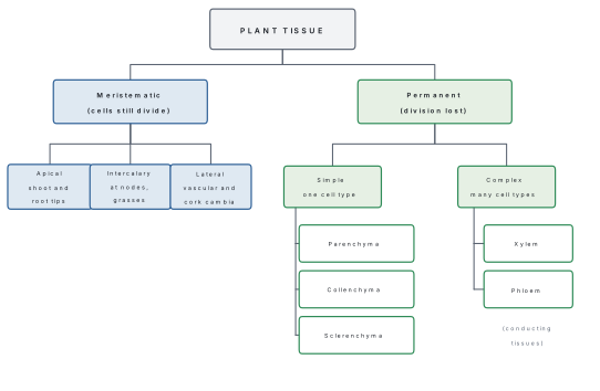
By the dividing power of the cells — this gives meristematic tissue (cells that retain the power to divide) and permanent tissue (cells that have lost it). This is the classification §1.2 develops.
By the number of cell types present — a simple tissue is made of only one kind of cell (parenchyma, collenchyma, sclerenchyma); a complex tissue is made of more than one kind of cell working as a unit (xylem, phloem).
The three tissue systems
Cut any organ across and the tissues fall into three concentric zones. This is Sachs' tissue system classification, and it is the fastest way to organise a labelled diagram.
| Tissue system | Tissues in it | Where | Main job |
|---|---|---|---|
| Epidermal (dermal) | epidermis, cuticle, stomata, trichomes, root hairs | outermost single layer | protection, control of water loss and gas exchange |
| Ground (fundamental) | cortex, endodermis, pericycle, pith, medullary rays, mesophyll | everything between epidermis and vascular tissue | photosynthesis, storage, support |
| Vascular (conducting) | xylem, phloem, cambium | in bundles or a ring | long-distance transport of water and food |
When you are asked to "describe the internal structure" of any organ, walk inwards in that order — epidermis, then ground tissue, then vascular tissue, then centre. You will never leave a layer out, and the answer reads like a section rather than a list.
1.2 Meristems and permanent tissues
Meristematic tissue
A meristem (Greek meristos, divisible) is a tissue of immature cells that retain the power of continuous division. Every permanent tissue in a plant was once meristematic. Because meristems are retained throughout life, a plant can keep growing for as long as it lives — open or indeterminate growth, which animals do not have.
Characteristics of meristematic cells. They are small, isodiametric and tightly packed, with no intercellular spaces. The wall is thin and made of cellulose. The cytoplasm is dense and the nucleus large and conspicuous. Vacuoles are absent or very small. Ergastic substances (starch, crystals) and plastids are absent or present only as proplastids. Metabolic rate is high.
Meristems are classified three ways. Read each classification as answering a different question.
(a) By origin — where the meristem came from.
- Promeristem (primordial meristem): the few cells at the very tip of a root or shoot from which all other meristems arise.
- Primary meristem: derived directly from the promeristem, present from the embryonic stage, and responsible for the primary body of the plant.
- Secondary meristem: arises later from permanent tissue that has redifferentiated and regained the power to divide — vascular cambium in the interfascicular region, cork cambium, wound cambium.
(b) By position — where the meristem sits.
- Apical meristem: at the tips of root and shoot. Causes primary growth, that is, increase in length.
- Intercalary meristem: a piece of apical meristem left behind between permanent tissues, typically at the base of an internode or leaf sheath. It is the reason a grass lawn regrows after mowing and a bamboo culm elongates so fast. Found in grasses, Equisetum, and in mint.
- Lateral meristem: cylinders running the length of the axis — the vascular cambium and the cork cambium (phellogen). Cause secondary growth, that is, increase in girth.
(c) By function — Haberlandt's classification of the apical meristem. The apical meristem is already sorted into three zones, each destined for one tissue system:
| Primary meristem | Gives rise to |
|---|---|
| Protoderm (dermatogen) | epidermis |
| Procambium (plerome, in part) | primary xylem, primary phloem, vascular cambium |
| Ground meristem (periblem + part of plerome) | cortex, endodermis, pericycle, pith, medullary rays |
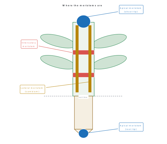
"Apical meristem increases length, lateral meristem increases girth" is true but incomplete as an answer. The examiner wants the consequence: apical meristem produces the primary body (primary growth), lateral meristem produces the secondary body (secondary growth). Monocots have apical and intercalary meristems but, with rare exceptions such as Dracaena and Yucca, no normal vascular cambium — which is exactly why a maize stem never thickens into a trunk.
Permanent tissue
Permanent cells have differentiated: they have taken a fixed shape, wall and function, and have lost the power to divide. Most are dead at maturity or become so. Two useful words:
Differentiation — a meristematic cell matures into a permanent cell. Dedifferentiation — a permanent cell regains the power to divide (this is how interfascicular cambium and cork cambium form). Redifferentiation — the products of a dedifferentiated cell mature again into permanent tissue.
Simple permanent tissues
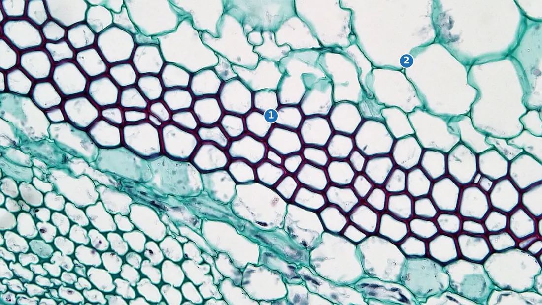
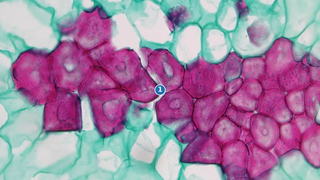
Parenchyma is the least specialised and most abundant tissue — the "packing" of the plant, and the tissue from which all others evolved. Cells are living, thin-walled, isodiametric or oval, with conspicuous intercellular spaces. It fills the cortex, pith, medullary rays and mesophyll. Functions: storage of food and water, and, because the cells stay alive and thin-walled, it is the seat of most metabolism. It retains some power to divide, which is why parenchyma can dedifferentiate into cambium and why a cutting can regenerate. Modifications:
- Chlorenchyma — parenchyma with chloroplasts; photosynthetic. The mesophyll of a leaf is chlorenchyma.
- Aerenchyma — parenchyma with very large air cavities; gives buoyancy and supplies oxygen to submerged parts. Characteristic of hydrophytes such as Hydrilla, Nymphaea and the rice root.
- Prosenchyma — elongated, pointed parenchyma giving some mechanical support.
- Idioblasts — isolated parenchyma cells storing oil, tannin or crystals.
Collenchyma is living mechanical tissue. The cells are elongated with unevenly thickened walls of cellulose, hemicellulose and a large amount of pectin, which is what makes it flexible rather than brittle. Intercellular spaces are usually absent. It occurs just below the epidermis in young dicot stems, in the petiole, and in the ridges of a Cucurbita or Helianthus stem — never in monocots and rarely in roots. Because the wall is not lignified, the cell can still stretch, so collenchyma supports an organ that is still growing. By the thickening pattern:
- Angular collenchyma — thickened at the corners where several cells meet (Datura, Solanum).
- Lamellar (plate) collenchyma — thickened on the tangential walls only (Helianthus stem).
- Lacunar (tubular) collenchyma — thickened around intercellular spaces (Cucurbita, Ambrosia).
Sclerenchyma is dead mechanical tissue. The wall is thick, uniformly lignified and pierced by simple pits; the lumen is narrow and the protoplast disappears at maturity. It gives rigidity, hardness and tensile strength. Two forms:
- Fibres — very long, narrow cells with pointed ends, occurring in bundles. Commercially these are the bast fibres: jute (Corchorus), flax (Linum), hemp (Cannabis), and in Nepal allo or Himalayan nettle (Girardinia diversifolia), whose phloem fibres are woven in Sankhuwasabha and Bajhang.
- Sclereids (stone cells) — short, irregular, extremely thick-walled cells. The grit in a guava or pear fruit, the shell of a walnut, the hard seed coat of a pulse, and the stiff core of a tea leaf are all sclereids. Types include brachysclereids, macrosclereids, osteosclereids, astrosclereids and trichosclereids.
Do not write "sclerenchyma is found only in old organs". It is found wherever finished, non-stretching strength is needed — including the hypodermis of a young monocot stem and the bundle caps of a young dicot stem. The reliable distinction is collenchyma supports a growing organ and is alive; sclerenchyma supports a mature organ and is dead.
Complex permanent tissues
Xylem conducts water and dissolved minerals upwards from root to leaf, and gives mechanical strength. It has four elements:
| Element | Living? | Structure | Function |
|---|---|---|---|
| Tracheids | dead | elongated, tapering, lignified, with bordered pits; end walls not perforated | conduction (in all vascular plants; the only element in gymnosperms) and support |
| Vessels (trachea) | dead | short, wide tubes placed end to end with perforated end walls, forming a continuous pipe | efficient conduction; characteristic of angiosperms |
| Xylem fibres (wood fibres) | dead | sclerenchymatous, thick-walled, narrow lumen | mechanical support |
| Xylem parenchyma | living | thin-walled, may store starch or tannin | storage and radial (lateral) conduction |
Phloem conducts prepared food — mainly sucrose — from source to sink, and unlike xylem the flow can be in either direction. Its four elements:
| Element | Living? | Structure | Function |
|---|---|---|---|
| Sieve tube elements | living but enucleate | cylindrical cells joined end to end by perforated sieve plates; cytoplasm peripheral | translocation of food |
| Companion cells | living | narrow, dense cytoplasm, prominent nucleus, connected to the sieve tube by plasmodesmata | control the sieve tube, whose own nucleus is gone; load sugar |
| Phloem fibres (bast fibres) | dead | sclerenchymatous | support; the only hard element of phloem |
| Phloem parenchyma | living | thin-walled | storage of food, resin and latex |
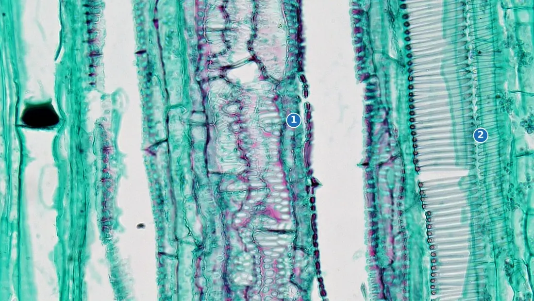
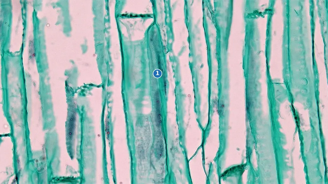
Protoxylem and metaxylem. Xylem formed first, while the organ is still elongating, is protoxylem; its vessels are narrow with annular or spiral lignification so they can be stretched. Xylem formed later is metaxylem, with wider vessels and scalariform, reticulate or pitted thickening. Where the protoxylem lies relative to the centre of the organ is a diagnostic feature:
- Exarch — protoxylem towards the periphery, metaxylem towards the centre. Found in roots.
- Endarch — protoxylem towards the centre, metaxylem towards the periphery. Found in stems.
"Roots are EXtroverts, stems are ENDoverts." Ex-arch in the root, end-arch in the stem. One word in an answer on root anatomy that most candidates forget.
Special (secretory) tissues are sometimes counted as a third category: laticiferous tissue (latex vessels of Euphorbia, Calotropis, Ficus and the rubber plant Hevea) and glandular tissue (nectaries, oil glands of Citrus peel, resin canals of Pinus, digestive glands of Drosera).
1.3 Anatomy of dicot and monocot root
Cut a young root across, a few centimetres behind the tip, and you get the primary structure. Work inwards.
Epiblema (rhizodermis). The outermost layer, a single row of thin-walled cells with no cuticle and no stomata — a root must absorb water, so it cannot afford to waterproof itself. Some cells extend into unicellular root hairs, which is why the layer is also called the piliferous layer.
Cortex. A broad zone of thin-walled parenchyma with conspicuous intercellular spaces, storing starch. There is no chlorenchyma and, normally, no collenchyma.
Endodermis. The innermost layer of the cortex, a single ring of barrel-shaped cells fitted together without spaces. Their radial and tangential walls carry a band of suberin and lignin, the Casparian strip, which is impermeable to water. Water therefore cannot slip past the endodermis through the wall; it is forced through the living protoplast, and the root gains control over what enters the stele. Opposite the protoxylem groups, a few endodermal cells remain thin-walled — these are the passage cells or transfusion cells, and water crosses there.
Pericycle. A single layer (occasionally more) of parenchyma just inside the endodermis. It is the most important layer to name, because lateral roots arise from the pericycle — they are endogenous, arising deep inside the parent root, whereas branches of a stem arise exogenously from the surface. In a dicot the pericycle also gives rise to a part of the vascular cambium and to the cork cambium.
Vascular tissue. Xylem and phloem lie in separate, alternating, radial strips on different radii. This radial arrangement is the single most reliable sign that you are looking at a root. The xylem is exarch.
Xylem and phloem on the same radius with cambium between them means stem. Xylem and phloem on alternate radii means root. Decide that first; then count the xylem groups to decide dicot or monocot.
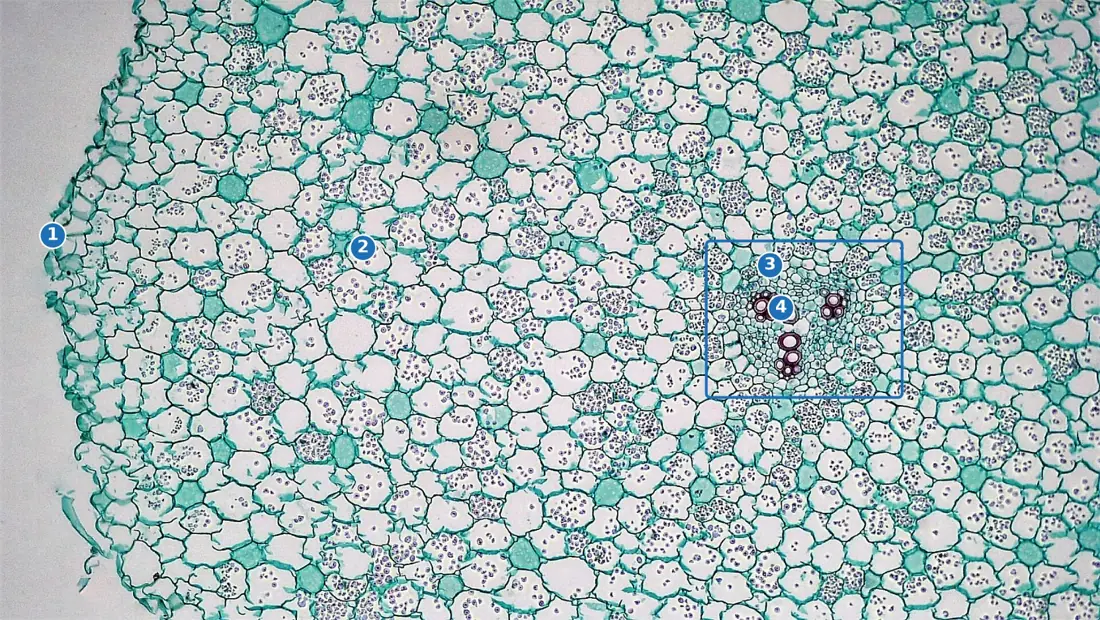
Dicot root. The number of xylem groups is small — usually two to six (diarch, triarch, tetrarch, pentarch, hexarch). Ranunculus is commonly tetrarch. The centre of the stele is occupied by xylem, so the pith is absent or very small. Conjunctive tissue, the parenchyma lying between the xylem and phloem strips, later becomes part of the cambium. Because a dicot root has a cambium in waiting, it is capable of secondary growth.
Monocot root. The same ground plan, but the numbers change. The xylem groups are many — more than six, often twenty or more, so the root is polyarch. The bundles are pushed out into a ring near the periphery, and the centre is filled by a large, well-developed pith of parenchyma. Conjunctive tissue is often sclerenchymatous. There is no cambium, so a monocot root shows no secondary growth.
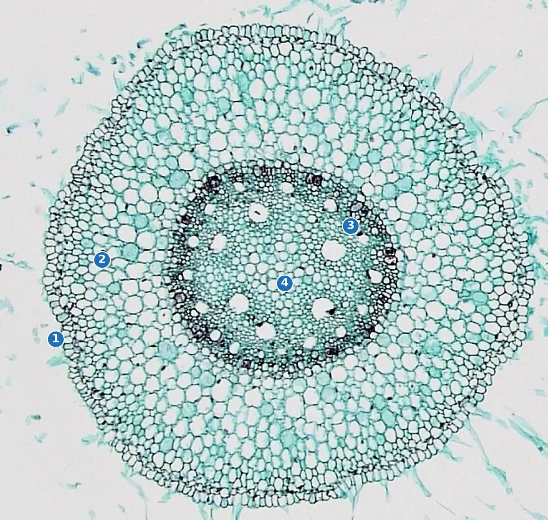
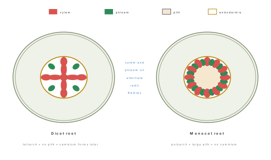
| Feature | Dicot root | Monocot root |
|---|---|---|
| Xylem groups | 2–6 (di- to hexarch) | many, more than 6 (polyarch) |
| Pith | absent or very small | large and well developed |
| Conjunctive tissue | parenchymatous | usually sclerenchymatous |
| Cambium | develops later from conjunctive tissue and pericycle | absent |
| Secondary growth | present | absent |
| Cortex | comparatively narrow | comparatively wide |
| Example | Helianthus, Cicer, Ranunculus | Zea mays, Oryza, Allium |
Everything above the double line in that table is shared: both have epiblema with root hairs, both have endodermis with Casparian strips and passage cells, both have a pericycle, both have radial bundles, and in both the xylem is exarch.
1.4 Anatomy of dicot and monocot stem
A stem is built for support, conduction and often photosynthesis, and its anatomy shows it.
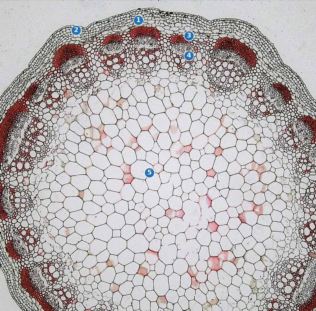
Dicot stem (Helianthus, sunflower)
Epidermis — one layer, with a cuticle, a few stomata and often multicellular trichomes. Unlike the root, the stem is above ground and must limit water loss.
Hypodermis — several layers of collenchyma, often thickest under the ridges of the stem. This is what makes a young sunflower stem ridged and flexible.
General cortex — chlorenchyma outside, ordinary parenchyma inside; resin canals occur in Helianthus.
Endodermis — the innermost cortical layer, loaded with starch grains, so it is called the starch sheath. A Casparian strip is not usually visible.
Pericycle — semilunar patches of sclerenchyma sitting like caps on the outer face of each vascular bundle, alternating with parenchyma. These bundle caps are a strong diagnostic feature.
Vascular bundles — arranged in a single ring, limited in number, all of the same size, and wedge-shaped. Each bundle is conjoint (xylem and phloem in one bundle), collateral (phloem outside, xylem inside, on the same radius), and open (a strip of cambium lies between them). The xylem is endarch.
Pith and medullary rays — a large central parenchymatous pith, with strips of parenchyma running radially between adjacent bundles. Those strips are the medullary (pith) rays, and the part of them lying between the bundles is where the interfascicular cambium will appear.
Monocot stem (Zea mays, maize)
The differences are large enough that the two sections are never confused once you know them.
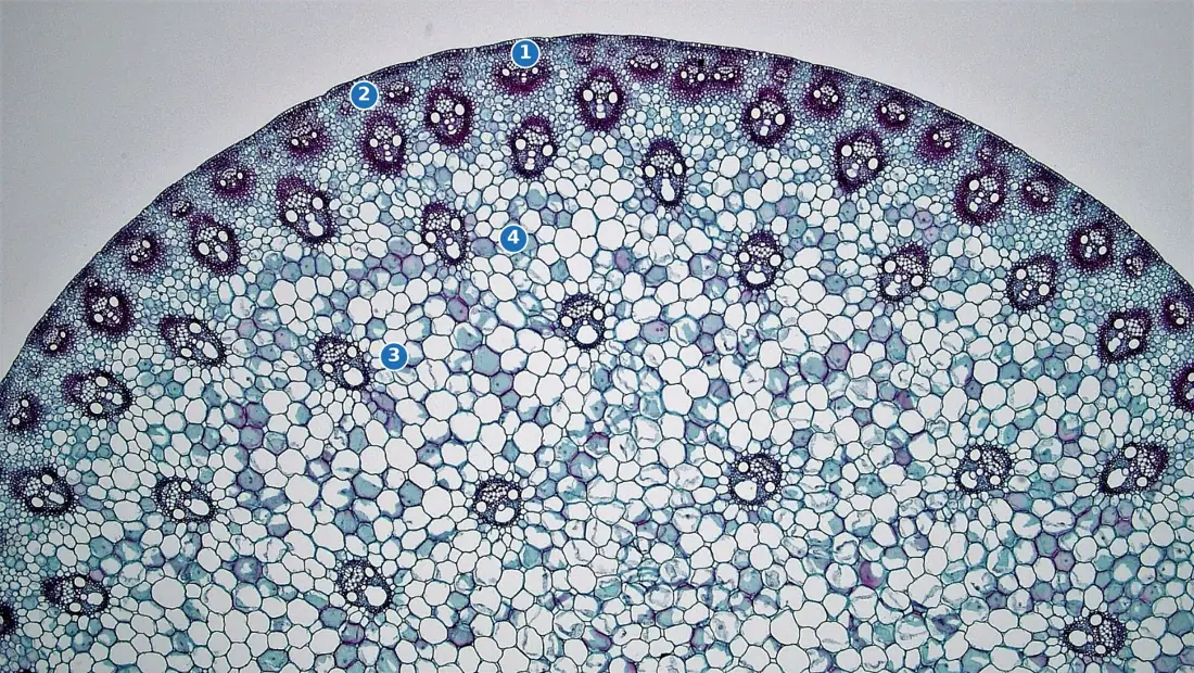
Epidermis — one layer with a thick cuticle; stomata present.
Hypodermis — sclerenchyma, not collenchyma, two to three layers thick. A maize stem is stiff rather than flexible.
Ground tissue — there is no differentiation into cortex, endodermis, pericycle and pith. The whole mass inside the hypodermis is one continuous parenchymatous ground tissue.
Vascular bundles — scattered throughout the ground tissue (an atactostele), numerous, and of unequal size: the bundles near the periphery are small and crowded, those towards the centre are large and widely spaced. Each bundle is conjoint, collateral and closed — there is no cambium, so a monocot stem cannot grow in girth in the ordinary way. The xylem is endarch.
Bundle sheath — each bundle is surrounded by a sheath of sclerenchyma. Inside, the xylem is arranged in a characteristic "Y" or "V" shape: two large pitted metaxylem vessels form the arms, protoxylem lies below them, and the lowest protoxylem vessel disintegrates to leave a water-filled cavity, the lysigenous cavity or protoxylem lacuna. Phloem lies above, consisting of sieve tubes and companion cells only — phloem parenchyma is absent in a monocot stem.
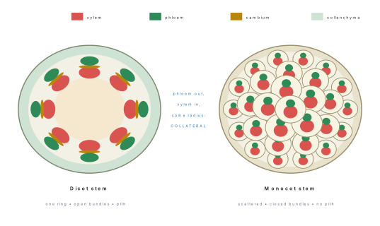
| Feature | Dicot stem | Monocot stem |
|---|---|---|
| Hypodermis | collenchymatous | sclerenchymatous |
| Ground tissue | differentiated into cortex, endodermis, pericycle, pith | undifferentiated, continuous |
| Bundle arrangement | a single ring (eustele) | scattered (atactostele) |
| Bundle number and size | few, all alike | many, unequal — small outside, large inside |
| Bundle type | conjoint, collateral, open | conjoint, collateral, closed |
| Bundle sheath | absent (bundle caps of pericyclic sclerenchyma instead) | present, sclerenchymatous |
| Phloem parenchyma | present | absent |
| Water-containing cavity | absent | present (lysigenous cavity) |
| Medullary rays | present | absent |
| Secondary growth | present | absent |
Types of vascular bundle — the vocabulary
You will be asked to name a bundle, not merely describe it.
- Radial — xylem and phloem on different radii, alternating. Roots.
- Conjoint — xylem and phloem in the same bundle. Stems and leaves.
- Collateral — phloem on the outer side only (most stems).
- Bicollateral — phloem on both the outer and inner sides of the xylem. Family Cucurbitaceae (Cucurbita, Luffa), and Solanum.
- Concentric — one tissue completely surrounds the other.
- Amphivasal (leptocentric) — xylem surrounds phloem. Dracaena, Yucca.
- Amphicribral (hadrocentric) — phloem surrounds xylem. Ferns, and the bundles in some fruits.
- Open — cambium present between xylem and phloem, secondary growth possible.
- Closed — no cambium, no secondary growth.
Amphi-VAS-al = VAScular tissue, i.e. xylem, on the outside. Amphi-CRIB-ral — cribrum is Latin for a sieve, so sieve tubes (phloem) on the outside. Two marks that hinge on one syllable.
1.5 Anatomy of dicot and monocot leaf
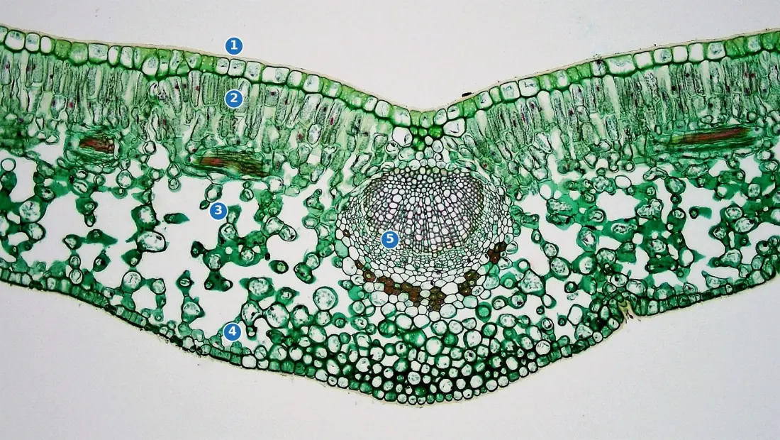
A typical dicot leaf is held horizontally, so its upper surface gets far more light than its lower. Its anatomy is correspondingly two-faced, or dorsiventral. A typical monocot leaf such as grass or maize stands more or less vertically, so both surfaces are lit equally, and its anatomy is alike on both sides — isobilateral.
Dorsiventral (dicot) leaf
Upper epidermis — a single layer with a thick cuticle and very few or no stomata. Lower epidermis — a thinner cuticle and many stomata. A leaf with stomata mainly on the lower surface is hypostomatic, which reduces water loss because the lower surface is cooler and shaded.
Mesophyll — the chlorenchyma between the two epidermes, and here it is differentiated into two zones:
- Palisade parenchyma — one to three layers of elongated, closely packed cylindrical cells just below the upper epidermis, standing on end with their long axes at right angles to the surface, and packed with chloroplasts. Most photosynthesis happens here.
- Spongy parenchyma — loosely arranged, irregular cells below the palisade, with large intercellular spaces continuous with the substomatal cavity. Fewer chloroplasts; this is the tissue that handles gas exchange.
Vascular bundles — the veins. Each is conjoint, collateral and closed, with xylem towards the upper (adaxial) side and phloem towards the lower (abaxial) side — a point worth stating, because it follows directly from the leaf being a flattened extension of the stem. Larger veins have a bundle sheath of parenchyma, and the midrib bundle sits in a ridge of collenchyma and parenchyma. Venation is reticulate.
Isobilateral (monocot) leaf
Epidermis on both surfaces — both cuticularised, both bearing stomata in roughly equal numbers (the leaf is amphistomatic). The guard cells are dumb-bell shaped, not kidney-shaped as in a dicot. In the upper epidermis of grasses, groups of large, thin-walled, highly vacuolated bulliform (motor) cells occur at intervals; when the plant loses water these lose turgor and the leaf rolls inwards, cutting transpiration. You can see this in a maize field on a hot Terai afternoon.
Mesophyll — not differentiated. There is no palisade and no spongy zone; the whole mesophyll is uniform chlorenchyma with small intercellular spaces.
Vascular bundles — arranged in a parallel row, of unequal size (large bundles alternating with small), each surrounded by a sclerenchymatous bundle sheath which often extends as a girder to both epidermes. Venation is parallel.
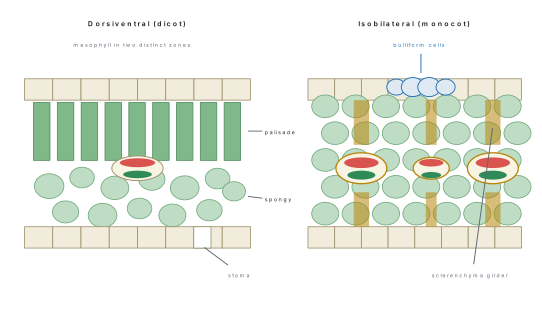
| Feature | Dorsiventral (dicot) leaf | Isobilateral (monocot) leaf |
|---|---|---|
| Orientation | horizontal | more or less vertical |
| Stomata | mostly on the lower surface (hypostomatic) | equal on both surfaces (amphistomatic) |
| Guard cells | kidney (bean) shaped | dumb-bell shaped |
| Mesophyll | differentiated into palisade and spongy | uniform, undifferentiated |
| Bulliform cells | absent | present in the upper epidermis |
| Bundle sheath | parenchymatous | sclerenchymatous, often with girders |
| Venation | reticulate | parallel |
| Example | Mangifera, Helianthus, Ficus | Zea mays, Oryza, Cynodon |
Problem. You are given a stained transverse section. It shows a single-layered epidermis with a thick cuticle on one side only, a hypodermis of cells thickened at their corners, a ring of eight wedge-shaped bundles each with a strip of thin cells between the xylem and phloem, and a wide central parenchyma. Identify the organ and the class, giving your reasons.
Solution. 1. Root or stem? The xylem and phloem lie in the same bundle on the same radius, not alternating. Therefore it is a stem, not a root. 2. Cuticle and corner-thickened hypodermis. The cuticle confirms an aerial organ; corner thickening is angular collenchyma, which occurs in dicot stems and never in monocots. 3. A ring of bundles, all of similar size. Scattered unequal bundles would mean a monocot; one ring means a dicot. 4. A strip of thin cells between xylem and phloem is the cambium, so the bundle is open — again a dicot, and the stem is capable of secondary growth. 5. Wide central parenchyma is the pith, present in a dicot stem, absent in a monocot stem.
Answer. A transverse section of a young dicot stem, showing conjoint, collateral, open vascular bundles in a single ring.
1.6 Secondary growth of dicot stem
A sunflower stem a metre tall can stand on its primary tissues. A Shorea robusta (sal) thirty metres tall cannot. As a woody plant adds height and leaves, the primary body can neither support the load nor carry the water the crown demands, so the plant adds girth — and the tissues that do that are the two lateral meristems: the vascular cambium and the cork cambium.
Secondary growth is the increase in the thickness (girth) of a plant axis brought about by the activity of lateral meristems. It occurs in dicot and gymnosperm stems and roots, and is normally absent in monocots and in leaves.
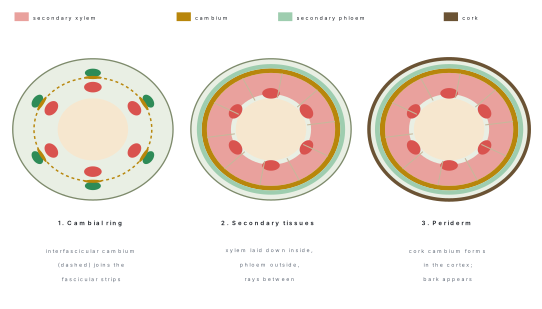
Step 1 — Formation of the complete cambial ring
Look again at the primary dicot stem. Inside each vascular bundle, between the xylem and phloem, there is already a strip of cambium: the fascicular (intrafascicular) cambium. Between the bundles there is only medullary-ray parenchyma.
Now those ray cells lying in line with the fascicular strips dedifferentiate and become meristematic. This newly formed meristem is the interfascicular cambium. The two kinds join end to end, and the result is a continuous cambial ring encircling the pith.
Step 2 — Activity of the cambial ring
The ring is one cell thick, and its cells divide tangentially (by periclinal divisions), cutting off daughter cells on both faces:
- cells cut off towards the inside mature into secondary xylem — the wood;
- cells cut off towards the outside mature into secondary phloem — the bast.
Far more xylem is produced than phloem, which is why a tree trunk is overwhelmingly wood. The consequences follow in a fixed order, and the examiner wants them in that order:
- The ring is pushed steadily outwards as wood accumulates inside it.
- The primary phloem is crushed and obliterated against the cortex; the primary xylem survives, squeezed against the pith at the centre.
- Certain cambial cells stay parenchymatous and produce radial files of living cells running through the wood — the secondary medullary rays — which carry water and food radially and store starch.
- The cortex and epidermis, which cannot stretch indefinitely, eventually rupture; the plant must make a new protective layer, which is Step 3.
Annual rings
In a place with a marked growing season, the cambium does not work at a constant rate.
- In spring, water is plentiful and growth is rapid. The cambium cuts off xylem with wide vessels and thin walls — this is spring wood or early wood, and it looks light in colour and low in density.
- In autumn (and through a dry or cold season), activity slows. The xylem has narrow vessels and thick walls — autumn wood or late wood, which looks dark and dense.
One band of spring wood plus the following band of autumn wood is one annual ring or growth ring, and it represents one year's growth. Counting the rings on a cut stump therefore gives the age of the tree, a technique called dendrochronology; the widths also record good and bad years, so rings are used to reconstruct past climate.
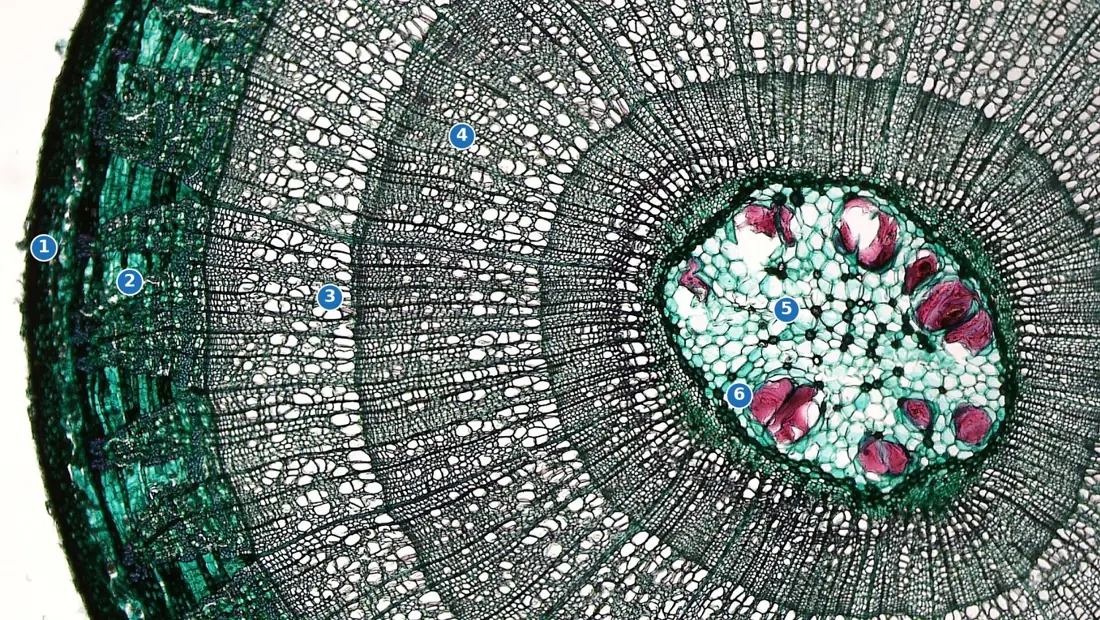
Annual rings are only reliable where the seasons are sharply contrasted. In the hot, wet Terai and in the tropics generally, the cambium may work almost all year and the rings are faint or absent; a drought in mid-season can also produce a false annual ring, and a very bad year can produce none at all. So write "counting annual rings gives the age of a tree in regions with a distinct growing season" — the qualification is worth the mark.
Heartwood and sapwood
As a trunk ages, the older wood at the centre stops conducting. Its parenchyma dies, tannins, resins, gums and oils are deposited in the cells, and neighbouring parenchyma cells balloon into the vessels through the pits as tyloses, plugging them. The result is heartwood (duramen) — dark, hard, durable, resistant to insects and decay, and purely mechanical in function. It is the timber that is commercially valuable: sal, sisau (Dalbergia sissoo) and teak are prized for their heartwood.
The outer, lighter, water-conducting zone is the sapwood (alburnum). It contains living parenchyma and functioning vessels and is the only part of the wood that actually carries the transpiration stream.
Step 3 — The cork cambium and bark
While the vascular cambium works inside, the outer tissues are being torn apart. A second lateral meristem, the cork cambium or phellogen, arises by dedifferentiation of cells in the hypodermis or outer cortex (in the root, from the pericycle). It too divides tangentially:
- outwards it cuts off phellem (cork), whose walls become impregnated with suberin; the cells die, and the layer is impermeable to water and gases;
- inwards it cuts off phelloderm (secondary cortex), a living parenchymatous layer.
Periderm = phellogen + phellem + phelloderm, taken together. Bark = all the tissues lying outside the vascular cambium, that is, secondary phloem + periderm and whatever remains of the cortex and epidermis. A bark formed early in the season and containing living secondary phloem is soft bark; the dead, flaking outer bark of an old trunk is rhytidome. If it comes away as a complete hollow cylinder it is ring bark (Betula, birch); if in irregular flakes, scale bark (Psidium, guava).
Cork would suffocate the stem if it were unbroken, so at certain points the phellogen produces loosely arranged, rounded, thin-walled cells with large intercellular spaces instead of compact cork. These break through the surface as raised lens-shaped pores, the lenticels, and they are the route by which a woody stem exchanges gases. Look at the trunk of a Dalbergia or a mango and you can see them with the naked eye.
Cork's commercial value comes from exactly these properties — light, compressible, impermeable, chemically inert. Bottle cork is the phellem of Quercus suber, the cork oak.
Problem. Arrange the following in the order in which they occur in a dicot stem, and state which meristem is responsible for each: formation of lenticels; crushing of primary phloem; dedifferentiation of medullary ray cells; deposition of tyloses; formation of secondary xylem.
Solution. 1. Dedifferentiation of medullary ray cells — ray parenchyma in line with the fascicular cambium becomes meristematic and forms the interfascicular cambium, completing the cambial ring. Meristem: none yet — this is the event that creates one. 2. Formation of secondary xylem — the completed vascular cambium cuts off xylem on its inner face. This begins immediately and continues for the life of the tree. 3. Crushing of primary phloem — as secondary xylem accumulates, the cambial ring is pushed outwards and the primary phloem, which lies outside it, is compressed and obliterated. Vascular cambium. 4. Formation of lenticels — the cork cambium (phellogen) arises in the cortex and, at intervals, produces loose complementary tissue that ruptures the epidermis. This must come after secondary growth has begun to stretch the cortex. Cork cambium. 5. Deposition of tyloses — only in old wood, when central xylem ceases to conduct and becomes heartwood. This is the last event of the five. No meristem; it is a change in existing xylem parenchyma.
Answer. 3 → 5 → 2 → 1 → 4 in the order the question lists them; that is: dedifferentiation of ray cells, secondary xylem, crushing of primary phloem, lenticels, tyloses.
Problem. A coconut palm is twenty metres tall, yet its stem has no annual rings and its diameter at the top is almost the same as at the base. Explain anatomically, and say how a palm nevertheless achieves its girth.
Solution. 1. A monocot vascular bundle is conjoint, collateral and closed — there is no cambium between the xylem and phloem. 2. There is also no interfascicular region of the normal kind, because the bundles are scattered through undifferentiated ground tissue rather than arranged in a ring; there is no line of ray parenchyma along which an interfascicular cambium could form. 3. With no vascular cambium, no secondary xylem can be produced, so there is no wood and therefore no spring-wood/autumn-wood alternation and no annual rings. 4. The palm instead achieves its girth by primary thickening: a zone of meristem just below the shoot apex enlarges the stem to its final diameter before it elongates. This is why the trunk is a near-uniform cylinder — each segment was made at full width and then simply pushed up. 5. A few monocots (Dracaena, Yucca, Agave) do show anomalous secondary growth from a cambium that arises outside the bundles in the ground tissue and produces whole new amphivasal bundles, not ordinary wood.
Answer. No vascular cambium, hence no secondary xylem and no annual rings; girth comes from a primary thickening meristem, and the exceptions (Dracaena, Yucca) use an anomalous cambium.
Secondary growth in the dicot root
The principle is the same but the ring has to be assembled differently, because in a root the xylem and phloem are on alternating radii.
- Conjunctive parenchyma lying just inside each phloem strip becomes meristematic. These arcs are the first pieces of cambium.
- Cells of the pericycle lying outside each protoxylem group also dedifferentiate and join the arcs.
- Because the arcs curve inside the phloem but bulge outwards at the protoxylem, the completed ring is at first wavy in outline.
- The portions inside the phloem work faster, pushing outwards, and the ring soon becomes circular.
- Thereafter it behaves exactly as in the stem: secondary xylem inside, secondary phloem outside, and a cork cambium — here arising from the pericycle — forms the periderm. The epiblema and cortex are shed.
Chapter summary
- A tissue is a group of cells of common origin doing one job. Tissues are meristematic (still dividing) or permanent (differentiated), and simple (one cell type) or complex (several). The three tissue systems are epidermal, ground and vascular.
- Meristems are classified by origin (promeristem, primary, secondary), by position (apical → length, intercalary → grasses regrow, lateral → girth) and by function (protoderm → epidermis, procambium → vascular tissue, ground meristem → cortex and pith).
- Simple permanent tissues: parenchyma (living, thin-walled, storage; variants chlorenchyma, aerenchyma), collenchyma (living, pectin-rich, unevenly thickened, flexible support in growing dicot organs — angular, lamellar, lacunar), sclerenchyma (dead, lignified, rigid — fibres and sclereids).
- Complex tissues: xylem = tracheids + vessels + fibres + parenchyma (upward, one way); phloem = sieve tubes + companion cells + fibres + parenchyma (both ways). Protoxylem is exarch in roots, endarch in stems.
- Root: epiblema without cuticle, wide cortex, endodermis with Casparian strips and passage cells, pericycle giving endogenous lateral roots, radial bundles, exarch xylem. Dicot 2–6 xylem groups with little or no pith and later a cambium; monocot polyarch with a large pith and no cambium.
- Stem: cuticle and stomata, collenchymatous hypodermis (dicot) or sclerenchymatous (monocot), conjoint collateral bundles, endarch xylem. Dicot — one ring, open, pith and medullary rays present. Monocot — scattered, closed, sclerenchymatous bundle sheath, lysigenous cavity, no phloem parenchyma, no medullary rays.
- Leaf: dorsiventral (dicot) has palisade + spongy mesophyll, stomata mainly below, kidney-shaped guard cells, reticulate venation; isobilateral (monocot) has uniform mesophyll, stomata on both faces, dumb-bell guard cells, bulliform cells, sclerenchymatous sheaths and parallel venation.
- Secondary growth of a dicot stem: interfascicular cambium forms from medullary-ray cells and joins the fascicular cambium into a cambial ring; it cuts secondary xylem inwards and secondary phloem outwards, crushing the primary phloem. Spring wood + autumn wood = one annual ring. Old central wood becomes heartwood (tyloses, tannins); the conducting outer zone is sapwood.
- The cork cambium (phellogen) makes phellem (cork, suberised) outwards and phelloderm inwards; the three together are the periderm. Bark is everything outside the vascular cambium, and lenticels are its breathing pores.
Practice questions
Group A — Multiple choice (1 mark each)
- Xylem and phloem lying on alternate radii is characteristic of [1]aa dicot stemba monocot stemca rootda dorsiventral leaf
- The Casparian strip is found in the [1]aepiblemabpericyclecendodermisdconjunctive tissue
- Bulliform cells occur in the [1]aupper epidermis of a grass leafblower epidermis of a dicot leafcmesophyll of a maize leafdhypodermis of a dicot stem
- Interfascicular cambium is formed by the dedifferentiation of [1]apericycle cellsbmedullary ray cellscendodermal cellsdphloem parenchyma
- A vascular bundle in which the phloem completely surrounds the xylem is called [1]aamphivasalbamphicribralcbicollateraldradial
- Cork is impermeable to water because its walls are impregnated with [1]aligninbcutincsuberindpectin
Answers to Group A
1. (c) — radial bundles are the defining feature of a root; stems and leaves have conjoint bundles.
2. (c) — the strip of suberin and lignin lies on the radial and tangential walls of the endodermis.
3. (a) — large thin-walled motor cells in the upper epidermis of grasses; they lose turgor and roll the leaf.
4. (b) — ray parenchyma lying in line with the fascicular strips becomes meristematic.
5. (b) — cribrum = sieve, so amphicribral means phloem outside. Amphivasal is the reverse.
6. (c) — suberin. Lignin stiffens sclerenchyma, cutin forms the cuticle, pectin softens collenchyma.
Group B — Short answer (4 marks each)
- Distinguish between collenchyma and sclerenchyma under four headings. [4]
- Describe the structure and function of the endodermis and pericycle of a dicot root. [4]
- What is an annual ring? Explain how spring wood and autumn wood differ, and state one limitation of using annual rings to age a tree in Nepal. [4]
- Give four anatomical differences between a dorsiventral and an isobilateral leaf. [4]
- Define bark. Distinguish between periderm and bark, and explain the function of lenticels. [4]
- Name the four elements of xylem and state one function of each. [4]
Answers to Group B
1. Table of four rows. Cell state — collenchyma living, sclerenchyma dead. Wall material — cellulose, hemicellulose and pectin (collenchyma) versus lignin (sclerenchyma). Thickening — uneven, at corners or on particular walls, versus uniform and heavy with a narrow lumen. Function and location — flexible support in a still-growing dicot organ (hypodermis, petiole) versus rigid support in a mature organ (fibres, sclereids, bundle caps).
3. Definition, then the contrast: spring wood has wide, thin-walled vessels and is light-coloured and less dense; autumn wood has narrow, thick-walled vessels and is dark and dense. One of each = one year. Limitation: in the Terai and the tropics the cambium may work almost continuously, so the rings are faint or absent, and a mid-season drought can lay down a false ring — so ring counts are reliable only where the growing season is sharply defined.
6. Tracheids — dead, tapering, bordered pits; conduction and support, and the only conducting element in gymnosperms. Vessels — dead, wide tubes with perforated end walls; efficient conduction, characteristic of angiosperms. Xylem fibres — dead, sclerenchymatous; mechanical support. Xylem parenchyma — living; storage of starch and tannin, and radial conduction.
Group C — Long answer (8 marks each)
- (a) Draw a labelled transverse section of a young dicot stem. [4](b) Compare the dicot stem with a monocot stem under the headings hypodermis, ground tissue, arrangement of bundles, bundle type and capacity for secondary growth. [4]
- (a) Describe, in order, the formation of the cambial ring and the production of secondary tissues in a dicot stem. [5](b) Explain how the cork cambium arises and what it produces, and define periderm and bark. [3]
- (a) Draw a labelled transverse section of a monocot root and state four features that distinguish it from a dicot root. [5](b) Lateral roots are said to be endogenous while stem branches are exogenous. Explain what this means and why it matters. [3]
Answer outline to Group C question 2
(a) Five stages, in this order and no other. (i) The fascicular (intrafascicular) cambium already lies between the xylem and phloem of each bundle. (ii) Medullary-ray parenchyma in line with those strips dedifferentiates to form the interfascicular cambium. (iii) The two join into a continuous cambial ring, one cell thick. (iv) The ring divides tangentially, cutting off secondary xylem on its inner face and secondary phloem on its outer face, with far more xylem than phloem. Some initials remain parenchymatous and produce secondary medullary rays. (v) The ring is pushed outwards; the primary phloem is crushed while the primary xylem persists against the pith. With time the central wood becomes heartwood and the outer wood remains conducting sapwood, and the spring-wood/autumn-wood alternation shows as annual rings.
(b) The cork cambium (phellogen) arises by dedifferentiation of hypodermal or outer cortical cells (in a root, from the pericycle), because the epidermis and cortex cannot stretch to match the expanding stele and are rupturing. It divides tangentially, cutting off phellem (cork) outwards — suberised, dead, waterproof — and phelloderm (secondary cortex) inwards, living parenchyma. Periderm = phellogen + phellem + phelloderm. Bark = everything outside the vascular cambium, that is, the secondary phloem together with the periderm and the crushed remains of cortex and epidermis. Gas exchange continues through lenticels.
Practise more: Biology question bank