Unit 6 · Zoology · 8 teaching hours · 28 min read
Animal Tissues
What the syllabus asks for 4
- Epithelial tissue
- Connective tissue
- Muscular tissue
- Nervous tissue
A tissue is a group of similar cells, together with their intercellular material, that share a common origin and do one job. The multicellular animal body is built from only four of them — epithelial, connective, muscular and nervous. Every organ you meet in the Human Biology unit, from the stomach wall to the eyeball, is these four tissues stacked in different proportions. Learn the four properly now and the rest of Zoology becomes description rather than memorisation.
Three things, every year. A labelled diagram of a named tissue as it looks under the microscope — the neuron, the T.S. of bone, the three muscle types. A difference table — tendon vs ligament, cartilage vs bone, striated vs cardiac vs smooth muscle. And a "where is it found and why" question, which wants structure linked to function, not a list. Practise drawing; a correct diagram with six labels is often the whole four marks.
6.1 Epithelial tissue
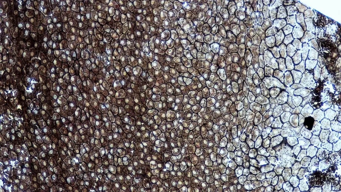
Epithelium is a sheet of closely packed cells, with almost no intercellular matrix, resting on a non-cellular basement membrane, which covers the free surfaces of the body and lines its cavities, ducts and tubes.
Epithelium is the only tissue that arises from all three germ layers: the epidermis of the skin and the lining of the mouth and anus are ectodermal, the lining of the gut and its glands endodermal, and the lining of blood vessels, kidney tubules and coelom mesodermal.
Its general characters follow from its job of forming a boundary:
- Cells are packed tightly and joined by cell junctions (tight junctions, desmosomes), so the sheet is continuous and leak-proof.
- Intercellular matrix is negligible.
- The sheet has polarity — a free (apical) surface and a basal surface cemented to the basement membrane, a thin glycoprotein layer secreted partly by the epithelium and partly by the connective tissue below.
- It is avascular. No blood vessel enters an epithelium; food and oxygen diffuse in from the capillaries of the connective tissue beneath.
- It has a very high power of regeneration, because it is the layer that gets worn away.
Epithelia are classified first by the number of cell layers, then by the shape of the cells at the free surface.
Compound (stratified) epithelium
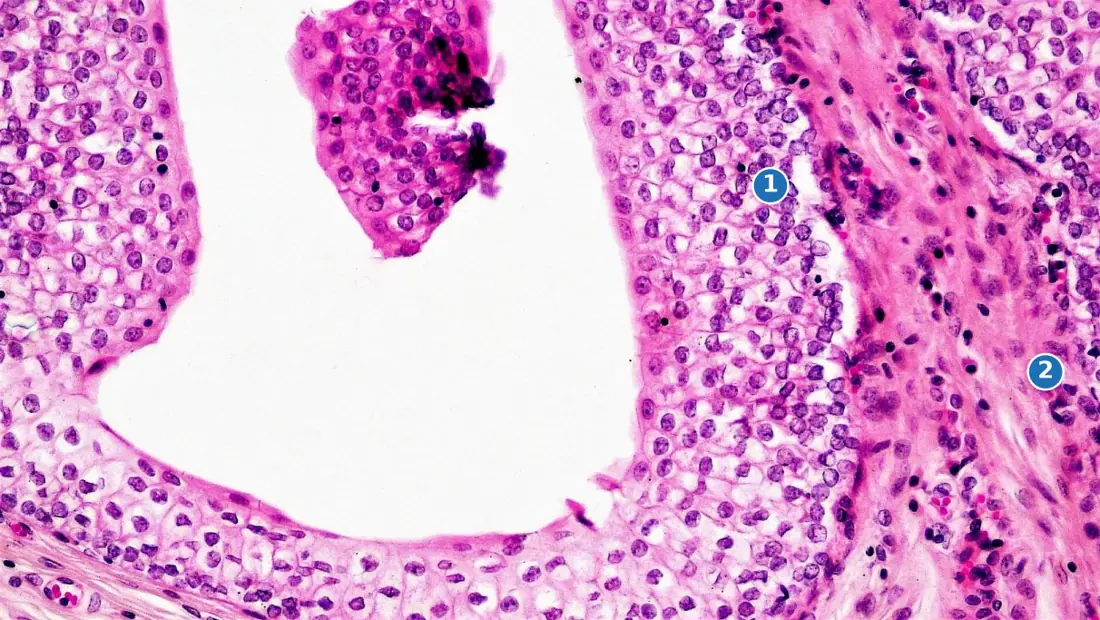
When the epithelium must resist wear, it becomes many cells thick. Only the deepest layer — the germinative or Malpighian layer — touches the basement membrane and divides; its daughter cells are pushed upwards, flatten, die and are shed. In the epidermis of the skin the dying cells fill with the protein keratin, giving a dry, waterproof, keratinised stratified squamous epithelium. In the mouth, oesophagus and vagina the surface stays moist and the epithelium is non-keratinised.
Transitional epithelium (urothelium) lines the ureter and urinary bladder. It has no true stratum corneum, and its cells slide over one another so the sheet can stretch: relaxed it looks 4–5 cells thick with dome-shaped surface cells, distended it looks 2–3 cells thick and flat.
Glandular epithelium
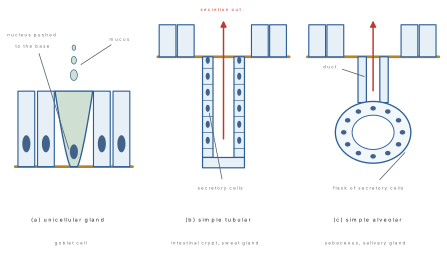
When epithelial cells specialise for secretion they sink into the connective tissue below and form a gland. A single cell doing this is a unicellular gland — the goblet cell of the gut and trachea. Many cells doing it together make a multicellular gland, tubular if the secretory part is a tube (intestinal crypts, sweat glands) or alveolar/acinar if it is a flask (salivary, sebaceous).
Glands are grouped by where the secretion goes — exocrine glands release through a duct, endocrine glands have no duct and pour hormones into the blood — and by how the cell releases it.
| Mode | How the cell secretes | Example |
|---|---|---|
| Merocrine | by exocytosis; the cell stays intact | salivary gland, pancreas |
| Apocrine | apical part of the cell pinches off with the secretion | mammary gland |
| Holocrine | the whole cell disintegrates and becomes the secretion | sebaceous gland |
| Type | Shape at free surface | Chief location | Main function |
|---|---|---|---|
| Simple squamous | flat, tile-like, central bulge | alveoli, Bowman's capsule, endothelium of vessels | diffusion, filtration |
| Simple cuboidal | cube, round central nucleus | kidney tubules, thyroid follicles, gland ducts | absorption, secretion |
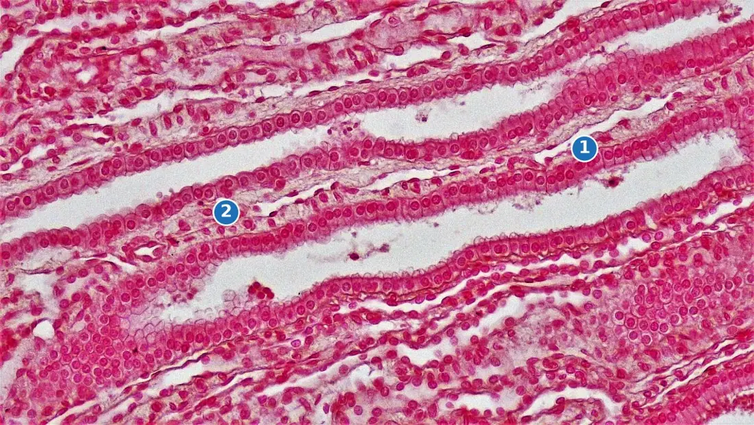
| Simple columnar | tall, nucleus near the base | stomach, small intestine, gall bladder | secretion, absorption |
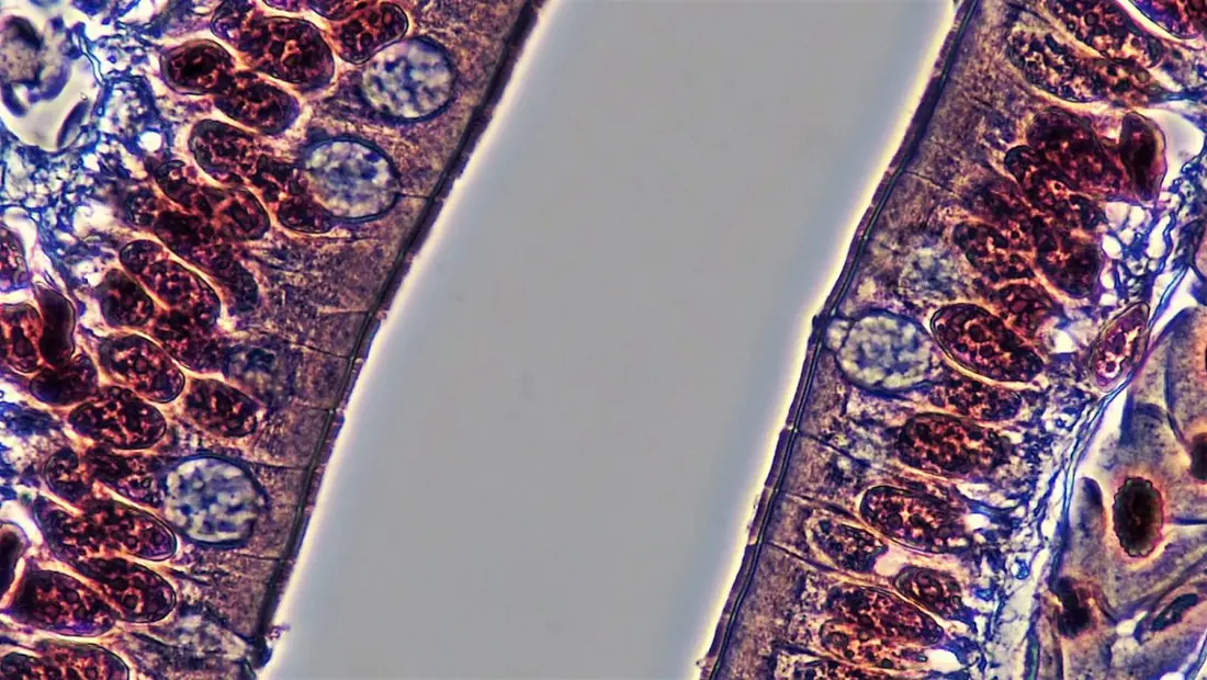
| Ciliated columnar | tall, with cilia | oviduct, bronchioles, ventricles of brain | moves mucus, ovum, CSF |
| Pseudostratified | tall and short cells, nuclei at two levels | trachea, nasal cavity, male urethra | protection, mucus movement |
| Stratified squamous | many layers, flat and dead at the top | epidermis, oesophagus, vagina, cornea | protection against wear |
| Transitional | 2–5 layers, cells slide | ureter, urinary bladder | allows stretching |
| Glandular | secretory, sunk into the tissue | gut lining, all glands | secretion |
A pseudostratified epithelium is one cell layer thick. Every cell reaches the basement membrane, but the cells are of different heights, so their nuclei sit at different levels and the sheet only looks layered. A stratified epithelium has real layers and only the deepest touches the basement membrane.
Problem. In a photomicrograph of the lining of the small intestine taken at a magnification of \times 1000, one columnar cell measures 40\ \text{mm} from base to free surface and 8\ \text{mm} across. (a) Find its real height and width in micrometres. (b) How many such cells lie side by side along 1\ \text{mm} of the intestinal lining?
Solution. Magnification is defined as
(a) Height:
Width: 8/1000 = 0.008\ \text{mm} = 8\ \mu\text{m}. (Recall 1\ \text{mm} = 1000\ \mu\text{m}.)
(b) Each cell is 8\ \mu\text{m} wide and 1\ \text{mm} = 1000\ \mu\text{m}, so
A cell 40\ \mu\text{m} tall and only 8\ \mu\text{m} wide is five times taller than it is broad — that is exactly what "columnar" means.
6.2 Connective tissue
Connective tissue is a mesodermal tissue in which relatively few cells lie scattered in an abundant intercellular matrix of fibres and ground substance. It binds, supports, packs, transports and defends.
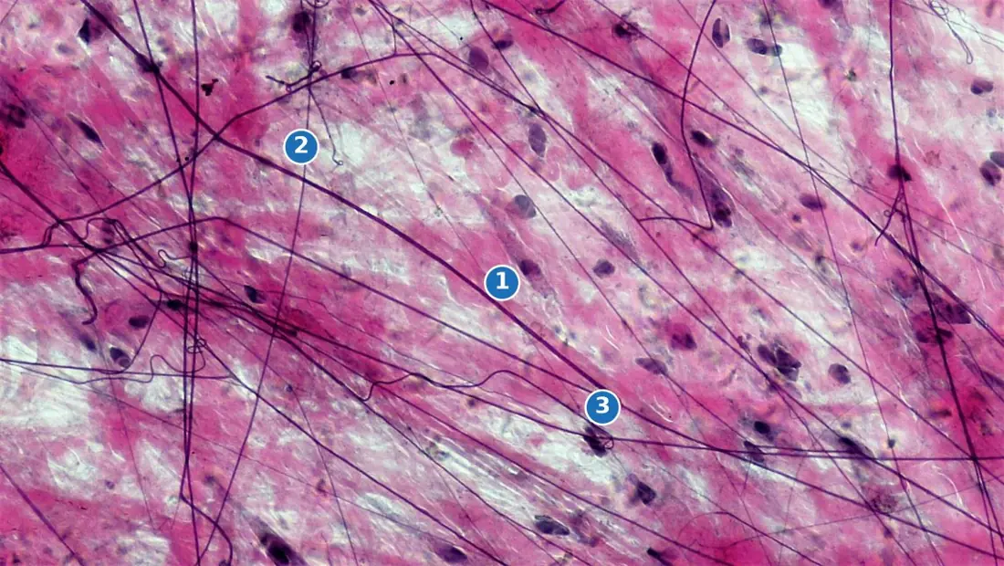
Connective tissue is the mirror image of epithelium: there the cells crowd together and the matrix is negligible, here the matrix does the work and the cells are far apart. Its properties are the properties of its matrix. Three kinds of fibre are woven into it:
- White collagen fibres — thick, wavy, non-branching bundles of the protein collagen. Very strong, inelastic. Boiling turns them into gelatin.
- Yellow elastic fibres — thinner, straight, branching fibres of elastin. They stretch to about 1.5 times their length and snap back.
- Reticular fibres — fine branching collagen, forming the mesh of lymph nodes, spleen and bone marrow.
The commonest cells are the fibroblast (spindle-shaped; makes the fibres and ground substance), the macrophage or histiocyte (amoeboid; engulfs bacteria and debris), the mast cell (granular; secretes heparin, which stops blood clotting inside vessels, and histamine, which causes inflammation and allergy), the plasma cell (secretes antibodies) and the adipocyte (stores fat).
| Group | Types | Distinguishing feature | Chief function |
|---|---|---|---|
| Loose connective | areolar, adipose, reticular | soft, cells many, matrix loose | packing, fat store, framework |
| Dense connective | tendon, ligament (regular); dermis (irregular) | fibres crowded and ordered | transmits or resists pull |
| Skeletal (supporting) | cartilage, bone | matrix solid | support, protection, movement |
| Fluid (vascular) | blood, lymph | matrix is liquid plasma | transport, defence |
Loose connective tissue
Areolar tissue is the universal packing material — under the skin, between muscles, around nerves, blood vessels and organs. Its loose, jelly-like matrix lets tissue fluid and white blood cells move freely, which is why it is also the main site of inflammation.
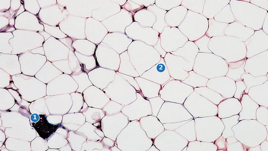
Adipose tissue is areolar tissue in which fat-storing cells have multiplied. Each adipocyte is almost filled by a single fat droplet, so the cytoplasm is a thin rim and the nucleus is squashed against one side — the "signet-ring" appearance. It stores energy, insulates against cold and cushions the kidneys, eyeballs and soles of the feet.
Dense connective tissue
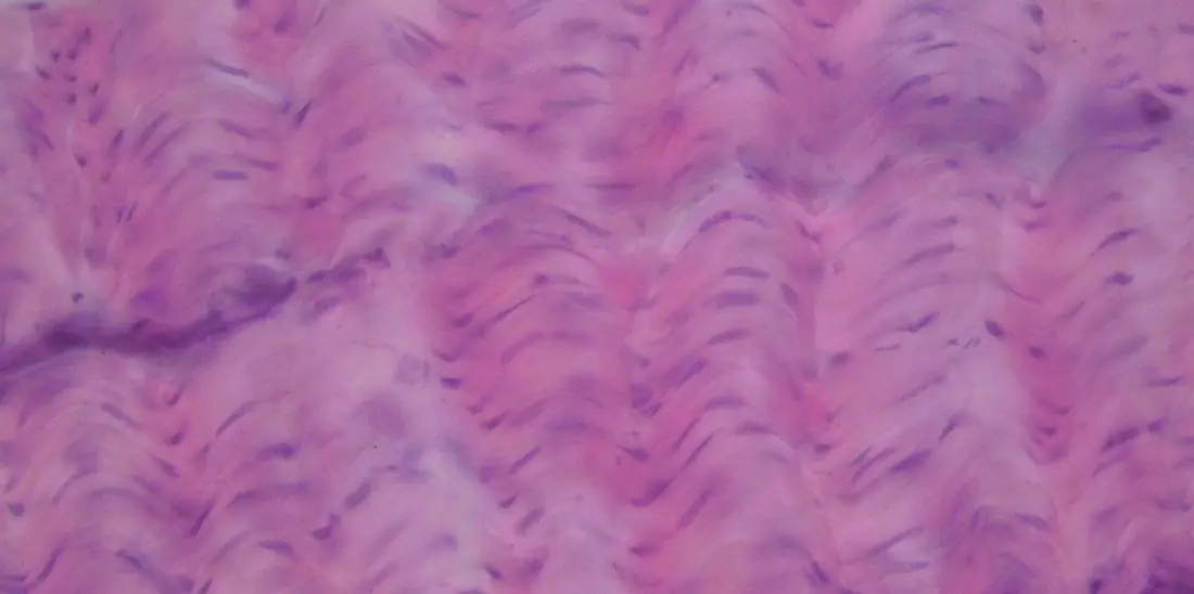
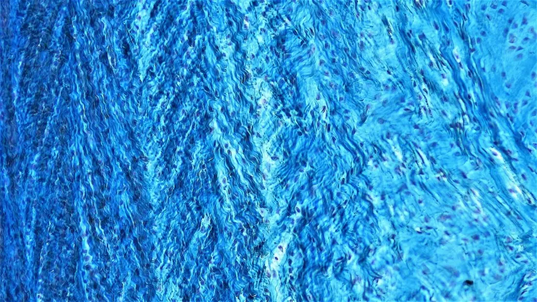
In dense tissue the fibres are packed so tightly that the cells are squeezed into rows between them. When the fibres all run one way the tissue is regular and resists pull along a single line — the tendon and the ligament. When they run in all directions it is irregular and resists pull from any direction — the dermis of the skin and the capsules of organs.
| Feature | Tendon | Ligament |
|---|---|---|
| Chief fibre | white collagen | yellow elastic |
| Arrangement | straight, parallel bundles | branching, interlacing |
| Elasticity | almost none — inextensible | elastic, stretches and recoils |
| Colour when fresh | glistening white | yellowish |
| Cells | fibroblasts in rows between bundles | fibroblasts scattered |
| Connects | muscle to bone | bone to bone at a joint |
| Blood supply | poor, so it heals slowly | poor |
Cartilage
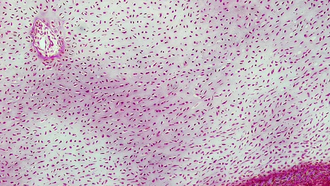
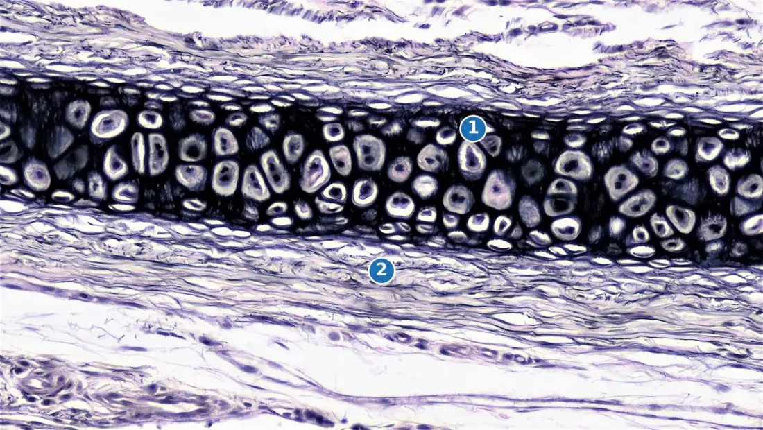
Cartilage is a solid but flexible skeletal tissue. Its cells, the chondrocytes, sit singly or in twos inside fluid-filled spaces called lacunae, embedded in a firm matrix of the protein chondrin. The whole mass is wrapped in a fibrous sheath, the perichondrium, from which it grows and through which it is fed — because cartilage itself has no blood vessels.
| Type | Matrix contains | Where found |
|---|---|---|
| Hyaline | chondrin only; clear, bluish, glassy | tracheal rings, nose tip, articular surfaces, embryonic skeleton |
| Elastic | a network of yellow elastic fibres | pinna of ear, epiglottis, eustachian tube |
| Fibrous (white) | dense bundles of collagen | intervertebral discs, pubic symphysis |
| Calcified | calcium salts deposited in the matrix | head of the humerus, suprascapula of frog |
Bone
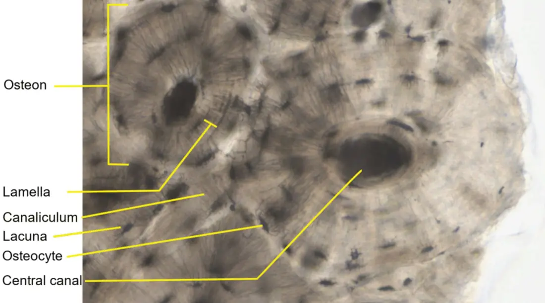
Bone is the hardest connective tissue. Its matrix, ossein, is about one-third organic protein — which gives toughness — and two-thirds inorganic salts, mainly calcium phosphate with some calcium carbonate, which give hardness. Bone cells, the osteocytes, lie in lacunae and keep in touch with one another and with the blood supply through hair-fine channels, the canaliculi.
In the compact bone of a mammal, this material is organised into cylinders. Each cylinder, an osteon or Haversian system, is a central Haversian canal carrying a blood capillary, a lymph vessel and a nerve, surrounded by 4–20 concentric lamellae of matrix with rings of lacunae between them. Haversian canals run lengthwise along the bone and are linked sideways by Volkmann's canals. The whole bone is sheathed in the periosteum.
| Feature | Cartilage | Bone |
|---|---|---|
| Matrix | chondrin (protein), soft and flexible | ossein + calcium salts, hard and rigid |
| Cells | chondrocytes, 1–4 per lacuna | osteocytes, 1 per lacuna |
| Canaliculi | absent | present, linking lacunae |
| Blood supply | absent (avascular) | rich, through Haversian canals |
| Covering | perichondrium | periosteum |
| Growth | interstitial and appositional | appositional only |
Because cartilage has no blood vessels, its cells are fed only by diffusion through the matrix from the perichondrium. That is why a torn knee cartilage heals far more slowly than a broken bone. Second point, often asked: well-formed Haversian systems are a feature of mammalian compact bone. The bone of the frog and other lower vertebrates is nourished by diffusion and lacks Haversian canals.
Fluid connective tissue
Blood is a connective tissue whose matrix is liquid. About 55% of it by volume is plasma — 90–92% water, with plasma proteins (albumin, globulin, fibrinogen), glucose, salts, hormones and wastes — and about 45% is the formed elements.
| Formed element | Normal count (per mm³) | Structure | Function |
|---|---|---|---|
| Erythrocyte (RBC) | 4.5–5.5 million | biconcave disc, 7–8 μm, no nucleus in mammals | carries O₂ and CO₂ as oxyhaemoglobin and carbaminohaemoglobin |
| Neutrophil | 4000–11000 WBC in total; 60–65% of them | 3–5 lobed nucleus, fine granules | phagocytosis of bacteria |
| Eosinophil | 2–3% of WBC | bilobed nucleus, coarse granules | allergy and parasite defence |
| Basophil | 0.5–1% of WBC | lobed nucleus, large dark granules | secretes heparin and histamine |
| Lymphocyte | 20–25% of WBC | large round nucleus, thin cytoplasm | antibodies and cell-mediated immunity |
| Monocyte | 6–8% of WBC | kidney-shaped nucleus, largest WBC | becomes a macrophage in tissues |
| Thrombocyte (platelet) | 150,000–400,000 | cell fragment, no nucleus | blood clotting |
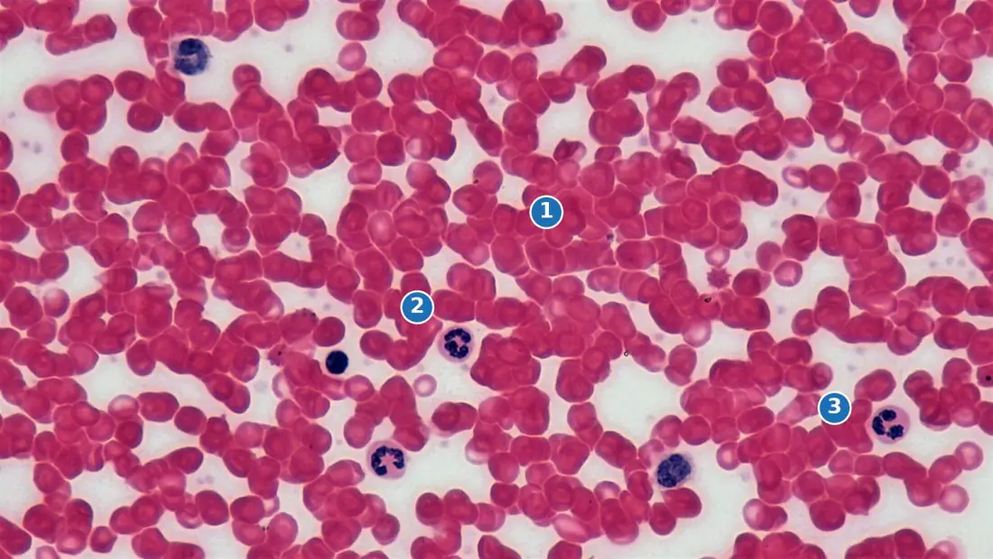
Problem. Blood is diluted 1 : 200 and loaded into a Neubauer counting chamber. The central ruled square is 1\ \text{mm} \times 1\ \text{mm}, divided into 400 small squares, and the depth under the cover-slip is 0.1\ \text{mm}. A student counts 500 red cells in 80 small squares. Find the RBC count per mm³ and say whether the person is anaemic.
Solution. Area of one small square:
Volume of blood over 80 small squares:
Cells per mm³ of the diluted blood:
Multiplying by the dilution factor 200 to get back to whole blood:
Five million per mm³ lies in the normal range of 4.5–5.5 million, so the person is not anaemic.
6.3 Muscular tissue
Muscular tissue is a mesodermal tissue made of elongated cells, the muscle fibres, whose cytoplasm is packed with the contractile protein filaments actin and myosin, so that the cell can shorten and generate force.
A muscle fibre has its own vocabulary: the cell membrane is the sarcolemma, the cytoplasm the sarcoplasm, the endoplasmic reticulum the sarcoplasmic reticulum (it stores the Ca²⁺ that triggers contraction), and the contractile threads running the length of the fibre are the myofibrils. About 40% of the body mass of an adult is muscle, in three kinds.
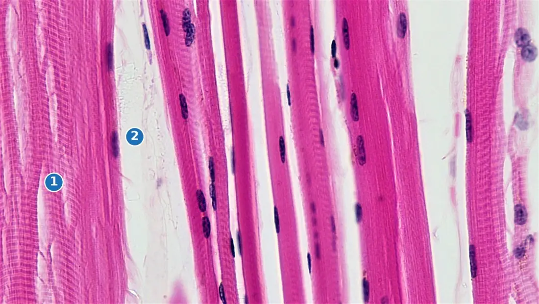
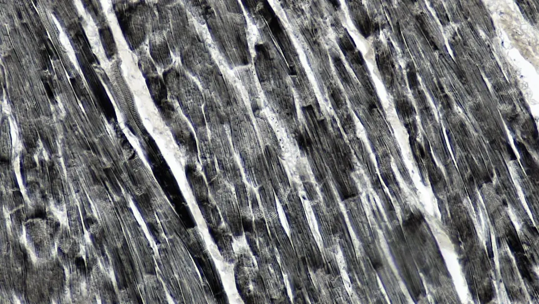
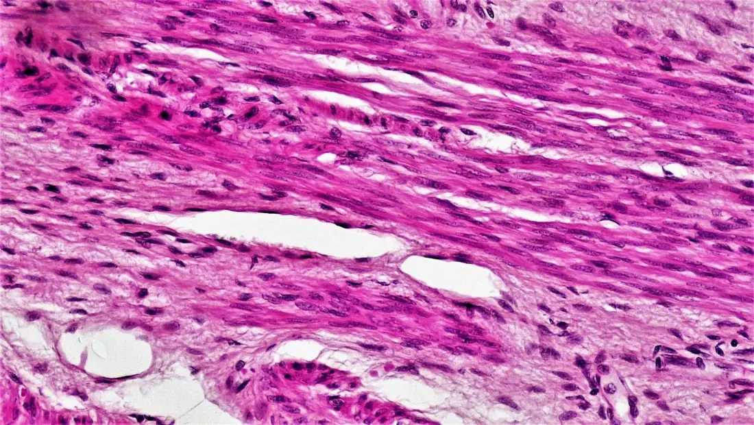
| Feature | Striated (skeletal) | Cardiac | Smooth (unstriated) |
|---|---|---|---|
| Shape of fibre | long, cylindrical, unbranched | cylindrical, branched | spindle-shaped (fusiform) |
| Striations | present, prominent | present, faint | absent |
| Nuclei | many, at the periphery | one (rarely two), central | one, central |
| Intercalated discs | absent | present | absent |
| Control | voluntary | involuntary | involuntary |
| Speed and fatigue | fast, powerful, tires quickly | moderate, rhythmic, never tires | slow, sustained, tires slowly |
| Location | attached to bones, tongue, diaphragm | wall of the heart only | gut, uterus, blood vessels, iris, bronchi |
The sarcomere
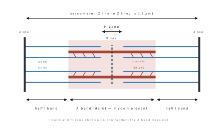
Under high power, a myofibril shows repeating light and dark bands. The dark A band (anisotropic) is the length of the thick myosin filaments; the light I band (isotropic) contains only thin actin filaments and is bisected by a dark Z line. The H zone in the middle of the A band contains myosin only, and is crossed by the M line.
A sarcomere is the segment of a myofibril between two successive Z lines. It is the functional (contractile) unit of striated muscle. At rest it is about 2.0–2.5 μm long.
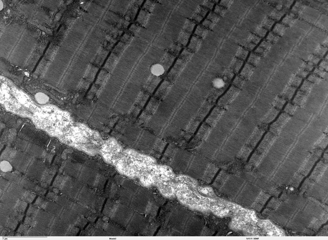
Problem. A myofibril in the biceps is 2.4\ \text{cm} long and its sarcomeres are each 2.4\ \mu\text{m} long at rest. (a) How many sarcomeres lie end to end along it? (b) If every sarcomere shortens to 1.8\ \mu\text{m} during a full contraction, what is the new length of the myofibril and the percentage shortening?
Solution.
(a) Convert to the same unit: 2.4\ \text{cm} = 24\ \text{mm} = 24\,000\ \mu\text{m}.
(b) New length = 10\,000 \times 1.8\ \mu\text{m} = 18\,000\ \mu\text{m} = 1.8\ \text{cm}.
Each sarcomere shortens by only 0.6\ \mu\text{m}, but because 10,000 of them are joined in series the whole fibril shortens by 0.6\ \text{cm}. That is how a change too small to see adds up to a visible movement.
6.4 Nervous tissue
Nervous tissue is ectodermal and is built from two kinds of cell: the neurons, which carry the messages, and the neuroglia, about ten times more numerous, which support, insulate, feed and defend them. Neurons are excitable — they respond to a stimulus by generating an electrical impulse — and conductive — they pass that impulse on. Mature neurons have lost the power to divide, which is why nerve damage is often permanent.
A typical motor neuron has three parts:
- Cyton (cell body or perikaryon) — contains the nucleus, and granular patches of rough endoplasmic reticulum with ribosomes called Nissl granules, which make the neuron's proteins. It has no centriole.
- Dendrites — many short, branched, tapering processes that carry impulses towards the cyton. They also contain Nissl granules.
- Axon — a single long process arising from the cone-shaped axon hillock that carries the impulse away from the cyton. It has no Nissl granules. It ends in fine branches, the telodendria, each tipped by a synaptic knob.
In a myelinated fibre the axon is wrapped in a fatty myelin sheath made by Schwann cells, covered outside by the neurilemma. The sheath is interrupted every millimetre or so at the nodes of Ranvier. The impulse jumps from node to node — saltatory conduction — which is why a myelinated fibre conducts up to a hundred times faster than a bare one.
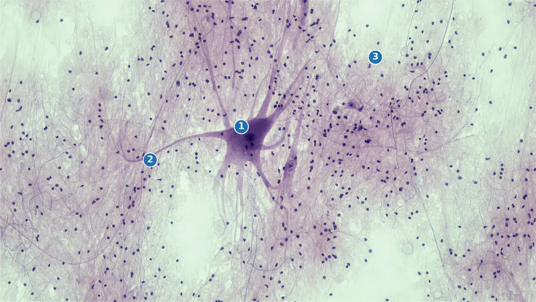
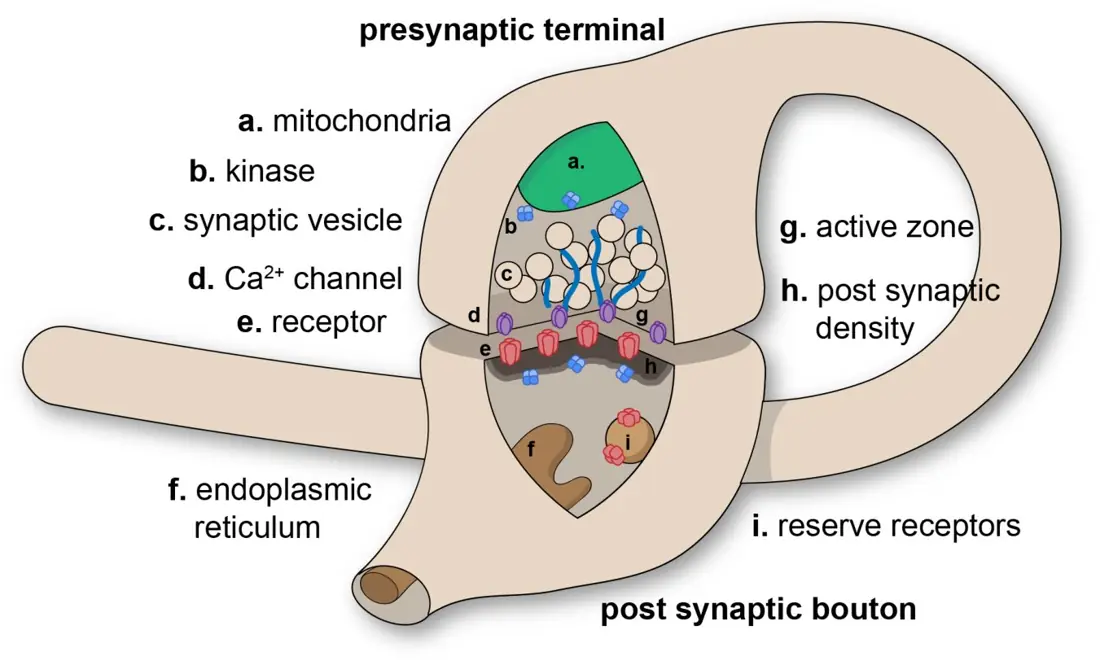
Dendrite = Delivers to the cell body. Axon carries away. At a synapse the vesicles are always on the axon side, so transmission across a synapse is strictly one-way.
Neurons are named by the number of processes leaving the cyton — unipolar (one process; in insect and embryonic nerve cells), bipolar (one axon and one dendrite; retina, olfactory epithelium) and multipolar (one axon and many dendrites; the commonest type, in the brain and spinal cord). By function they are sensory (afferent), motor (efferent) or association (interneurons).
The main neuroglia are astrocytes (anchor neurons to capillaries and help form the blood–brain barrier), oligodendrocytes (make myelin inside the brain and spinal cord), microglia (phagocytes of the nervous system) and ependymal cells (line the brain ventricles and circulate cerebrospinal fluid).
A synapse is the functional junction between the synaptic knob of one neuron and the dendrite or cyton of the next. The membranes do not touch; they are separated by a synaptic cleft about 20 nm wide. The impulse crosses chemically: it makes the knob release a neurotransmitter such as acetylcholine, which diffuses across the cleft and excites the next membrane.
Problem. You step on a thorn in a Chitwan forest and pull your foot back. The sensory path from the foot to the spinal cord is 0.90\ \text{m} and the motor path back to the leg muscle is 0.80\ \text{m}; both are myelinated fibres conducting at 85\ \text{m s}^{-1}. The reflex arc contains 2 synapses, each causing a delay of 0.5\ \text{ms}. (a) Find the total reflex time. (b) How long would the same journey take along a non-myelinated fibre conducting at 0.5\ \text{m s}^{-1}?
Solution.
(a) Total length of nerve fibre = 0.90 + 0.80 = 1.70\ \text{m}.
Synaptic delay = 2 \times 0.5 = 1.0\ \text{ms}. Total reflex time
(b) Along a bare fibre:
The myelin sheath makes the response about 160 times faster — quite literally the difference between a scratch and a deep wound.
The myelin sheath is the fatty insulating layer produced by the Schwann cell. The neurilemma is the thin outer living membrane of the Schwann cell itself, outside the myelin. Non-myelinated fibres still have a neurilemma — they simply lack myelin, and so have no nodes of Ranvier and conduct slowly.
| Feature | Myelinated fibre | Non-myelinated fibre |
|---|---|---|
| Myelin sheath | present | absent |
| Nodes of Ranvier | present | absent |
| Conduction | saltatory, fast (up to about 120 m s⁻¹) | continuous, slow (about 0.5–2 m s⁻¹) |
| Found in | cranial and spinal nerves, white matter | autonomic nerves, grey matter |
Chapter summary
- A tissue is a group of similar cells with a common origin doing one job; the four animal tissues are epithelial, connective, muscular and nervous.
- Epithelium: cells packed, matrix negligible, avascular, polar, on a basement membrane, high regeneration. Simple (squamous, cuboidal, columnar, ciliated, pseudostratified), compound (stratified squamous, transitional) or glandular.
- Connective tissue: few cells, abundant matrix of collagen, elastic and reticular fibres. Loose (areolar, adipose), dense (tendon = collagen, muscle→bone; ligament = elastin, bone→bone), skeletal (cartilage, bone) and fluid (blood, lymph).
- Cartilage: chondrocytes in lacunae in chondrin, avascular, covered by perichondrium. Bone: osteocytes in lacunae joined by canaliculi, matrix of ossein + calcium salts, organised into Haversian systems in mammals.
- Blood = 55% plasma + 45% formed elements. RBC 4.5–5.5 million per mm³, WBC 4000–11000 per mm³, platelets 150,000–400,000 per mm³.
- Muscle: striated (cylindrical, unbranched, many peripheral nuclei, voluntary), cardiac (branched, central nucleus, intercalated discs, involuntary, tireless), smooth (spindle, one central nucleus, no striations, involuntary).
- The sarcomere, from Z line to Z line, is the contractile unit; on contraction the I band and H zone shorten but the A band does not.
- A neuron has a cyton with Nissl granules, dendrites carrying impulses in, and one axon carrying them out. Myelin plus nodes of Ranvier give saltatory conduction; the synapse transmits chemically and in one direction only.
Practice questions
Group A — Multiple choice (1 mark each)
-
The tissue that arises from all three germ layers is [1]aconnectivebepithelialcmusculardnervous
-
Transitional epithelium is characteristic of the [1]atracheabsmall intestinecurinary bladderdoesophagus
-
Tendons are made chiefly of [1]ayellow elastic fibresbwhite collagen fibrescreticular fibresdchondrin
-
Which is absent in cartilage? [1]alacunaebchondrocytescblood vesselsdperichondrium
-
Intercalated discs occur in [1]askeletal musclebcardiac musclecsmooth muscledall three
-
During contraction of a sarcomere, the band whose length does not change is the [1]aI bandbH zonecA banddZ line
-
Nissl granules are absent from the [1]acytonbdendritecaxondnucleus
-
Heparin and histamine are secreted by the [1]afibroblastbmast cellcplasma celldosteocyte
Answers to Group A
1. (b) — epidermis is ectodermal, gut lining endodermal, endothelium mesodermal.
2. (c) — it must stretch as the bladder fills.
3. (b) — which is why a tendon is inextensible.
4. (c) — cartilage is avascular and is fed by diffusion from the perichondrium.
5. (b) — they are the junctions between adjacent cardiac fibres.
6. (c) — the A band equals the length of the myosin filament, which does not shorten.
7. (c) — the axon has no Nissl granules; this distinguishes it from a dendrite.
8. (b) — heparin prevents clotting inside vessels, histamine causes inflammation.
Group B — Short answer questions (4 marks each)
-
Define epithelial tissue and give four general characters of it. [4]
-
Distinguish between a tendon and a ligament under four heads. [4]
-
Draw a labelled T.S. of a Haversian system and name any four parts. [4]
-
Give four differences between cardiac muscle and smooth muscle. [4]
-
In a haemocytometer count, blood is diluted 1 : 200, and 420 red cells are counted in 80 small squares, each of area \frac{1}{400}\ \text{mm}^{2} under a depth of 0.1\ \text{mm}. Calculate the RBC count per mm³ and comment. [4]
-
Why is cartilage slow to heal while bone heals comparatively fast? [4]
Answers to Group B
2. Tendon — white collagen fibres, straight parallel bundles, inextensible, joins muscle to bone. Ligament — yellow elastic fibres, branching, elastic, joins bone to bone.
4. Cardiac: branched fibres, one central nucleus, intercalated discs present, found only in the heart, never fatigues. Smooth: spindle-shaped unbranched cells, one central nucleus, no discs, no striations, found in gut, uterus and blood vessels.
5. Volume over 80 small squares = 80 \times \frac{1}{400}\ \text{mm}^{2} \times 0.1\ \text{mm} = 0.02\ \text{mm}^{3}. Diluted count = 420/0.02 = 21\,000 per mm³. Whole blood = 21\,000 \times 200 = 4\,200\,000 = 4.2 million per mm³. This is below the normal range of 4.5–5.5 million, so the person is mildly anaemic.
6. Cartilage is avascular: its chondrocytes receive food and oxygen only by slow diffusion through the solid matrix from the perichondrium, and they divide slowly. Bone is richly supplied with blood through the Haversian and Volkmann's canals, and the periosteum contains osteoblasts that can lay down new matrix quickly, so a fracture unites in weeks.
Group C — Long answer questions (8 marks each)
-
(a) Classify epithelial tissue with one example and one location for each type. [5](b) Draw a labelled diagram of pseudostratified ciliated columnar epithelium and explain why it is not a stratified epithelium. [3]
-
(a) Describe the structure of a myelinated motor neuron with a labelled diagram. [5](b) What is a synapse? Explain how an impulse crosses it and why transmission is one-way. [3]
Answer outline to Group C question 2
(a) Cyton with nucleus, Nissl granules and no centriole; several branched dendrites conducting towards the cyton; a single axon arising at the axon hillock, covered by myelin made by Schwann cells, with neurilemma outside and nodes of Ranvier between successive Schwann cells; ending in telodendria tipped by synaptic knobs. Award marks for the labelled diagram.
(b) A synapse is the junction between the synaptic knob of one neuron and the dendrite or cyton of the next, separated by a cleft of about 20 nm. The arriving impulse opens Ca²⁺ channels in the knob; synaptic vesicles fuse with the presynaptic membrane and release acetylcholine, which diffuses across the cleft and binds receptors on the postsynaptic membrane, starting a new impulse. The enzyme cholinesterase then destroys the transmitter. Transmission is one-way because vesicles and transmitter exist only on the presynaptic side and receptors only on the postsynaptic side.
Practise more: Biology question bank