Unit 8 · Zoology · 28 teaching hours · 75 min read
Human Biology
What the syllabus asks for 8
- Digestive system
- Respiratory System
- Circulatory System
- Excretory System
- Nervous system
- Sense organs
- Endocrinology
- Reproductive System
Twenty-eight hours — more than a fifth of the whole Grade 12 course — are given to one animal, Homo sapiens. The reason is not vanity. The human body is the one animal whose physiology you will use every day: when a doctor reads your blood pressure, when a patient in Bharatpur is put on dialysis, when an adolescent is taught about the menstrual cycle. This unit works organ system by organ system. For each one, learn three things in order: the parts (gross anatomy, with a labelled diagram you can reproduce in ten minutes), the mechanism (what each part physically does), and the numbers (the normal values, because a value is what tells a clinician that something is wrong).
Because it is the largest unit, Section II almost always draws one of its two 8-mark long questions from here, plus two or three of its four 4-mark short questions. Long questions are nearly always "describe the structure of X with a labelled diagram and explain its working" — so the diagram is half the marks. Practise drawing the heart, the nephron, the brain and the reproductive systems until you can label them from memory.
8.1 Digestive system
Food as we eat it is made of polymers — starch, protein, triglyceride — whose molecules are far too large to cross a cell membrane. Digestion is the controlled hydrolysis of those polymers into absorbable monomers, and the digestive system is a 9-metre tube with glands attached that does the job in stages.
The human digestive system has two parts: the alimentary canal (a continuous tube from mouth to anus, about 8–10 m long in an adult) and the associated digestive glands (salivary glands, liver with gall bladder, and pancreas), which lie outside the tube and pour their secretions into it through ducts.
The alimentary canal
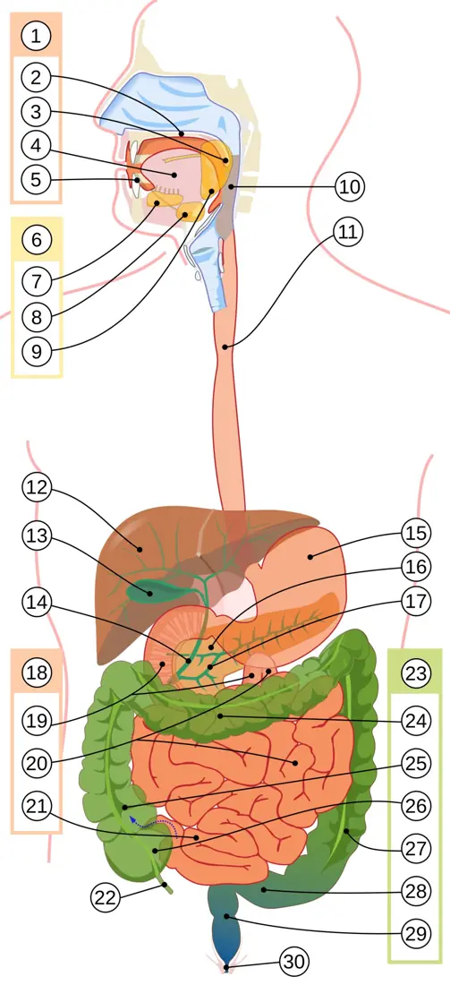
The canal is regionally specialised:
- Buccal cavity — mechanical breakdown by the teeth, lubrication and partial starch digestion by saliva, and rolling of the food into a bolus by the tongue. Swallowing (deglutition) pushes the bolus into the pharynx; the epiglottis closes the glottis so food does not enter the trachea.
- Oesophagus — a muscular tube about 25 cm long. It secretes no enzyme; it moves the bolus by peristalsis, a travelling wave in which the circular muscle behind the bolus contracts and that in front relaxes.
- Stomach — a J-shaped bag of capacity 1–1.5 L with three regions (cardiac, fundus, pyloric) and three muscle layers (an extra oblique layer for churning). Gastric glands in its mucosa contain mucous cells (mucus + bicarbonate), parietal (oxyntic) cells (HCl and intrinsic factor) and peptic (chief/zymogen) cells (pepsinogen, and prorennin in infants). The bolus becomes acidic semi-fluid chyme, released in spurts through the pyloric sphincter.
- Small intestine — about 6 m long, the main site of both digestion and absorption, in three parts: the C-shaped duodenum (receives bile and pancreatic juice), the coiled jejunum, and the longest part, the ileum.
- Large intestine — about 1.5 m: caecum with the vermiform appendix (a vestigial organ in humans, housing symbiotic bacteria), colon (ascending, transverse, descending, sigmoid) and rectum. No enzymes are secreted here; it absorbs water, some minerals and vitamins (K and some B vitamins made by gut bacteria) and forms faeces.
Mouth → pharynx → oesophagus → stomach → duodenum → jejunum → ileum → caecum → colon (ascending, transverse, descending, sigmoid) → rectum → anus. Three sphincters guard it: cardiac (oesophagus–stomach), pyloric (stomach–duodenum) and anal.
Teeth and the dental formula
Human teeth are thecodont (each root fixed in a bony socket), dicyphodont (two sets in a lifetime — 20 deciduous or milk teeth, replaced by 32 permanent teeth) and heterodont (four different types with different jobs).
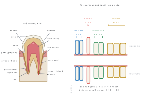
The permanent dental formula, written for one half of the upper jaw over one half of the lower jaw and doubled, is
and the deciduous (milk) dental formula, which has no premolars, is
Incisors cut, canines tear, premolars and molars crush and grind. The last molars ("wisdom teeth") erupt at 17–25 years.
The formula counts one half of each jaw only — that is why it is multiplied by 2. Writing \frac{2123}{2123} = 32 without the 2 outside is a marked error, and so is calling the milk formula "2(2 1 2 2)": milk teeth have no premolars, so it is 2 1 0 2, written simply as I2 C1 M2.
Digestive glands, enzymes and chemical digestion
Three sets of glands supply the tube. Salivary glands (three pairs: parotid, submandibular, sublingual) secrete about 1–1.5 L of saliva a day, pH 6.8, containing salivary amylase, lysozyme and mucin. The liver, the largest gland of the body (1.2–1.5 kg), secretes bile, stored and concentrated in the gall bladder. Bile contains no enzyme: its bile salts (sodium glycocholate and taurocholate) emulsify fat — they break large fat droplets into a fine suspension, hugely increasing the surface available to lipase — and its bicarbonate neutralises the acid chyme. The pancreas is a heterocrine gland; its exocrine part secretes pancreatic juice, the most complete digestive juice of all, containing enzymes for every class of food.
| Enzyme | Source (juice) | Substrate | Product | Optimum pH |
|---|---|---|---|---|
| Salivary amylase (ptyalin) | salivary glands (saliva) | starch, glycogen | maltose + dextrins | 6.8 |
| Pepsin (from pepsinogen) | peptic cells (gastric juice) | proteins | peptones, proteoses | 1.5–2.0 |
| Rennin (infants only) | peptic cells (gastric juice) | casein of milk | paracasein (curd) | 3.5 |
| Gastric lipase | gastric glands | emulsified fat | fatty acids + glycerol | 4–5 |
| Pancreatic amylase | pancreas (pancreatic juice) | starch, dextrins | maltose | 7.1 |
| Trypsin (from trypsinogen) | pancreas | proteins, peptones | dipeptides, peptides | 7.8–8.0 |
| Chymotrypsin | pancreas | proteins, peptones | peptides | 8.0 |
| Carboxypeptidase | pancreas | peptides (C-terminal) | amino acids | 7.2 |
| Pancreatic lipase (steapsin) | pancreas | emulsified triglycerides | fatty acids + monoglycerides | 8.0 |
| Nucleases (DNase, RNase) | pancreas | DNA, RNA | nucleotides | 7.0 |
| Enterokinase | intestinal glands | trypsinogen | trypsin (activation) | 8.0 |
| Maltase | brush border | maltose | glucose + glucose | 6.1–6.8 |
| Sucrase (invertase) | brush border | sucrose | glucose + fructose | 6.2 |
| Lactase | brush border | lactose | glucose + galactose | 6.0 |
| Dipeptidase, aminopeptidase | brush border | dipeptides, peptides | amino acids | 8.0 |
Note the logic of the table. Proteolytic enzymes are secreted as inactive zymogens (pepsinogen, trypsinogen, chymotrypsinogen) so that they cannot digest the gland that makes them; pepsinogen is activated by HCl, trypsinogen by enterokinase from the intestinal mucosa, and trypsin then activates all the rest. Note also the pH jump: pepsin needs pH 2, every intestinal enzyme needs pH about 8, which is why bile and pancreatic bicarbonate must neutralise the chyme first.
Carbohydrates → monosaccharides (glucose, fructose, galactose). Proteins → amino acids. Fats → fatty acids + glycerol/monoglycerides. Nucleic acids → nucleosides and nitrogenous bases. Only these cross the intestinal epithelium.
Absorption: the villus
Absorption is the passage of the end products from the lumen into the blood or lymph. It happens almost entirely in the small intestine, whose surface is amplified three times over — by circular folds, by finger-like villi (0.5–1.5 mm long, 20–40 per mm²), and by microvilli on each epithelial cell forming the brush border. The total absorptive area reaches about 200 m², roughly a badminton court packed inside your abdomen.
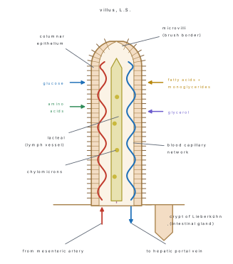
Glucose, galactose and amino acids are absorbed by active transport (against their gradient, using ATP and a Na⁺ co-transporter) into the blood capillaries, and travel by the hepatic portal vein to the liver before entering the general circulation. Fructose and some amino acids move by facilitated diffusion; water, most fatty acids, glycerol and fat-soluble vitamins by simple diffusion. Fatty acids and monoglycerides are re-esterified inside the epithelial cell, coated with protein to form chylomicrons, and enter the lacteal; lymph carries them through the thoracic duct into the left subclavian vein — bypassing the liver. Assimilation, the use of absorbed nutrients by cells, completes the process; undigested cellulose (roughage) and bacteria form the faeces, egested by the defaecation reflex.
8.2 Respiratory System
Every cell of the body oxidises glucose to make ATP, and every cell produces CO₂. Breathing (external respiration) is only the transport half of the job — moving O₂ from the air to the blood and CO₂ the other way. Cellular respiration, the chemistry, happens in the mitochondria.
The respiratory tract
Air passes through a conducting portion, which warms, moistens and filters it but takes no part in exchange, and a respiratory portion, where exchange occurs.
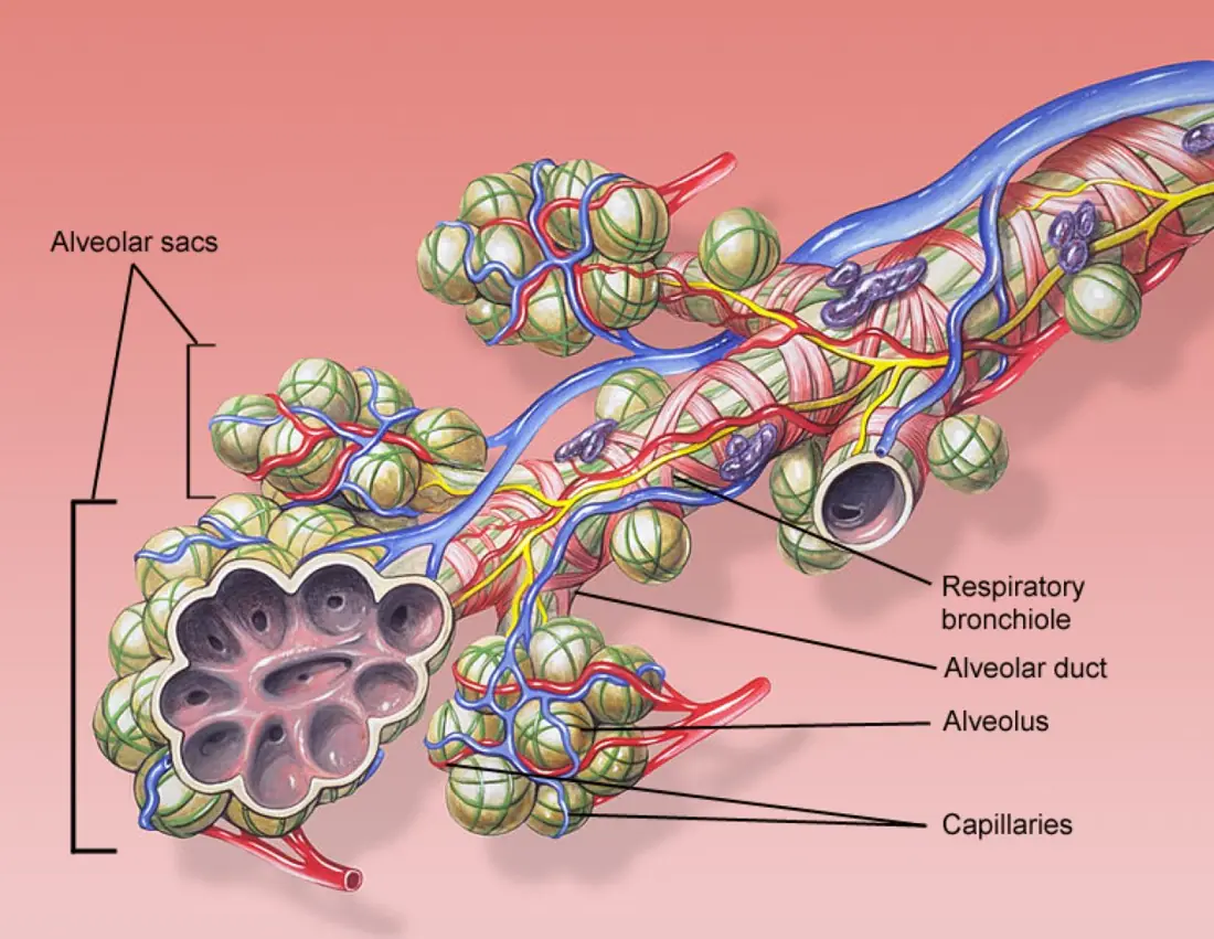
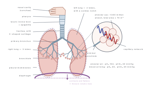
The conducting portion is: external nares → nasal chambers (lined by ciliated mucous epithelium; the turbinate bones or conchae make the air swirl so that dust sticks) → pharynx → larynx → trachea → two primary bronchi → secondary and tertiary bronchi → bronchioles → terminal bronchioles. The trachea is held open by about 16–20 C-shaped rings of hyaline cartilage, incomplete at the back so that a swallowed bolus can bulge into the space.
The respiratory portion begins at the respiratory bronchioles and ends in the alveoli — some 300 million thin-walled sacs giving a total surface of about 70 m². The respiratory membrane across which gases diffuse is astonishingly thin (less than 1 μm) and has only three layers: the squamous alveolar epithelium, a fused basement membrane, and the capillary endothelium.
Fick's law says diffusion rate ∝ (surface area × pressure difference) ÷ thickness. The lung maximises all three: 70 m² of area, a steep pO₂ gradient maintained by ventilation and by blood flow, and a membrane under 1 μm thick.
Mechanism of breathing
Breathing is entirely a pressure story, governed by Boyle's law: increase the volume of the thoracic cavity and the pressure inside falls below atmospheric, so air rushes in.
- Inspiration (active). The diaphragm contracts and flattens (increasing the vertical axis of the thorax); the external intercostal muscles contract and pull the ribs up and out (increasing the antero-posterior axis). Thoracic volume rises, intrapulmonary pressure falls about 1–3 mmHg below atmospheric, and roughly 500 ml of air enters.
- Expiration (normally passive). The diaphragm and external intercostals relax; the elastic recoil of the lungs and chest wall reduces the volume and raises intrapulmonary pressure 1–3 mmHg above atmospheric, driving air out. Forced expiration adds the internal intercostals and abdominal muscles.
A normal adult breathes 12–16 times a minute. Breathing is controlled by the respiratory rhythm centre in the medulla oblongata, moderated by the pneumotaxic centre in the pons, and by chemoreceptors near the aorta and carotid arteries that respond chiefly to a rise in CO₂ and H⁺ — not to a fall in O₂.
Lung volumes and capacities
A spirometer records the volume of air moved. A volume is a single measurement; a capacity is the sum of two or more volumes.

| Volume / capacity | Symbol | Definition | Normal value |
|---|---|---|---|
| Tidal volume | TV | air breathed in or out in one quiet breath | 500 ml |
| Inspiratory reserve volume | IRV | extra air taken in by a forced inspiration | 2500–3000 ml |
| Expiratory reserve volume | ERV | extra air expelled by a forced expiration | 1000–1100 ml |
| Residual volume | RV | air left after the most forceful expiration | 1100–1200 ml |
| Inspiratory capacity | IC | TV + IRV | 3500 ml |
| Expiratory capacity | EC | TV + ERV | 1600 ml |
| Functional residual capacity | FRC | ERV + RV | 2300 ml |
| Vital capacity | VC | TV + IRV + ERV (max out after max in) | 4600 ml |
| Total lung capacity | TLC | VC + RV | 5800 ml |
RV is the air you can never blow out, so a spirometer never sees it. It is found indirectly (helium dilution or nitrogen wash-out). Consequently TLC and FRC, which contain RV, also cannot be read straight off a spirometer trace.
Problem. A student breathes 15 times per minute with a tidal volume of 500 ml. The anatomical dead space is 150 ml. Calculate (a) the pulmonary ventilation rate and (b) the alveolar ventilation rate. (c) If she instead takes shallow breaths of 250 ml at 30 per minute, what happens to each?
Solution.
(a) Pulmonary ventilation = TV \times breathing rate
(b) Only the air beyond the dead space reaches the alveoli:
(c) Pulmonary ventilation = 250\times30 = 7500\ \text{ml min}^{-1} — unchanged. But alveolar ventilation = (250-150)\times 30 = 100\times30 = 3000\ \text{ml min}^{-1}, a fall of 43 %.
Conclusion. Deep slow breathing ventilates the alveoli far better than rapid shallow breathing, even when the total air moved is identical. This is why panting is an inefficient way to get oxygen.
Transport of gases
| Gas | Partial pressure (mmHg) | Atmospheric air | Alveoli | Deoxygenated blood | Oxygenated blood | Tissues |
|---|---|---|---|---|---|---|
| O₂ | pO₂ | 159 | 104 | 40 | 95 | 40 |
| CO₂ | pCO₂ | 0.3 | 40 | 45 | 40 | 45 |
Gases move only down these gradients, by simple diffusion. Oxygen is carried almost entirely (97 %) by haemoglobin as oxyhaemoglobin, only 3 % dissolved in plasma. One haemoglobin molecule binds four O₂ molecules, and the oxygen dissociation curve is sigmoid. A fall in pH, a rise in pCO₂ and a rise in temperature shift the curve to the right — the Bohr effect — so that haemoglobin unloads more oxygen exactly in the tissues that are working hardest.
Carbon dioxide is carried in three ways: about 70 % as bicarbonate (HCO₃⁻) in the plasma, after the reaction CO₂ + H₂O ⇌ H₂CO₃ ⇌ H⁺ + HCO₃⁻ inside the red cell, catalysed by carbonic anhydrase; about 20–25 % as carbamino-haemoglobin bound to the globin part; and about 7 % dissolved in plasma.
Problem. A healthy man has 15 g of haemoglobin per 100 ml of blood, and 1 g of haemoglobin combines with 1.34 ml of O₂ at full saturation. His total blood volume is 5 L. Find (a) the oxygen capacity of 100 ml of blood, (b) the total O₂ carried by haemoglobin in his body, and (c) the O₂ delivered to the tissues each minute if 5 ml of O₂ leaves every 100 ml of blood and cardiac output is 5 L min⁻¹.
Solution.
(a) 1.34 \times 15 = 20.1\ \text{ml of O}_2 per 100 ml of blood — the familiar "20 volumes per cent".
(b) 5000\ \text{ml} \div 100 = 50 units of 100 ml, so
(c) Oxygen delivery = cardiac output \times arterio-venous O₂ difference
which matches the resting oxygen consumption of an adult, about 250 ml min⁻¹.
Disorders of this system are common in Nepal's brick-kiln and city air: asthma (bronchiolar spasm and inflammation), emphysema (alveolar walls destroyed by smoking, so surface area collapses), bronchitis, pneumonia and occupational silicosis/asbestosis.
8.3 Circulatory System
A human being is too large for diffusion alone. Oxygen would take years to diffuse from the lung to the toe. The circulatory system solves this by bulk flow: a pump, a closed network of tubes, and a fluid — blood — that carries everything.
Blood
Blood is a fluid connective tissue. An adult has about 5–6 L, which is 6–8 % of body weight. Centrifuged, it separates into a straw-coloured plasma (55 %) and formed elements (45 %); the packed-cell fraction is the haematocrit.
| Component | Share / count | Key features | Main function |
|---|---|---|---|
| Plasma | 55 % of blood | 90–92 % water; 6–8 % proteins (albumin, globulins, fibrinogen); salts, glucose, urea, hormones | transport medium; osmotic balance; clotting; immunity |
| Erythrocytes (RBC) | 5.0–5.5 million mm⁻³ | biconcave discs, 7 μm, no nucleus in mammals, life 120 days, made in red bone marrow, destroyed in spleen ("graveyard of RBC") | carry O₂ and some CO₂ as haemoglobin (12–16 g dl⁻¹) |
| Leucocytes (WBC) | 6000–8000 mm⁻³ | nucleated, colourless; granulocytes: neutrophil 60–65 %, eosinophil 2–3 %, basophil 0.5–1 %; agranulocytes: lymphocyte 20–25 %, monocyte 6–8 % | defence — phagocytosis, antibodies, allergy, inflammation |
| Thrombocytes (platelets) | 1.5–3.5 lakh mm⁻³ | cell fragments from megakaryocytes, no nucleus, life 5–9 days | release thromboplastin; start blood clotting |
Clotting. Injury exposes tissue and platelets release thromboplastin, which with Ca²⁺ converts prothrombin → thrombin; thrombin converts the soluble plasma protein fibrinogen → fibrin, whose threads trap blood cells and form the clot. Vitamin K is needed by the liver to make prothrombin.
Blood groups. The ABO system depends on antigens (agglutinogens) on the RBC surface and antibodies (agglutinins) in the plasma. Mixing an antigen with its matching antibody makes the cells clump (agglutination), which can kill the recipient.
| Group | Antigen on RBC | Antibody in plasma | Can donate to | Can receive from |
|---|---|---|---|---|
| A | A | anti-B (b) | A, AB | A, O |
| B | B | anti-A (a) | B, AB | B, O |
| AB | A and B | none | AB only | A, B, AB, O — universal recipient |
| O | none | anti-A and anti-B | A, B, AB, O — universal donor | O only |
The Rh factor is a separate antigen (D antigen) present in about 95 % of Nepali people (Rh⁺). An Rh⁻ mother carrying an Rh⁺ foetus may be sensitised at the first delivery and her anti-Rh antibodies can destroy the red cells of a later Rh⁺ foetus — erythroblastosis foetalis, prevented by giving the mother anti-Rh (anti-D) immunoglobulin within 72 hours of delivery.
O⁻ blood has no A, B or D antigen, so it is safest in an emergency. But O plasma carries both anti-A and anti-B antibodies, so only small volumes (packed cells) are safe. Modern practice is always to cross-match.
Structure of the human heart
The heart is a hollow muscular organ the size of a closed fist (about 300 g), lying in the thoracic cavity between the lungs, tilted so its apex points down and to the left. It is enclosed in a double pericardium with pericardial fluid between the layers, and its wall has three coats: outer epicardium, thick middle myocardium of cardiac muscle, and inner endocardium.
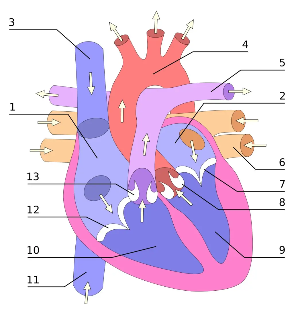
Four chambers. Two thin-walled atria (auricles) receive blood; two thick-walled ventricles pump it. The interatrial septum and interventricular septum separate the right (deoxygenated) side from the left (oxygenated) side, so the two never mix. The left ventricle has a wall about three times thicker than the right because it must push blood around the whole body, not just to the lungs.
Four valves, all of which open only one way so that blood cannot flow back:
| Valve | Position | Cusps | Closes at |
|---|---|---|---|
| Tricuspid | right atrium → right ventricle | 3 | start of ventricular systole (1st sound) |
| Bicuspid (mitral) | left atrium → left ventricle | 2 | start of ventricular systole (1st sound) |
| Pulmonary semilunar | right ventricle → pulmonary trunk | 3 pocket-like | end of ventricular systole (2nd sound) |
| Aortic semilunar | left ventricle → aorta | 3 pocket-like | end of ventricular systole (2nd sound) |
The cusps of the AV valves are anchored by tendinous cords, the chordae tendineae, to papillary muscles on the ventricle wall — they act like the strings of a parachute, stopping the cusps from turning inside-out when the ventricle contracts.
Double circulation
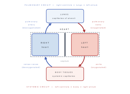
Because the blood passes through the heart twice in one complete circuit — once as it goes to the lungs and once as it goes to the body — the human circulation is called double circulation. It is also closed (blood stays inside vessels) and complete (oxygenated and deoxygenated blood never mix).
Two special routes matter:
- Coronary circulation — the heart muscle cannot use the blood inside its own chambers. Coronary arteries branch from the base of the aorta to supply the myocardium; their blockage causes a heart attack.
- Hepatic portal system — blood from the stomach, intestine, pancreas and spleen goes first to the liver by the hepatic portal vein, so that absorbed food is processed and toxins removed before reaching the rest of the body. A portal vein is any vein that starts in capillaries and ends in capillaries.
| Feature | Artery | Vein | Capillary |
|---|---|---|---|
| Direction | away from heart | towards heart | between the two |
| Wall | thick, elastic, muscular | thinner, less elastic | one cell thick (endothelium only) |
| Lumen | narrow | wide | very narrow (7–8 μm) |
| Valves | absent (except at the heart) | present, prevent backflow | absent |
| Blood | oxygenated (except pulmonary artery) | deoxygenated (except pulmonary vein) | — |
| Pressure | high (~120/80 mmHg) | low (~10 mmHg) | falls 35 → 15 mmHg |
The cardiac cycle
Cardiac muscle is myogenic: the beat starts inside the heart itself. The sino-atrial (SA) node, a patch of modified cardiac muscle in the wall of the right atrium near the opening of the superior vena cava, fires 70–75 times a minute and is therefore the pacemaker. The impulse spreads over both atria to the atrio-ventricular (AV) node, is delayed there by about 0.1 s (letting the atria empty), then travels down the bundle of His, its two branches, and the Purkinje fibres into the ventricle walls.
One cardiac cycle at 72 beats per minute lasts 0.8 s:
| Phase | Duration | What happens |
|---|---|---|
| Atrial systole | 0.1 s | atria contract, pushing the last ~30 % of blood into the ventricles; AV valves open |
| Ventricular systole | 0.3 s | ventricles contract; AV valves shut ("lubb", 1st sound); pressure rises; semilunar valves open and about 70 ml is ejected |
| Joint diastole | 0.4 s | all chambers relax; semilunar valves shut ("dupp", 2nd sound); ventricles fill passively |
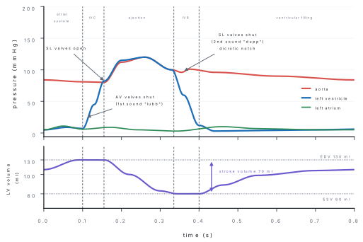
Problem. A patient at the Shahid Gangalal Heart Centre has a heart rate of 72 beats per minute, an end-diastolic volume of 130 ml and an end-systolic volume of 60 ml. Find (a) the stroke volume, (b) the cardiac output, (c) the ejection fraction, and (d) the cardiac output when exercise raises the heart rate to 150 min⁻¹ and the stroke volume to 110 ml.
Solution.
(a) SV = EDV - ESV = 130 - 60 = 70\ \text{ml}
(b) CO = SV \times HR = 70 \times 72 = 5040\ \text{ml min}^{-1} \approx 5.0\ \text{L min}^{-1}
(c) Ejection fraction = \dfrac{SV}{EDV}\times 100 = \dfrac{70}{130}\times100 = 53.8\ \% (normal is 55–70 %; below 40 % indicates heart failure).
(d) CO = 110 \times 150 = 16\,500\ \text{ml min}^{-1} = 16.5\ \text{L min}^{-1} — more than three times the resting value. This extra flow is the whole point of a trained heart.
Electrocardiogram (ECG)
The electrical changes that sweep the heart can be picked up from the skin. The standard trace of one beat has three deflections.
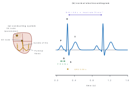
| Wave | Electrical event | Mechanical event that follows |
|---|---|---|
| P | depolarisation of the atria | atrial systole |
| QRS complex | depolarisation of the ventricles (atrial repolarisation is hidden inside it) | ventricular systole begins |
| T | repolarisation of the ventricles | ventricular systole ends |
Counting the R–R interval gives the heart rate directly: heart rate = 60 ÷ (R–R in seconds). A raised ST segment, an absent P wave or a widened QRS each point to a specific disease, which is why the ECG is the first test done in a suspected heart attack.
Blood pressure is the force blood exerts on the artery wall, measured with a sphygmomanometer over the brachial artery and written systolic/diastolic. Normal adult value is 120/80 mmHg; persistently above 140/90 mmHg is hypertension, a leading cause of stroke and kidney failure in Nepal.
Cardiac output has units of volume per time (L min⁻¹), not beats per minute. An athlete has a low resting heart rate (50 min⁻¹) but a normal cardiac output, because the stroke volume is large. Always write CO = SV \times HR.
8.4 Excretory System
Every reaction in the body leaves waste. Deamination of surplus amino acids in the liver produces ammonia (NH₃), which is extremely toxic and very soluble; the liver at once converts it to urea through the ornithine cycle. Because humans excrete nitrogen mainly as urea, we are ureotelic. Bony fish, which have unlimited water, excrete ammonia directly (ammonotelic); birds, reptiles and insects, which must save water, excrete almost insoluble uric acid (uricotelic).
| Organ | Substance excreted |
|---|---|
| Kidneys | urea, uric acid, creatinine, excess water, salts, drugs |
| Lungs | CO₂ and water vapour |
| Skin (sweat glands) | water, NaCl, traces of urea and lactic acid |
| Liver | bile pigments (bilirubin, biliverdin), cholesterol, excess metals |
| Large intestine | excess Ca²⁺, Fe²⁺ (this is egestion for undigested food) |
The urinary system and the kidney
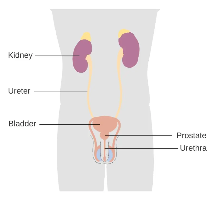
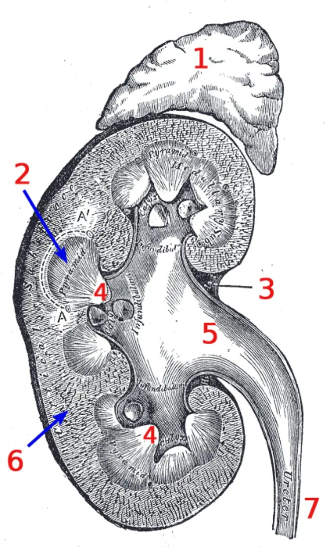
Each kidney is a dark-red bean about 11 cm × 6 cm × 3 cm weighing roughly 150 g, lying against the back wall of the abdomen at the level of the last thoracic and first three lumbar vertebrae. The right kidney sits slightly lower because the liver is above it. A section shows, from outside in:
- Fibrous capsule — tough protective coat.
- Cortex — the outer granular zone containing all the renal corpuscles.
- Medulla — 8–18 striated renal pyramids; each pyramid ends in a renal papilla. Cortical tissue dipping between pyramids forms the renal columns of Bertini.
- Pelvis — the funnel that collects urine. Each papilla drains into a minor calyx; minor calyces join into 2–3 major calyces, and these into the pelvis, which narrows into the ureter.
The nephron
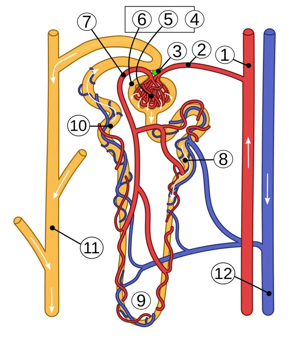
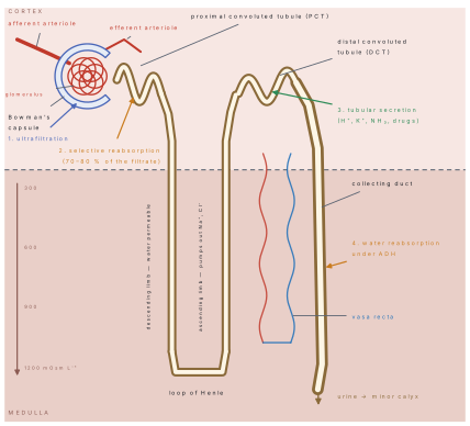
The nephron is the structural and functional unit of the kidney; each kidney has about 1 million of them. A nephron has two parts:
- Malpighian body (renal corpuscle) — the glomerulus, a knot of about 50 capillaries, cupped inside the double-walled Bowman's capsule.
- Renal tubule — proximal convoluted tubule (PCT) → loop of Henle (descending + ascending limb) → distal convoluted tubule (DCT) → collecting duct.
About 85 % are cortical nephrons, with short loops that barely enter the medulla. The remaining 15 % are juxtamedullary nephrons, whose long loops plunge deep into the medulla; these are the ones that make concentrated urine.
The blood supply is unusual. The afferent arteriole entering the glomerulus is wider than the efferent arteriole leaving it. Blood therefore piles up inside the glomerulus and the pressure there stays high — this is what drives filtration. The efferent arteriole then breaks up again into peritubular capillaries around the PCT and DCT, and into the hairpin vasa recta around the loop of Henle. Two capillary beds in series like this make a portal system (the renal portal system of the kidney).
Urine formation
Step 1 — Ultrafiltration. Blood pressure in the glomerulus forces water and all small solutes through three layers (capillary endothelium → basement membrane → podocytes of the capsule) into the capsular space. Blood cells and plasma proteins are too big and stay behind. The driving force is the net filtration pressure:
where P_{gc} is glomerular capillary hydrostatic pressure, P_{cs} the hydrostatic pressure of the fluid already in the capsule and \pi_{gc} the colloid osmotic (oncotic) pressure of the plasma proteins. The glomerular filtration rate (GFR) in a healthy adult is 125 ml min⁻¹, that is about 180 litres per day of filtrate — yet only 1–1.5 L leaves as urine, so about 99 % is reabsorbed.
Step 2 — Selective reabsorption. Useful substances are taken back into the peritubular blood.
| Region | Reabsorbed | Mechanism |
|---|---|---|
| PCT (70–80 % of filtrate) | all glucose and amino acids, 70 % Na⁺ and water, HCO₃⁻ | active transport + osmosis; microvilli give a huge surface |
| Descending limb | water only | osmosis into the hypertonic medulla |
| Ascending limb | Na⁺, Cl⁻ (pumped out); impermeable to water | active transport |
| DCT | Na⁺ (under aldosterone), water (under ADH) | selective, hormone-controlled |
| Collecting duct | water (under ADH), urea | osmosis |
Step 3 — Tubular secretion. The tubule cells actively add H⁺, K⁺, NH₃, creatinine and many drugs (penicillin, aspirin) from the blood into the filtrate. Secretion of H⁺ and NH₃ is how the kidney keeps blood pH near 7.4.
The ascending limb pumps Na⁺ and Cl⁻ out but will not let water follow. Salt therefore piles up in the medullary interstitium while the descending limb loses water into it. Because the two limbs run in opposite directions side by side, a small difference at each level is multiplied along the loop, building a gradient from 300 mOsm L⁻¹ at the cortex to 1200 mOsm L⁻¹ at the papilla. The vasa recta run counter-current too, so they remove reabsorbed water without washing the salt away. The collecting duct then passes through this gradient, and water leaves it by osmosis — which is how urine four times more concentrated than blood can be produced.
Problem. Renal blood flow is 1200 ml min⁻¹ and the haematocrit is 45 %. If the glomerular filtration rate is 125 ml min⁻¹, find (i) the renal plasma flow, (ii) the filtration fraction, and (iii) the volume filtered in 24 hours.
Solution. (i) Plasma is the non-cell fraction, so plasma = (100 − 45) % = 55 % of blood.
(ii) The filtration fraction is the part of that plasma which actually filters:
(iii) Volume filtered per day:
About one-fifth of the plasma entering the glomerulus is filtered; the other four-fifths carries the plasma proteins on into the efferent arteriole. Of the 180 L filtered, roughly 178.5 L is reabsorbed and about 1.5 L is voided.
Control of kidney function, and disorders
- ADH (vasopressin) from the posterior pituitary makes the DCT and collecting duct permeable to water. Dehydration → more ADH → less, more concentrated urine. No ADH → diabetes insipidus, up to 20 L of dilute urine a day.
- Renin–angiotensin–aldosterone system. A fall in blood pressure is sensed by the juxtaglomerular apparatus (JGA), where the afferent arteriole touches the DCT. It releases renin → angiotensin II → vasoconstriction and release of aldosterone from the adrenal cortex → Na⁺ and water reabsorption in the DCT → blood pressure restored.
- ANF (atrial natriuretic factor) from a stretched atrial wall does the opposite: it causes vasodilation and salt loss, lowering blood pressure.
Micturition. Urine trickles down the ureters by peristalsis and is stored in the urinary bladder. Stretch receptors fire when about 300–400 ml has collected; the reflex centre in the sacral spinal cord relaxes the internal sphincter while the detrusor muscle contracts. The external sphincter is under voluntary control, which is why micturition can be delayed.
Normal urine is pale yellow (from urochrome), slightly acidic (pH ≈ 6), 95 % water, with about 2 % urea, 0.05 % uric acid and 0.075 % creatinine, plus salts. Glucose, protein, blood cells or ketone bodies in urine are always abnormal.
| Disorder | Cause | Sign |
|---|---|---|
| Uraemia | kidney failure; urea retained in blood | blood urea ≫ 40 mg/dl |
| Renal calculi (stones) | crystals of calcium oxalate/uric acid in the pelvis | severe loin pain, blood in urine |
| Glomerulonephritis | inflammation of the glomeruli after infection | protein and blood in urine |
| Diabetes mellitus | insulin deficiency | glucose in urine (glycosuria) |
| Diabetes insipidus | lack of ADH | huge volume of dilute urine |
Kidney failure is treated by haemodialysis — blood is pumped past a semi-permeable cellophane membrane bathed in dialysing fluid that has the same composition as normal plasma except that it contains no urea, so urea diffuses out down its concentration gradient while glucose and salts stay in. The permanent cure is renal transplantation, for which tissue matching is essential.
A common exam error is to say "the kidney filters 1.5 L a day". The kidney filters 180 L a day and reabsorbs 178.5 L of it. Also, glucose is normally 100 % reabsorbed in the PCT; it appears in urine only when the plasma glucose exceeds the renal threshold of about 180 mg/dl and the carriers saturate.
8.5 Nervous system
The nervous system is the body's fast control system: it works in milliseconds, by electrical impulses along fixed wires, and its effects stop as soon as the impulse stops. It is divided into the central nervous system (CNS) — brain and spinal cord — and the peripheral nervous system (PNS) — 12 pairs of cranial nerves and 31 pairs of spinal nerves. The PNS in turn has a somatic (voluntary, skeletal muscle) division and an autonomic (involuntary) division, which is itself split into the sympathetic ("fight or flight", thoraco-lumbar, noradrenaline) and the parasympathetic ("rest and digest", cranio-sacral, acetylcholine) systems.
The neuron
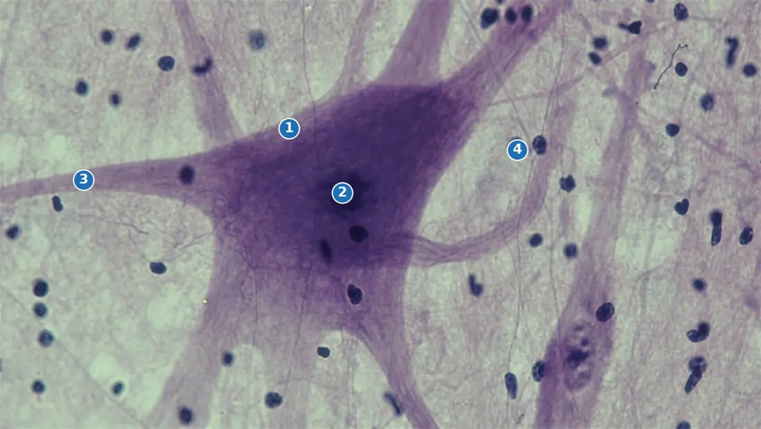
A neuron never divides after birth. Its cell body (cyton) contains the nucleus and dark-staining Nissl granules (rough ER) that make the neurotransmitter; dendrites carry impulses towards the cell body; the single axon carries them away. In myelinated neurons, Schwann cells wrap the axon in an insulating fatty myelin sheath, broken every ~1 mm at a node of Ranvier. Depending on their job, neurons are sensory (afferent), motor (efferent) or interneurons (relay).
The nerve impulse
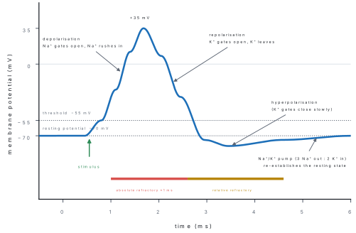
At rest the axon membrane is polarised: the Na⁺/K⁺ pump throws out 3 Na⁺ for every 2 K⁺ it takes in, and the membrane leaks K⁺ freely but Na⁺ hardly at all, so the inside is about −70 mV with respect to the outside. A stimulus that depolarises a patch to the threshold of −55 mV throws open the voltage-gated Na⁺ channels; Na⁺ floods in and the potential shoots to about +35 mV (depolarisation). The Na⁺ gates then shut and K⁺ gates open, so K⁺ leaves and the inside becomes negative again (repolarisation), briefly overshooting to about −80 mV (hyperpolarisation). During the absolute refractory period of roughly 1 ms no second impulse is possible at all — this is what fixes the maximum firing frequency and makes the impulse travel one way only.
A stimulus below threshold produces no impulse; any stimulus at or above threshold produces an action potential of exactly the same size. A stronger stimulus does not give a bigger impulse — it gives more impulses per second and recruits more neurons. That is how the brain codes intensity.
In an unmyelinated axon the impulse creeps along at 0.5–2 m s⁻¹. In a myelinated axon current can only enter at the nodes of Ranvier, so the impulse leaps from node to node — saltatory conduction — reaching 100–120 m s⁻¹ in the thickest human fibres, and using far less ATP.
At a synapse the impulse cannot jump the 20 nm cleft electrically. Ca²⁺ entering the knob makes vesicles fuse with the pre-synaptic membrane and release a neurotransmitter (acetylcholine at the neuromuscular junction, also noradrenaline, dopamine, GABA, serotonin). It diffuses across, binds receptors on the post-synaptic membrane and starts a new impulse; the enzyme cholinesterase then destroys it so the synapse resets. Because vesicles are only on one side, a synapse conducts in one direction only.
Problem. A nerve is stimulated at the elbow and again at the wrist of a patient in Bharatpur. The muscle responds 4.0 ms after the wrist stimulus and 9.5 ms after the elbow stimulus. The two stimulating points are 33 cm apart. Find the conduction velocity of the nerve, and say whether the fibre is myelinated.
Solution. The delay at the neuromuscular junction is the same for both stimuli, so subtracting removes it:
60 m s⁻¹ is far above the 0.5–2 m s⁻¹ of unmyelinated fibres, so this is a myelinated fibre conducting saltatorily. (Values below about 40 m s⁻¹ in a motor nerve suggest demyelination.)
The brain
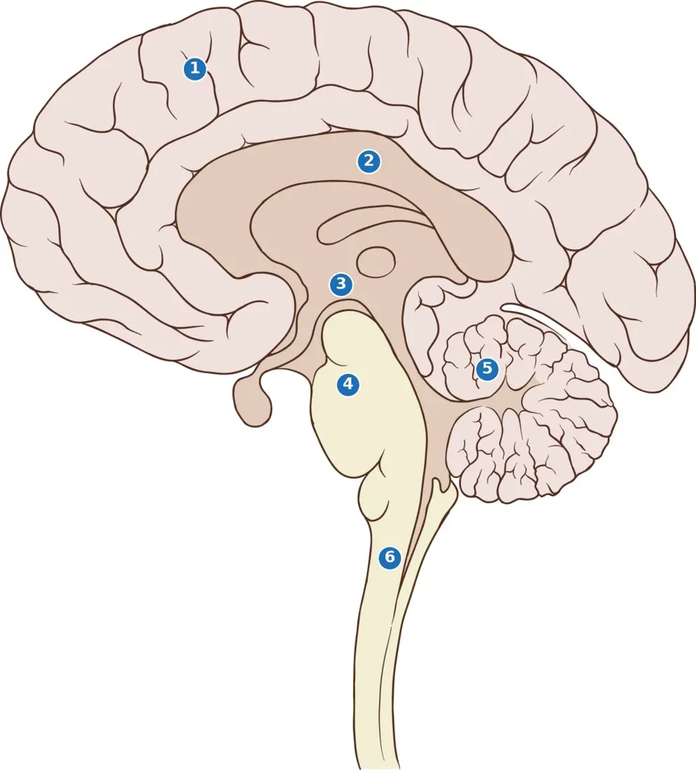
The brain weighs about 1.35 kg, floats in cerebrospinal fluid and is wrapped in three meninges (dura, arachnoid, pia mater). Its parts:
| Part | Position | Main functions |
|---|---|---|
| Cerebrum (2 hemispheres) | forebrain, largest part | intelligence, memory, speech, will, all voluntary action; sensory perception |
| Corpus callosum | between the hemispheres | band of nerve fibres joining left and right |
| Thalamus | forebrain floor | relay station for all sensory impulses except smell |
| Hypothalamus | below the thalamus | temperature, hunger, thirst, sleep, emotion; controls the pituitary |
| Mid-brain | brainstem | reflex centres for sight and hearing (pupil, head-turning) |
| Cerebellum | hindbrain, behind | balance, posture, coordination of muscle action |
| Pons Varolii | hindbrain | bridge between the two halves of the cerebellum; helps control breathing |
| Medulla oblongata | lowest, joins spinal cord | involuntary vital centres: heartbeat, breathing, blood pressure, swallowing, vomiting |
The spinal cord runs from the medulla to the first lumbar vertebra inside the vertebral column. In section, its grey matter (cell bodies) is central and butterfly-shaped, while the white matter (myelinated tracts) is outside — the opposite arrangement to the brain.
The reflex arc
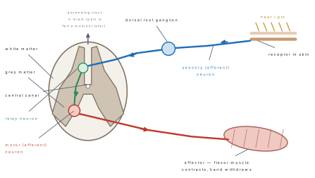
A reflex action is a rapid, automatic, involuntary response to a stimulus. The path the impulse takes is the reflex arc, and it has five parts:
receptor → sensory neuron → CNS (relay neuron) → motor neuron → effector
Touching a hot tawa takes the impulse from the skin receptor along the sensory neuron (whose cell body sits in the dorsal root ganglion) into the dorsal horn of the spinal cord, across a relay neuron, out along the motor neuron through the ventral root, to the flexor muscle — all in about 20 ms. A branch also runs up the ascending tract to the brain, which is why you feel the pain only after your hand has already moved. Inborn (unconditioned) reflexes such as blinking, sneezing and the knee jerk are present from birth; conditioned reflexes, like Pavlov's dogs salivating at a bell, are learnt and need the cerebral cortex.
The brain is informed, but it is not consulted. Marks are lost for writing that the impulse "goes to the brain and comes back" — in a spinal reflex the decision is taken in the spinal cord. Note too that the knee jerk is monosynaptic: there is no relay neuron, the sensory neuron synapses directly with the motor neuron.
8.6 Sense organs
The five sense organs are the eye (photoreceptor), ear (phono- and statoreceptor), nose (olfactoreceptor), tongue (gustatoreceptor) and skin (thermo-, tango- and algesireceptor).
The eye
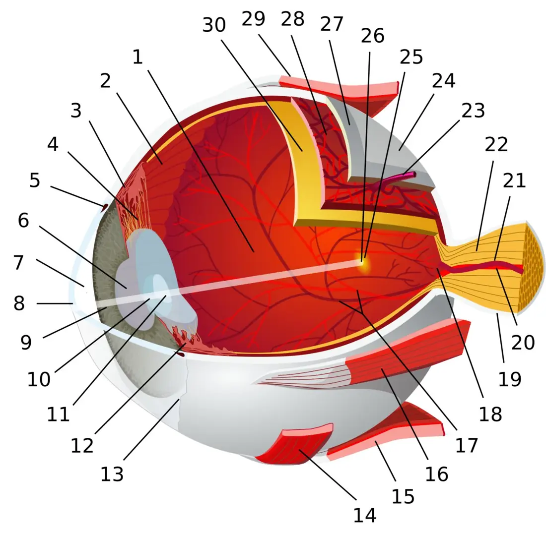
The eyeball has three coats — the tough sclera outside (transparent in front as the cornea), the pigmented, blood-rich choroid, and the light-sensitive retina. The retina carries about 120 million rods (pigment rhodopsin; dim light, black-and-white vision) and about 6–7 million cones (pigment iodopsin; bright light, colour). Cones are packed most densely at the yellow spot (fovea centralis), which gives the sharpest image; where the optic nerve leaves there are no receptors at all, giving the blind spot. The lens is held by suspensory ligaments from the ciliary body; when the ciliary muscle contracts the ligaments slacken and the elastic lens becomes more convex, focusing near objects — this is accommodation. Vitamin A deficiency destroys rhodopsin and causes night blindness, a real public-health problem in the Terai.
Common defects of the eye follow from the geometry. In myopia (short sight) the eyeball is too long, so the image of a distant object falls in front of the retina; a concave (diverging) lens corrects it. In hypermetropia (long sight) the eyeball is too short and the image would fall behind the retina; a convex (converging) lens corrects it. Presbyopia is the stiffening of the lens with age, astigmatism is an unevenly curved cornea, and a cataract is a lens that has gone opaque — treated by replacing it with an artificial one.
The ear
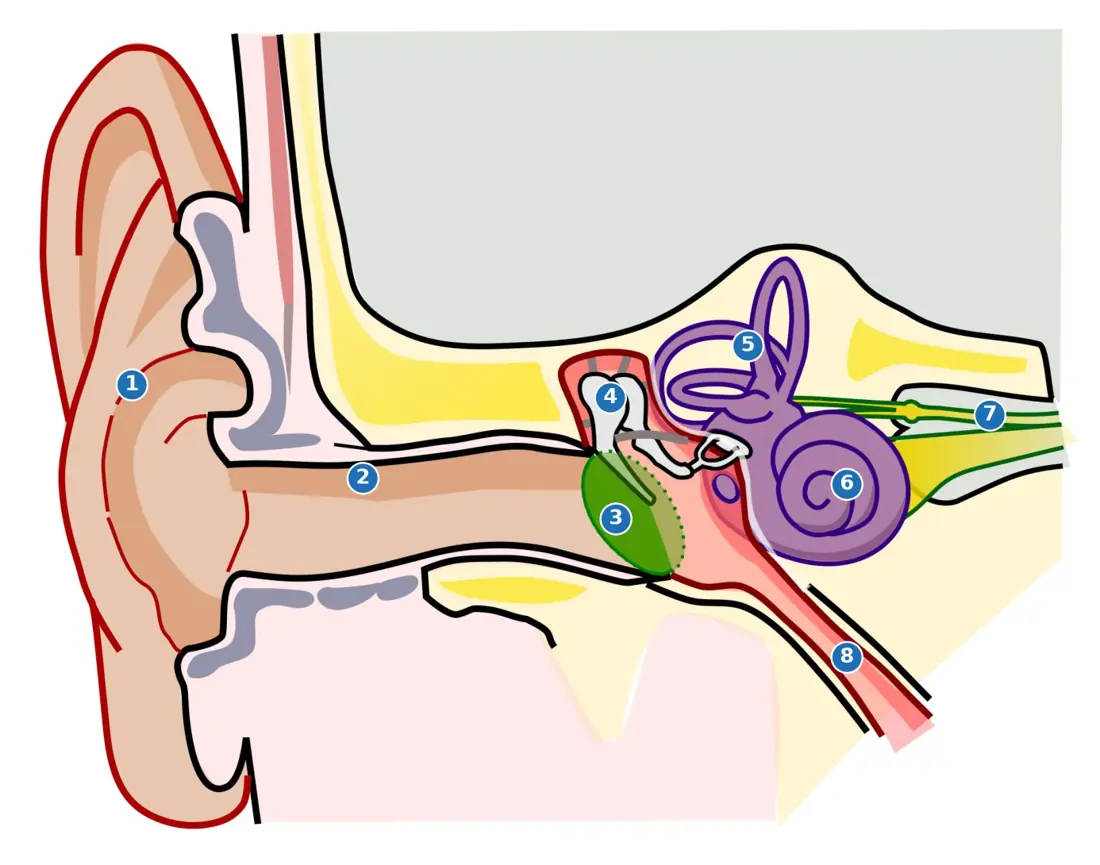
Sound waves collected by the pinna travel down the auditory canal and vibrate the tympanic membrane. The three ear ossicles — malleus → incus → stapes, the smallest bones in the body — act as levers that amplify the vibration about 20 times and pass it through the oval window into the fluid of the cochlea. Inside the cochlea, the organ of Corti on the basilar membrane carries hair cells that convert the fluid wave into impulses, which the auditory nerve carries to the temporal lobe. The round window lets the fluid move; the Eustachian tube connects the middle ear to the pharynx and equalises air pressure on the two sides of the ear drum (which is why the ears "pop" on the drive up to Muktinath). The three semicircular canals, set in three planes at right angles, detect rotation (dynamic balance), while the utriculus and sacculus with their otoliths detect the position of the head (static balance). Humans hear 20 Hz to 20 000 Hz.
| Sense organ | Receptor | Stimulus detected |
|---|---|---|
| Eye | rods and cones of the retina | light (photoreceptor) |
| Ear | organ of Corti; cristae and maculae | sound; rotation and gravity |
| Nose | olfactory epithelium | chemicals in air (smell) |
| Tongue | taste buds (sweet, sour, salt, bitter, umami) | chemicals in solution |
| Skin | Meissner, Pacinian, Krause, Ruffini, free nerve endings | touch, pressure, cold, heat, pain |
8.7 Endocrinology
The endocrine system is the body's slow but far-reaching control system. Endocrine (ductless) glands pour their secretion, a hormone, straight into the blood, which carries it to distant target organs that have matching receptors. Hormones are needed in minute amounts, are not stored in the target, and are destroyed after use — so they must be secreted continuously.
| Feature | Nervous system | Endocrine system |
|---|---|---|
| Message carried by | electrical impulse along a neuron | chemical hormone in blood |
| Route | fixed "wire" to one target | whole bloodstream, any cell with the receptor |
| Speed of response | milliseconds | seconds to hours |
| Duration of effect | very brief | long, sometimes lifelong (growth) |
| Control of | muscles and glands | metabolism, growth, reproduction |
| Feedback | reflex arc | negative feedback loops |
Chemically, hormones are of three kinds: peptides/proteins (insulin, GH, ADH), steroids (cortisol, aldosterone, testosterone, oestrogen — made from cholesterol) and amino-acid derivatives (thyroxine, adrenaline). Because steroids are lipid-soluble they pass straight through the cell membrane and act on the genes; peptide hormones cannot, so they bind a surface receptor and work through a second messenger such as cyclic AMP.
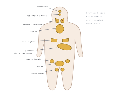
Hypothalamus and the hypophysis (pituitary gland)
The hypophysis or pituitary is a pea-sized gland, about 0.5 g, hanging by a stalk (the infundibulum) from the floor of the hypothalamus and resting in the sella turcica, a hollow in the sphenoid bone. It has two parts of completely different origin:
- The adenohypophysis (anterior lobe) develops from Rathke's pouch, an upgrowth of the embryonic mouth roof. It is true glandular tissue and makes its own hormones.
- The neurohypophysis (posterior lobe) is a downgrowth of the brain. It makes nothing: it only stores and releases two hormones manufactured in the hypothalamus.
The hypothalamus controls the anterior lobe chemically, through a private blood route called the hypothalamo-hypophyseal portal system — capillaries in the hypothalamus drain into veins that break up into capillaries again in the anterior lobe. Hypothalamic neurons pour releasing hormones (GnRH, TRH, CRH, GHRH) and inhibiting hormones (somatostatin, PIH) into this portal blood in nanogram amounts, and the anterior lobe responds. The posterior lobe is controlled neurally instead: the hormones ADH and oxytocin are made in the supraoptic and paraventricular nuclei of the hypothalamus and travel down the axons themselves.
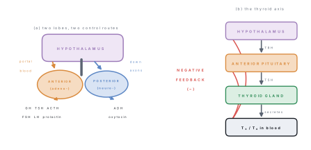
Hormones of the anterior lobe. Six, and NEB expects all six.
- Growth hormone (GH, somatotropin) — promotes growth of the long bones and muscles, protein synthesis and fat breakdown. Hyposecretion in childhood gives pituitary dwarfism (small but normally proportioned, normal intelligence); hypersecretion in childhood gives gigantism; hypersecretion after the epiphyses have closed gives acromegaly — thickened jaw, hands and feet.
- Thyroid-stimulating hormone (TSH) — drives the thyroid to make T₃ and T₄.
- Adrenocorticotropic hormone (ACTH) — drives the adrenal cortex to make glucocorticoids.
- Follicle-stimulating hormone (FSH) — ripens the Graafian follicle in the ovary; starts spermatogenesis in the testis.
- Luteinising hormone (LH), called ICSH in the male — triggers ovulation and forms the corpus luteum; stimulates Leydig cells to make testosterone.
- Prolactin (LTH) — starts and maintains milk secretion after childbirth.
The pars intermedia secretes melanocyte-stimulating hormone (MSH), which disperses melanin granules; in humans it is nearly vestigial. Because the anterior lobe controls the thyroid, adrenal cortex and gonads, it is traditionally called the master gland — although the hypothalamus is the real master above it.
Hormones of the posterior lobe. Both are small nonapeptides (9 amino acids).
- ADH (vasopressin) makes the DCT and collecting duct permeable to water, concentrating the urine (§8.4). Deficiency causes diabetes insipidus: 10–20 litres of dilute, tasteless urine a day and raging thirst.
- Oxytocin contracts the smooth muscle of the uterus during labour and causes milk ejection from the mammary glands. It is the textbook example of positive feedback — stretching of the cervix triggers oxytocin, which contracts the uterus, which stretches the cervix more, until the baby is delivered.
Thyroid gland
The thyroid is the largest purely endocrine gland, about 25 g, made of two lobes on either side of the trachea joined across the front by a bridge, the isthmus. Its tissue is a mass of hollow follicles filled with a jelly called colloid, which stores the hormone bound to the protein thyroglobulin — the only endocrine gland that stockpiles its product outside its cells.
The follicle cells trap iodide from the blood and build thyroxine (T₄, tetraiodothyronine) and triiodothyronine (T₃). T₃ is several times more active but is made in smaller amount. Their job is to set the basal metabolic rate: they raise oxygen consumption and heat production in almost every cell, speed up carbohydrate and fat oxidation, and are essential for normal growth and brain development in the infant. They are often called the personality hormone because a deficiency dulls mental activity. An adult needs about 150 µg of iodine a day.
Scattered between the follicles are the parafollicular (C) cells, which secrete calcitonin — this lowers blood calcium by inhibiting the release of calcium from bone. It opposes parathormone.
A goitre is simply a swollen thyroid, and it happens in both directions. In endemic (simple) goitre there is too little dietary iodine, so T₄ cannot be made; T₄ never rises, TSH is never switched off by feedback, and the gland enlarges as it is flogged by TSH — the hormone level is low. In Graves' disease (exophthalmic goitre) the gland is enlarged and overactive, with a racing pulse, weight loss despite a good appetite and bulging eyeballs. Hill districts of Nepal were historically iodine-deficient, which is why salt is iodised.
| Thyroid disorder | Direction | Main features |
|---|---|---|
| Simple (endemic) goitre | Iodine lack, low T₄ | Swollen neck, sluggishness |
| Cretinism | Hyposecretion in infancy | Stunted growth, retarded mental development, protruding tongue |
| Myxoedema (Gull's disease) | Hyposecretion in adult | Puffy face, dry skin, slow pulse, low BMR, weight gain, feels cold |
| Graves' disease | Hypersecretion | High BMR, weight loss, rapid heart, tremor, exophthalmos |
Parathyroid glands
Four small glands — about 0.05 g each — are embedded in pairs on the back surface of the thyroid lobes. They secrete parathormone (PTH, Collip's hormone), the main regulator of blood calcium, which is normally held at 9–11 mg per 100 ml. PTH raises blood Ca²⁺ in three ways: it activates osteoclasts so bone releases calcium; it increases calcium reabsorption (and phosphate excretion) in the kidney tubule; and it activates vitamin D, which increases calcium absorption from the intestine.
Hyposecretion drops blood calcium and causes hypocalcaemic tetany — sustained, painful spasms of the muscles of the hands, feet and larynx, which can be fatal. Hypersecretion strips calcium out of the skeleton, leaving bones soft and easily broken, and deposits it as kidney stones.
Adrenal (suprarenal) glands
A pair of caps, one on the upper pole of each kidney, each about 5 g. Each is really two glands in one, with different embryonic origins:
The cortex (outer, mesodermal, essential for life) has three zones, from outside in:
| Zone | Hormone class | Example | Action |
|---|---|---|---|
| Zona glomerulosa | Mineralocorticoid | Aldosterone | Na⁺ and water reabsorbed, K⁺ excreted in DCT/collecting duct → raises blood volume and BP |
| Zona fasciculata | Glucocorticoid | Cortisol | Gluconeogenesis from protein and fat, raises blood glucose, anti-inflammatory, suppresses immunity |
| Zona reticularis | Sex corticoid | Androgens | Small amounts; pubic and axillary hair |
The medulla (inner, ectodermal, derived from neural crest — effectively a modified sympathetic ganglion) secretes adrenaline (epinephrine), about 80 %, and noradrenaline, about 20 %. These are the emergency hormones of fight, flight and fright: heart rate and force rise, blood pressure rises, bronchioles and pupils dilate, blood is shunted from the gut and skin to the skeletal muscles, and glycogenolysis floods the blood with glucose. The medulla is not essential for life — the sympathetic nerves can do the same work.
| Adrenal disorder | Cause | Features |
|---|---|---|
| Addison's disease | Cortical hyposecretion | Bronze pigmentation of skin, low blood Na⁺ and BP, weakness, weight loss |
| Cushing's syndrome | Glucocorticoid hypersecretion | Moon face, buffalo hump, trunk obesity with thin limbs, high blood glucose |
| Conn's syndrome | Aldosterone hypersecretion | Na⁺ retention, K⁺ loss, high blood pressure |
Islets of Langerhans (endocrine pancreas)
The pancreas is a heterocrine (mixed) gland: the acini are exocrine and pour pancreatic juice into the duodenum (§8.1), while about 1–2 % of its mass is scattered as one to two million ductless clumps, the islets of Langerhans.
| Islet cell | Share | Hormone | Effect on blood glucose |
|---|---|---|---|
| β (beta) | ~70 % | Insulin | Lowers it — glucose enters cells, glycogenesis in liver and muscle, lipogenesis |
| α (alpha) | ~20 % | Glucagon | Raises it — glycogenolysis and gluconeogenesis in the liver |
| δ (delta) | ~5 % | Somatostatin | Inhibits both of the above |
| F (PP) | ~2 % | Pancreatic polypeptide | Damps pancreatic secretion |
Insulin (Banting and Best, 1921) is a protein of 51 amino acids in two chains joined by disulphide bridges. Fasting blood glucose is normally 70–110 mg per 100 ml; insulin and glucagon are antagonists that hold it there.
Diabetes mellitus follows a lack of insulin or a failure of the tissues to respond to it. Blood glucose climbs past the renal threshold of 180 mg/100 ml, so glucose appears in the urine (glycosuria) and drags water with it osmotically. The classical signs are the three P's: polyuria (excessive urine), polydipsia (excessive thirst) and polyphagia (excessive hunger), with weight loss. Because glucose cannot be used, fat is burned instead and ketone bodies accumulate, giving acidosis. Type 1 (IDDM) is autoimmune destruction of β cells, begins young and needs injected insulin; Type 2 (NIDDM) is insulin resistance, begins in middle age and is linked to obesity and inactivity.
Gonads, pineal body and thymus
- Testis — the interstitial cells of Leydig, stimulated by LH/ICSH, secrete testosterone: it drives spermatogenesis and the male secondary sexual characters (beard, deep voice, muscular build). Sertoli cells secrete inhibin, which feeds back to damp FSH.
- Ovary — the ripening Graafian follicle secretes oestrogen (female secondary sexual characters, repair of the uterine lining); the corpus luteum secretes progesterone, the "pregnancy hormone", which makes the endometrium secretory and holds a pregnancy; relaxin loosens the pubic symphysis at birth.
- Pineal body (epiphysis cerebri) — a tiny gland on the roof of the diencephalon secreting melatonin, which governs the daily (circadian) sleep–wake rhythm, pigmentation and the timing of puberty — it inhibits the gonads until puberty. Secretion is high in darkness.
- Thymus — behind the sternum; secretes thymosin, under which T-lymphocytes mature. It is large in the child and atrophies after puberty, which is why immunity weakens with age.
Some organs that are not glands also secrete hormones: the stomach (gastrin), the duodenum (secretin, cholecystokinin), the kidney (erythropoietin, which drives red-cell production) and the heart (atrial natriuretic factor, which lowers blood pressure).
| Gland | Hormone | Target | Principal effect |
|---|---|---|---|
| Hypothalamus | Releasing/inhibiting hormones | Anterior pituitary | Switch each pituitary hormone on or off |
| Anterior pituitary | GH | Bones, muscles | Growth, protein synthesis |
| TSH | Thyroid | T₃/T₄ secretion | |
| ACTH | Adrenal cortex | Glucocorticoid secretion | |
| FSH | Ovary / testis | Follicle ripening; spermatogenesis | |
| LH (ICSH) | Ovary / testis | Ovulation, corpus luteum; testosterone | |
| Prolactin | Mammary gland | Milk secretion | |
| Posterior pituitary | ADH | Kidney tubule | Water reabsorption |
| Oxytocin | Uterus, mammary gland | Labour contractions, milk ejection | |
| Thyroid | T₄ / T₃ | All cells | Raise BMR; growth and brain development |
| Calcitonin | Bone | Lowers blood Ca²⁺ | |
| Parathyroid | Parathormone | Bone, kidney, gut | Raises blood Ca²⁺ |
| Adrenal cortex | Aldosterone | Kidney tubule | Na⁺ retention, K⁺ loss, raises BP |
| Cortisol | Liver, immune cells | Raises blood glucose, anti-inflammatory | |
| Adrenal medulla | Adrenaline | Heart, vessels, liver | Emergency response, raises glucose and BP |
| Pancreas (β) | Insulin | Liver, muscle, fat | Lowers blood glucose |
| Pancreas (α) | Glucagon | Liver | Raises blood glucose |
| Testis | Testosterone | Whole body | Male secondary sexual characters |
| Ovary | Oestrogen | Whole body | Female secondary sexual characters |
| Progesterone | Uterus | Maintains pregnancy | |
| Pineal | Melatonin | Brain, gonads | Circadian rhythm, delays puberty |
| Thymus | Thymosin | T-lymphocytes | Maturation of T cells |
Blood glucose is raised by glucagon, adrenaline, cortisol and GH, but lowered by insulin alone. That is why the loss of one hormone — insulin — causes a disease as serious as diabetes mellitus: there is no substitute for it.
8.8 Reproductive System
Every other system in this chapter keeps one body alive. The reproductive system keeps the species alive, and it is the only system that is not needed for the individual's own survival. In humans reproduction is sexual, viviparous (the young develop inside the mother) and dioecious (the sexes are in separate individuals), with internal fertilisation.
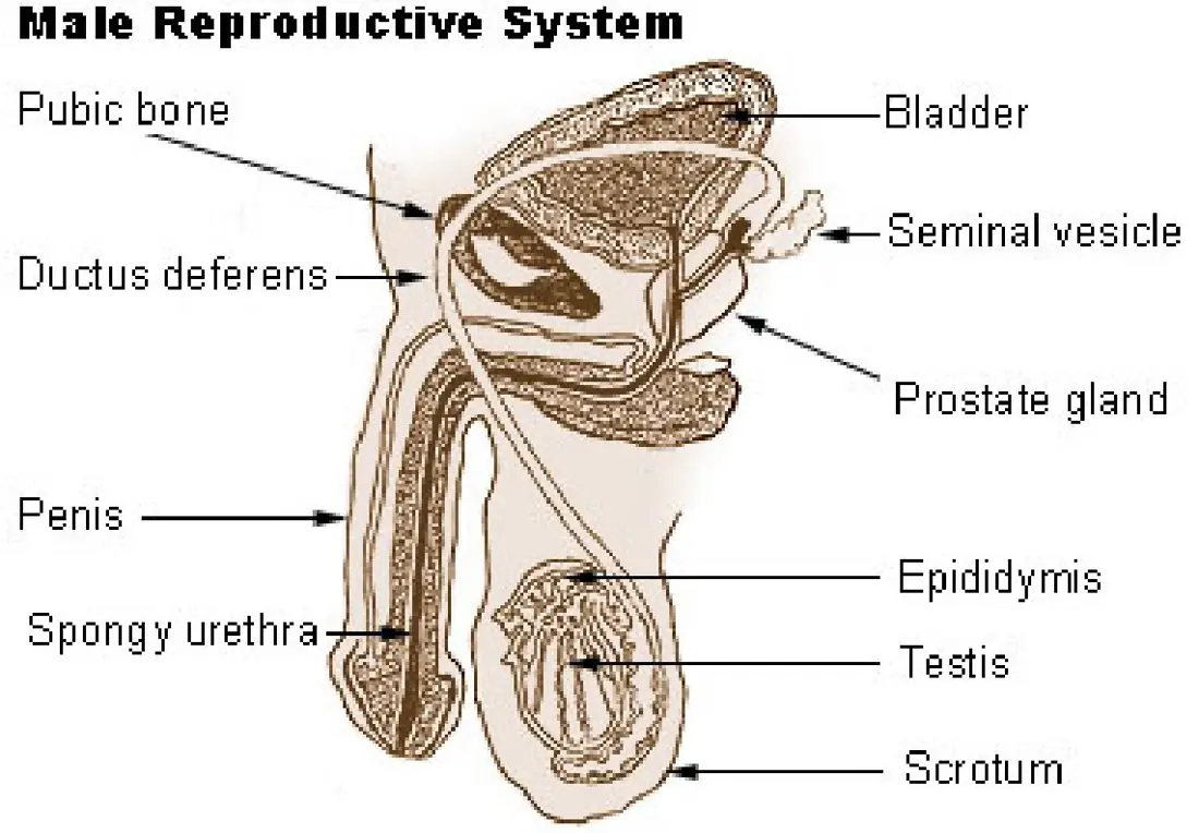
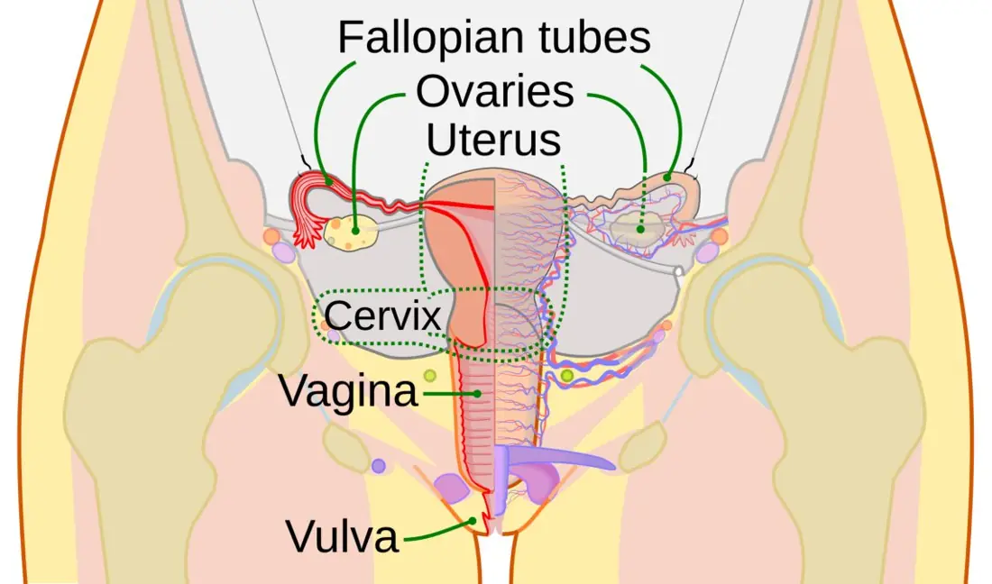
Male reproductive system
Testes. A pair of oval glands, about 4 × 2.5 cm, held outside the abdomen in the scrotum — a skin sac that keeps them 2–2.5 °C below body temperature, because spermatogenesis fails at core temperature. Each testis is divided into about 250 lobules, and each lobule holds one to three tightly coiled seminiferous tubules, the site of sperm formation. Two kinds of cell line the tubule: germinal cells, which divide to make sperms, and tall Sertoli (nurse) cells, which nourish the developing sperms and secrete inhibin. In the connective tissue between the tubules lie the interstitial cells of Leydig, which make testosterone.
Duct system. Seminiferous tubules → rete testis → vasa efferentia → epididymis (a 6-metre coiled tube on the back of the testis where sperms mature and are stored) → vas deferens → ejaculatory duct → urethra, which passes through the penis and is a shared passage for urine and semen.
Accessory glands. The paired seminal vesicles contribute about 60 % of the semen, a fluid rich in fructose that fuels the sperm tail. The prostate adds a thin alkaline milky fluid, about 30 %, which neutralises the acidity of the vagina. The two Cowper's (bulbourethral) glands add a lubricating mucus. An ejaculation is 2–5 ml of semen carrying roughly 200–300 million sperms.
The sperm is about 60 µm long and has four parts: the head, mostly nucleus, capped by the enzyme-filled acrosome that dissolves a path through the egg coverings; the neck, with the centrioles; the middle piece, packed with a spiral of mitochondria that supply ATP; and the whip-like tail (flagellum) that drives it forward.
Female reproductive system
Ovaries. A pair of almond-shaped glands, about 3 cm long, in the pelvic cavity. They are both gamete-producing and endocrine. A newborn girl already has about 1–2 million primary oocytes; only some 400 will ever be ovulated.
Fallopian tubes (oviducts). Two 10–12 cm tubes. The funnel-shaped infundibulum next to the ovary carries finger-like fimbriae that sweep the released egg in; the wide ampulla that follows is where fertilisation normally happens; the narrow isthmus opens into the uterus. Cilia and muscular waves move the egg along.
Uterus (womb). A hollow, pear-shaped muscular organ the size of a fist, with a fundus, body and a narrow neck, the cervix, opening into the vagina. Its wall has three coats: the outer perimetrium, the thick muscular myometrium that contracts at birth, and the inner glandular endometrium, which is rebuilt and shed every month and into which the embryo implants.
The menstrual cycle
At menarche (about 11–14 years) a girl's reproductive tract begins a repeating 28-day cycle which continues, except during pregnancy, until menopause at about 45–50. Day 1 of the cycle is the first day of bleeding.
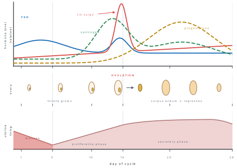
The cycle has four phases, and each is driven by the hormones plotted in the figure.
| Phase | Days | Hormones | What happens |
|---|---|---|---|
| Menstrual | 1–5 | All four low | The corpus luteum of the last cycle has died, so progesterone collapses; the endometrium breaks down and is shed as menstrual flow (50–80 ml of blood, mucus and tissue) |
| Proliferative (follicular) | 6–13 | FSH ↑, then oestrogen ↑ | FSH ripens one Graafian follicle, which secretes oestrogen; the endometrium is rebuilt and thickens |
| Ovulatory | 14 | LH surge | High oestrogen triggers a sudden burst of LH; the follicle ruptures and the secondary oocyte is released |
| Secretory (luteal) | 15–28 | Progesterone ↑ | The empty follicle becomes the yellow corpus luteum, whose progesterone makes the endometrium thick, glandular and ready to receive an embryo |
If fertilisation does not occur, the corpus luteum degenerates into the corpus albicans by about day 26, progesterone falls, and menstruation begins again. If fertilisation does occur, the young embryo secretes hCG (human chorionic gonadotropin), which keeps the corpus luteum alive — so progesterone stays high and there is no menstruation. This is exactly what a pregnancy test detects in urine.
The oocyte survives only about 24 hours after ovulation, but sperms can survive 3–5 days in the female tract. The fertile period is therefore roughly days 10–17 of a 28-day cycle, not a single day. And the luteal phase is the fixed 14 days — it is the follicular phase that varies from woman to woman, which is why "day 14" is only an average.
Gametogenesis, fertilisation and gestation
Gametogenesis is meiotic, so each gamete is haploid (n = 23).
| Spermatogenesis | Oogenesis | |
|---|---|---|
| Site | Seminiferous tubules | Ovary |
| Begins | At puberty | In foetal life, completed after puberty |
| Product of one diploid cell | 4 functional sperms | 1 ovum + 3 polar bodies |
| Cytoplasm division | Equal | Unequal — the ovum keeps it all |
| Duration | About 64–74 days, continuous | One ovum per month, interrupted for years |
| Ends | At death (slows with age) | At menopause |
Meiosis in the female is halted twice: the primary oocyte stops in prophase I until ovulation, and the secondary oocyte stops in metaphase II and only finishes meiosis if a sperm enters.
Fertilisation takes place in the ampulla of the fallopian tube, normally within 24 hours of ovulation. The acrosome releases hyaluronidase and other enzymes that digest a path through the corona radiata and zona pellucida; the moment one sperm fuses with the oocyte membrane, the cortical reaction hardens the zona and blocks all other sperms (preventing polyspermy). The oocyte completes meiosis II, the two haploid nuclei fuse, and the diploid zygote is formed.
The zygote divides as it travels down the tube, becoming a solid morula and then a hollow blastocyst, which implants in the endometrium about 6–7 days after fertilisation. The chorion of the embryo and the endometrium of the mother together build the placenta, across which oxygen and nutrients diffuse in and CO₂ and nitrogenous waste diffuse out — the two bloodstreams come close but never mix. The placenta is also a temporary endocrine gland, making hCG, oestrogen and progesterone.
Gestation lasts about 280 days (40 weeks) counted from the first day of the last menstrual period. Birth (parturition) is triggered by a foetal–maternal hormone cascade in which oxytocin from the posterior pituitary contracts the myometrium in a positive-feedback loop. After delivery, prolactin starts milk secretion and oxytocin ejects it; the first milk, colostrum, is thin and yellowish and is rich in antibodies (IgA) that give the newborn passive immunity.
Problem. A girl reaches menarche at 13 and menopause at 49. If she ovulates once every 28 days and has two pregnancies, each removing 10 months of cycling, estimate the number of ova she releases in her lifetime. Compare this with the 1 million primary oocytes present at birth.
Solution. Total reproductive span = 49 − 13 = 36 years = 36 × 365 = 13 140 days. Time lost to pregnancy = 2 × 10 months = 20 months = 20 × 30 = 600 days. Cycling time = 13 140 − 600 = 12 540 days.
Fraction of the original stock used:
Fewer than one oocyte in two thousand is ever ovulated; the rest degenerate by atresia. This is why a female is born with all the oocytes she will ever have, yet never runs short of them before menopause.
Chapter summary
- Digestion is chemical hydrolysis by enzymes, each with its own optimum pH: salivary amylase 6.8, pepsin 1.5–2.0, pancreatic and intestinal enzymes 7–8. Absorption happens in the ileum across villi, which raise the surface area to about 250 m².
- Breathing moves air; respiration releases energy. Tidal volume 500 ml, vital capacity 4500 ml, total lung capacity 5800 ml, residual volume 1200 ml. Pulmonary ventilation = tidal volume × breathing rate ≈ 500 × 12 = 6000 ml min⁻¹.
- Gas exchange is pure diffusion across the 0.5 µm alveolar wall over about 70 m² of surface. Oxygen travels as oxyhaemoglobin (1 g Hb carries 1.34 ml O₂); most CO₂ travels as bicarbonate.
- Cardiac output = \text{stroke volume} \times \text{heart rate} = 70 \times 72 \approx 5000 ml min⁻¹. The cardiac cycle lasts 0.8 s (atrial systole 0.1 s, ventricular systole 0.3 s, joint diastole 0.4 s); normal BP is 120/80 mmHg. The heart beats myogenically, from the SA node.
- Urine formation = ultrafiltration + selective reabsorption + tubular secretion. Net filtration pressure = 55 - (15 + 30) = 10 mmHg; GFR = 125 ml min⁻¹ = 180 L day⁻¹, of which more than 99 % is reabsorbed to leave 1–1.5 L of urine.
- A nerve impulse is a self-propagating reversal of membrane potential from −70 mV to +35 mV, restored by the Na⁺–K⁺ pump (3 Na⁺ out : 2 K⁺ in). Conduction is saltatory and up to 100 times faster in myelinated fibres. Conduction velocity = distance ÷ time.
- Hormones are chemical messengers carried in the blood: slow to start, long to act, controlled by negative feedback through the hypothalamo-hypophyseal axis. Insulin alone lowers blood glucose (normal 70–110 mg per 100 ml); glucagon, adrenaline, cortisol and GH raise it.
- The menstrual cycle is 28 days: menses 1–5, proliferative 6–13, ovulation at 14 on the LH surge, secretory 15–28 under progesterone from the corpus luteum. Fertilisation occurs in the ampulla; gestation is 280 days.
Practice questions
Group A — Multiple choice (1 mark each)
- The enzyme that works best at pH 1.5–2.0 is [1]asalivary amylasebpepsinctrypsindintestinal lipase
- Which lung volume cannot be measured with a spirometer? [1]atidal volumebinspiratory reserve volumecvital capacitydresidual volume
- If the stroke volume is 80 ml and the heart rate is 75 beats per minute, the cardiac output is [1]a4 L min⁻¹b5 L min⁻¹c6 L min⁻¹d7.5 L min⁻¹
- Ultrafiltration of blood takes place in the [1]aproximal convoluted tubulebglomerulus and Bowman's capsulecloop of Henledcollecting duct
- The resting membrane potential of a neuron is about [1]a+35 mVb−55 mVc−70 mVd−90 mV
- The blind spot of the retina contains [1]aonly rodsbonly conescboth rods and conesdneither rods nor cones
- Hyposecretion of thyroxine in an infant causes [1]amyxoedemabcretinismcGraves' diseasedtetany
- In a normal 28-day cycle, fertilisation of the ovum usually occurs in the [1]aovarybampulla of the fallopian tubecuterusdcervix
Answers to Group A
1. (b) — pepsin is the only enzyme of the strongly acidic gastric juice.
2. (d) — residual volume never leaves the lungs, so no spirometer can collect it.
3. (c) — 80 \times 75 = 6000 ml min⁻¹ = 6 L min⁻¹.
4. (b) — the pressure difference across the glomerular capillaries drives filtration into Bowman's capsule.
5. (c) — −70 mV inside relative to outside; −55 mV is the threshold, not the resting value.
6. (d) — the optic disc is where the optic nerve leaves, so it has no photoreceptors at all.
7. (b) — in the infant it stunts body and brain growth (cretinism); the adult form is myxoedema.
8. (b) — the wide ampulla of the oviduct, within about 24 hours of ovulation.
Group B — Short answer (4 marks each)
- Draw a labelled diagram of an intestinal villus and explain three ways in which it is adapted for absorption. [4]
- Distinguish between breathing and respiration. Define tidal volume and vital capacity, giving their normal values in an adult. [4]
- An athlete breathes 20 times a minute with a tidal volume of 600 ml. The anatomical dead space is 150 ml. Calculate (i) the pulmonary ventilation and (ii) the alveolar ventilation, both in litres per minute. [4]
- Describe the events of one cardiac cycle and give the duration of each of its three phases. [4]
- In a patient the renal plasma flow is 600 ml min⁻¹ and the GFR is 120 ml min⁻¹. Calculate the filtration fraction. If 99 % of the filtrate is reabsorbed, what volume of urine is formed in 24 hours? [4]
- What is a reflex action? Draw a labelled diagram of the reflex arc of the knee-jerk reflex. [4]
- Two points on a nerve 12 cm apart are stimulated in turn; the muscle responds after 1.0 ms and 2.0 ms respectively. Calculate the conduction velocity in m s⁻¹ and state, with a reason, whether the fibre is myelinated. [4]
- Name the disorders caused by the hyper- and hyposecretion of thyroxine and of growth hormone, and give one sign of each. [4]
Answers to Group B
1. Diagram as in Figure 3. Adaptations: (i) each villus is 0.5–1 mm long and covered in microvilli, giving a brush border that raises the absorptive area to about 250 m²; (ii) the epithelium is one cell thick, so the diffusion path is very short; (iii) a dense blood capillary network carries glucose and amino acids away while a central lacteal carries fats, so the concentration gradient is never allowed to fall.
2. Breathing is the mechanical movement of air in and out of the lungs; respiration is the enzyme-controlled oxidation of food inside the cell to release ATP. Breathing is physical, involves no enzymes and happens in the lungs; respiration is chemical, needs enzymes and happens in every cell. Tidal volume = volume of air breathed in or out in one quiet breath ≈ 500 ml. Vital capacity = maximum volume that can be expelled after the deepest possible inspiration = IRV + TV + ERV ≈ 4500 ml (about 3500 ml in women).
3. (i) Pulmonary ventilation = \text{TV} \times \text{rate} = 600 \times 20 = 12\,000 ml min⁻¹ = 12 L min⁻¹. (ii) Only 600 - 150 = 450 ml of each breath reaches the alveoli, so alveolar ventilation = 450 \times 20 = 9000 ml min⁻¹ = 9 L min⁻¹. The 3 L min⁻¹ difference is air that merely fills the conducting airways and takes no part in gas exchange.
4. One cycle lasts 0.8 s at 75 beats per minute. Atrial systole (0.1 s): atria contract, pushing the last 30 % of blood through the open AV valves into the ventricles. Ventricular systole (0.3 s): ventricles contract, AV valves shut (first sound, "lubb"), pressure rises isovolumetrically until the semilunar valves open and about 70 ml is ejected into the aorta and pulmonary trunk. Joint diastole (0.4 s): ventricles relax, semilunar valves shut (second sound, "dupp"), AV valves open and the chambers refill passively.
5. Filtration fraction = \dfrac{\text{GFR}}{\text{RPF}} \times 100 = \dfrac{120}{600} \times 100 = \mathbf{20\,\%}. Filtrate in 24 h = 120 \times 60 \times 24 = 172\,800 ml = 172.8 L. Urine = 1\,\% of this = 0.01 \times 172.8 = \mathbf{1.73\ L\ day^{-1}}, which is in the normal range of 1–1.5 L.
6. A reflex action is an immediate, involuntary, stereotyped response to a stimulus, carried out over a fixed nerve pathway without waiting for the brain's decision. Diagram as in Figure 15: stretch receptor in the quadriceps tendon → sensory (afferent) neuron → dorsal root ganglion → spinal cord grey matter → motor (efferent) neuron through the ventral root → quadriceps muscle (effector), which contracts and extends the leg. The knee-jerk is monosynaptic — there is no relay neuron.
7. The extra 12 cm of nerve added 1.0 ms to the latency, so
This is far above the 0.5–2 m s⁻¹ of unmyelinated fibres, so the fibre is myelinated: the impulse jumps from node to node (saltatory conduction). Taking the difference between two stimulation points cancels out the synaptic and muscle delay, which is why two points are used rather than one.
8. Thyroxine — hypersecretion: Graves' disease (exophthalmic goitre), sign = protruding eyeballs with rapid pulse and weight loss; hyposecretion: myxoedema in the adult, sign = puffy dry skin and low BMR, or cretinism in the infant, sign = stunted growth with mental retardation. Growth hormone — hypersecretion before the epiphyses close: gigantism, sign = abnormal height; after they close: acromegaly, sign = enlarged jaw, hands and feet; hyposecretion in childhood: pituitary dwarfism, sign = very short stature with normal body proportions and normal intelligence.
Group C — Long answer (8 marks each)
- Draw a labelled diagram of the human heart in longitudinal section and describe the course of blood through the double circulation. A person has a stroke volume of 75 ml and a heart rate of 80 beats per minute; calculate the cardiac output, and find the cardiac reserve if the maximum output during exercise is 25 L min⁻¹. [8]
- Describe the structure of a nephron with a labelled diagram and explain the three steps of urine formation. Why is glucose absent from the urine of a healthy person but present in an untreated diabetic? [8]
- Describe the structure of a myelinated neuron. With the help of a graph of membrane potential against time, explain the origin and conduction of a nerve impulse, naming the value of the resting potential, the threshold and the peak. [8]
- Describe the menstrual cycle of a human female under the headings menstrual, proliferative, ovulatory and secretory phases, and explain the hormonal control of each. What happens to the corpus luteum if fertilisation occurs? [8]
Answers to Group C
1. Diagram as in Figures 6 and 7. Structure: four chambers — two thin-walled atria and two thick-walled ventricles, the left ventricle wall being about three times thicker because it pumps to the whole body. The right atrium and ventricle are separated by the tricuspid valve, the left pair by the bicuspid (mitral) valve; both are anchored by chordae tendineae to papillary muscles so they cannot turn inside out. Semilunar valves guard the openings of the pulmonary artery and the aorta. The interventricular septum prevents mixing. Double circulation: blood passes through the heart twice in one complete round. Pulmonary circuit: right atrium → tricuspid valve → right ventricle → pulmonary semilunar valve → pulmonary artery → lungs (CO₂ out, O₂ in) → pulmonary veins → left atrium. Systemic circuit: left atrium → bicuspid valve → left ventricle → aortic semilunar valve → aorta → body tissues → venae cavae → right atrium. The advantage is that oxygenated and deoxygenated blood never mix and full pressure can be delivered to the body without damaging the delicate lung capillaries. Calculation: \text{CO} = \text{SV} \times \text{HR} = 75 \times 80 = 6000 ml min⁻¹ = 6 L min⁻¹. Cardiac reserve = 25 - 6 = \mathbf{19\ L\ min^{-1}}, i.e. the heart can raise its output roughly four-fold during exercise.
2. Diagram as in Figure 11. A nephron is the structural and functional unit of the kidney; there are about 1 million in each kidney. It consists of a Malpighian body (glomerulus + Bowman's capsule) in the cortex, followed by the proximal convoluted tubule, the loop of Henle that dips into the medulla, the distal convoluted tubule, and the collecting duct. (i) Ultrafiltration. Blood enters the glomerulus through a wide afferent arteriole and leaves by a narrower efferent one, so hydrostatic pressure is high. Net filtration pressure = 55 - (15 + 30) = 10 mmHg drives water and all small solutes into the capsule; blood cells and plasma proteins are held back. GFR = 125 ml min⁻¹ = 180 L day⁻¹. (ii) Selective reabsorption. The PCT returns 70–80 % of the filtrate — all the glucose and amino acids by active transport, Na⁺ actively and water osmotically. The loop of Henle builds the medullary salt gradient (300 → 1200 mOsm L⁻¹) by counter-current multiplication, and the DCT and collecting duct reabsorb the last water under ADH. (iii) Tubular secretion. H⁺, K⁺, ammonia, creatinine and drugs are actively added from the blood into the DCT, which also regulates blood pH. More than 99 % of the 180 L is reabsorbed, leaving 1–1.5 L of urine. Glucose: in a healthy person the PCT carriers reabsorb all the filtered glucose, so none remains. In an untreated diabetic there is too little insulin, so blood glucose rises above the renal threshold of 180 mg per 100 ml; the carriers are saturated and the excess glucose passes on into the urine (glycosuria), dragging water with it osmotically and causing polyuria.
3. Diagram as in Figures 12 and 13. Structure: a cyton (cell body) containing the nucleus and Nissl granules; many short branched dendrites that receive impulses; and one long axon that carries the impulse away. The axon is wrapped in a fatty myelin sheath laid down by Schwann cells, interrupted every 1–2 mm at the nodes of Ranvier; it ends in synaptic knobs holding neurotransmitter vesicles. Resting state: the Na⁺–K⁺ pump throws 3 Na⁺ out for every 2 K⁺ in, and the membrane leaks K⁺ outward, leaving the inside at −70 mV — polarised. Depolarisation: a stimulus that brings the membrane to the threshold of −55 mV opens voltage-gated Na⁺ channels; Na⁺ floods in and the potential shoots up to about +35 mV. Below threshold nothing happens at all — the all-or-none law. Repolarisation: Na⁺ channels close, K⁺ channels open and K⁺ leaves, restoring the negative inside, usually overshooting to about −80 mV (hyperpolarisation) before the pump restores the resting value. Refractory period: for about 1 ms the membrane cannot be re-excited, which fixes the maximum firing rate and keeps the impulse travelling one way only. Conduction: the local current from an active point depolarises the next point to threshold, so the impulse regenerates itself along the fibre without losing strength. In myelinated fibres only the nodes can depolarise, so the impulse jumps from node to node — saltatory conduction, up to 100–120 m s⁻¹ against 0.5–2 m s⁻¹ in unmyelinated fibres.
4. Graph as in Figure 21. The cycle averages 28 days, counted from the first day of bleeding. Menstrual phase (days 1–5). The corpus luteum of the previous cycle has degenerated, so oestrogen and progesterone fall sharply. The endometrium can no longer be maintained; it breaks down and is shed with 50–80 ml of blood. Proliferative / follicular phase (days 6–13). FSH from the anterior pituitary ripens one Graafian follicle, which secretes oestrogen. Oestrogen repairs and thickens the endometrium and restores its glands and blood vessels. Ovulatory phase (day 14). The peak of oestrogen acts by positive feedback on the pituitary to produce a sudden LH surge. The follicle ruptures and the secondary oocyte is released into the fallopian tube. Secretory / luteal phase (days 15–28). Under LH the ruptured follicle becomes the corpus luteum, which secretes progesterone (with some oestrogen). Progesterone makes the endometrium thick, soft, glandular and richly supplied with blood — ready for implantation — and inhibits FSH and LH so no new follicle develops. If fertilisation occurs, the implanted embryo secretes hCG, which keeps the corpus luteum alive for about three months. Progesterone therefore stays high, the endometrium is not shed, menstruation stops and the pregnancy is maintained until the placenta takes over hormone production. If fertilisation does not occur, the corpus luteum degenerates into the corpus albicans around day 26, hormone levels collapse, and the next menstrual phase begins.
Practise more: Biology question bank