Unit 1 · Botany · 15 teaching hours · 38 min read
Biomolecules and Cell Biology
What the syllabus asks for 3
- Biomolecules [4]: carbohydrates, proteins, lipids, nucleic acids, minerals, enzymes, water
- Cell [8]: prokaryotic and eukaryotic cell; cell wall; cell membrane; mitochondria; plastids; endoplasmic reticulum; golgi apparatus; lysosomes; ribosomes; nucleus; chromosomes; cilia and flagella; cell inclusions
- Cell division [3]: cell cycle; amitosis; mitosis; meiosis
Every living thing, from a Spirogyra filament in a Kathmandu pond to a blue pine on a Manang hillside, is built from the same short list of molecules and organised into the same basic unit — the cell. This unit does two jobs. First it names the molecules of life and says what each one does. Then it takes the cell apart organelle by organelle, and finally shows how one cell becomes two. Nearly everything in the remaining Botany units — photosynthesis, respiration, reproduction, genetics — is an application of what is set up here.
Three things come almost every year: a labelled diagram (plant cell, animal cell, mitochondrion or chloroplast) with 6–8 correct labels; a difference table (prokaryote vs eukaryote, plant vs animal cell, mitosis vs meiosis, DNA vs RNA); and a stage-by-stage description of mitosis or meiosis. Practise drawing, not just reading.
1.1 Biomolecules
A biomolecule is any organic molecule made by a living cell. Ninety-nine per cent of the atoms in protoplasm are just four elements — carbon, hydrogen, oxygen and nitrogen — and they are assembled into four great families: carbohydrates, proteins, lipids and nucleic acids. Three of these four are polymers: long chains built by joining small repeating units (monomers) with the loss of a water molecule at each join. That single reaction, condensation, builds almost everything; running it backwards with water, hydrolysis, takes it apart.
| Family | Monomer | Bond formed | Typical example | Chief role |
|---|---|---|---|---|
| Carbohydrate | monosaccharide | glycosidic | starch, cellulose | fuel and structure |
| Protein | amino acid | peptide | enzymes, keratin | catalysis and structure |
| Lipid | glycerol + fatty acid | ester | triglyceride, lecithin | energy store, membranes |
| Nucleic acid | nucleotide | phosphodiester | DNA, RNA | information |
Carbohydrates
Carbohydrates have the general formula Cₓ(H₂O)ᵧ — literally "hydrates of carbon". They are classified by how many sugar units they contain.
- Monosaccharides are single sugar units, sweet and soluble. Trioses have 3 carbons, pentoses 5 (ribose C₅H₁₀O₅, deoxyribose C₅H₁₀O₄) and hexoses 6 (glucose, fructose and galactose, all C₆H₁₂O₆). Glucose is the universal respiratory fuel.
- Oligosaccharides contain 2–10 units. The disaccharides matter most: sucrose (glucose + fructose, the transport sugar of plants), maltose (glucose + glucose, from starch digestion) and lactose (glucose + galactose, milk sugar).
- Polysaccharides are large, insoluble and tasteless. Starch, (C₆H₁₀O₅)ₙ, is the plant food store and is a mixture of unbranched amylose and branched amylopectin. Glycogen is the animal and fungal store. Cellulose forms the plant cell wall; its β-1,4 links make a straight, tough chain that human enzymes cannot digest. Chitin builds fungal walls and insect cuticle, and inulin is stored in Dahlia tubers.
A reducing sugar has a free aldehyde or ketone group and so reduces Benedict's or Fehling's solution, giving a brick-red precipitate of Cu₂O on heating. All monosaccharides, and maltose and lactose, are reducing. Sucrose is non-reducing — it must first be hydrolysed with dilute acid before it responds.
Proteins
A protein is a polymer of amino acids. Every amino acid has the same skeleton — a central carbon carrying an amino group (–NH₂), a carboxyl group (–COOH), a hydrogen atom and a variable side chain R. Twenty different R groups occur in proteins; nine of them cannot be made by the human body and must come from food, and these are the essential amino acids.
Amino acids join by a peptide bond (–CO–NH–) formed by condensation between the –COOH of one and the –NH₂ of the next. Two amino acids give a dipeptide, many give a polypeptide.
Protein structure is described at four levels:
| Level | What it is | Held by | Example |
|---|---|---|---|
| Primary | the sequence of amino acids | peptide bonds | insulin chain |
| Secondary | local coiling: α-helix or β-pleated sheet | hydrogen bonds | keratin, silk fibroin |
| Tertiary | the whole chain folded into a 3-D shape | H-bonds, ionic, disulphide, hydrophobic | myoglobin, all enzymes |
| Quaternary | two or more folded chains together | same as tertiary | haemoglobin (2α + 2β) |
Heat, extremes of pH and heavy-metal ions break the weak bonds holding the tertiary shape. The chain unfolds, the protein loses its function and usually becomes insoluble — this is denaturation, and for most proteins it cannot be reversed. Boiling an egg and curdling milk with lemon juice are the same process. Proteins give a violet colour with the biuret reagent.
Problem. (a) A polypeptide contains 150 amino acid residues in a single unbranched chain. How many peptide bonds does it contain, and how many water molecules were released when it was made? (b) Glycine has relative molecular mass 75 and alanine 89. Find the relative molecular mass of the dipeptide glycylalanine.
Solution.
(a) Each peptide bond joins two neighbouring residues, so a chain of n residues has n - 1 bonds:
One water molecule is lost per bond formed, so 149 molecules of water were released.
(b) Condensation removes one water molecule (RMM 18):
The dipeptide has a relative molecular mass of 146.
Lipids
Lipids are the fatty substances of the cell: insoluble in water, soluble in ether, chloroform and benzene. A triglyceride — the commonest lipid — is one molecule of glycerol esterified with three fatty acids, with three molecules of water removed.
Fatty acids whose hydrocarbon chains carry no C=C double bond are saturated (palmitic, stearic acid); they pack tightly and are solid at room temperature — ghee, butter. Chains with one or more double bonds are unsaturated (oleic, linoleic acid); the kinks stop them packing, so they are liquid oils — mustard oil, sunflower oil.
Other important lipids are phospholipids (one fatty acid replaced by a phosphate group; the basis of every membrane), glycolipids, steroids (cholesterol in animals, ergosterol in fungi) and waxes (cutin and suberin, which waterproof plant surfaces).
Lipids release about 39 kJ per gram on oxidation, against about 17 kJ per gram for carbohydrate or protein — more than twice as much, because lipid carbon is far less oxidised to begin with. Lipids are also stored without water, so a gram of fat carries no dead weight. That is why seeds such as mustard and sesame store oil, not starch. Lipids give a red colour with Sudan III.
Nucleic acids
Nucleic acids carry information. Their monomer, the nucleotide, has three parts: a phosphate group, a pentose sugar and a nitrogenous base. Sugar plus base alone is a nucleoside; add the phosphate and it becomes a nucleotide. Nucleotides join through phosphodiester bonds between the 3′ carbon of one sugar and the 5′ carbon of the next.
The bases are of two kinds: purines (adenine and guanine) have two rings, pyrimidines (cytosine, thymine and uracil) have one.
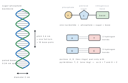
James Watson and Francis Crick proposed the structure of DNA in 1953, using Rosalind Franklin's X-ray photographs and Erwin Chargaff's analyses. Their model has these features, all of which are examinable:
- Two polynucleotide strands wound into a right-handed double helix about a common axis.
- The strands are antiparallel — one runs 5′→3′, the other 3′→5′.
- The backbones are on the outside, the bases point inwards and lie flat, perpendicular to the axis.
- Pairing is specific and complementary: adenine pairs with thymine by two hydrogen bonds, guanine with cytosine by three. A purine always pairs with a pyrimidine, so the helix has a constant width of 2 nm.
- Adjacent base pairs are 0.34 nm apart and each complete turn is 3.4 nm, so there are 10 base pairs per turn.
| Feature | DNA | RNA |
|---|---|---|
| Sugar | deoxyribose | ribose |
| Bases | A, G, C, T | A, G, C, U |
| Strands | double (usually) | single (usually) |
| Location | nucleus, mitochondria, plastids | nucleus and cytoplasm |
| Amount in a cell | constant | varies with activity |
| Function | stores the genetic message | expresses it (mRNA, tRNA, rRNA) |
Problem. A sample of double-stranded DNA contains 22 % adenine. Calculate the percentage of each of the other three bases, and find the ratio \dfrac{A+T}{G+C}.
Solution. Chargaff's rule for double-stranded DNA is A = T and G = C.
Since A = 22\ \%, thymine T = 22\ \%, so A + T = 44\ \%.
The four bases must total 100 %, so G + C = 100 - 44 = 56\ \%, and because G = C,
Hence A = T = 22\ \%, G = C = 28\ \%, and
In single-stranded RNA there is no pairing, so A need not equal U. A question that gives you "RNA with 30 % adenine" is not asking you to set U = 30\ \%.
Minerals
Besides the four main elements, a plant needs at least fourteen mineral elements absorbed from the soil as ions. They are divided by the quantity needed, not by importance — a micronutrient deficiency kills just as surely as a macronutrient one.
| Class | Elements | Example of role | Deficiency sign |
|---|---|---|---|
| Macronutrients | N, P, K, Ca, Mg, S | N in amino acids and nucleotides | general yellowing, stunting |
| Mg is the central atom of chlorophyll | yellowing between veins of old leaves | ||
| Ca as calcium pectate in the middle lamella | death of growing points | ||
| K regulates stomatal opening | scorched leaf margins | ||
| Micronutrients | Fe, Mn, Zn, Cu, B, Mo, Cl, Ni | Fe in cytochromes and ferredoxin | yellowing between veins of young leaves |
| Mo in nitrate reductase | pale, cupped leaves |
Enzymes
An enzyme is a biological catalyst — a globular protein that speeds up a specific reaction by lowering its activation energy, and is itself unchanged at the end. Wilhelm Kühne coined the word in 1878; James Sumner crystallised the first enzyme, urease, in 1926 and proved that enzymes are proteins. (A few RNA molecules, called ribozymes, are also catalytic, so "all enzymes are proteins" is no longer strictly true.)
Properties worth learning as a list:
- Needed in trace amounts; not used up.
- Highly specific — usually one enzyme, one substrate.
- Work fastest at an optimum temperature, about 37 °C in mammals and 25–35 °C in most plants. Above this the protein denatures and activity falls sharply.
- Work fastest at an optimum pH — pepsin at pH 2, salivary amylase at pH 6.8, trypsin at pH 8.
- Activity rises with substrate concentration until all active sites are occupied, then levels off.
- Many need a cofactor: a metal activator (Mg²⁺, Zn²⁺), a loosely bound coenzyme (NAD, FAD, coenzyme A — most are vitamin derivatives) or a tightly bound prosthetic group (haem).
- Reversible in action and named by adding -ase to the substrate (sucrase, lipase, protease).
The substrate binds to a small pocket called the active site. Emil Fischer's lock-and-key model (1894) treats the site as rigid; Daniel Koshland's induced-fit model (1958) — the accepted one — says the site changes shape slightly as the substrate arrives, closing round it like a hand round a ball.
Ox – Tr – Hy – Ly – Is – Li: Oxidoreductases (transfer H or electrons), Transferases (transfer a group), Hydrolases (split with water), Lyases (split without water, forming a double bond), Isomerases (rearrange), Ligases (join two molecules using ATP).
Water
Water is 65–90 % of the fresh mass of protoplasm, and the odd properties that follow from its polar, hydrogen-bonded structure are the reason life works at all.
- Excellent solvent. Its high dielectric constant pulls ionic compounds apart, so all cell reactions take place in solution.
- High specific heat capacity (4.18 J g⁻¹ K⁻¹). A large body of water — or a leaf — changes temperature only slowly, buffering the cell against sudden heat.
- High latent heat of vaporisation (about 2260 J g⁻¹). Transpiration and sweating therefore remove a great deal of heat for a little water lost.
- Cohesion and adhesion. Hydrogen bonding holds the water column in the xylem together, which is how water reaches the top of a Shorea robusta tree.
- Maximum density at 4 °C, so ice floats and a Rara-lake fish survives the winter under the ice rather than in it.
- Reactant. Water is split in the light reaction of photosynthesis and is consumed in every hydrolysis.
1.2 Cell
Robert Hooke saw and named "cells" in a slice of cork in 1665; Anton van Leeuwenhoek was the first to see living cells. In 1838–39 Matthias Schleiden and Theodor Schwann proposed the cell theory, and Rudolf Virchow completed it in 1855 with omnis cellula e cellula — every cell arises from a pre-existing cell.
- All living organisms are composed of one or more cells.
- The cell is the basic structural and functional unit of life.
- All cells arise from pre-existing cells by division.
Problem. (a) A microscope has a 10× eyepiece and a 45× objective. What is the total magnification? (b) On a photomicrograph taken at ×600, a chloroplast measures 2.7 mm. Find its actual length in micrometres.
Solution.
(a) Magnifications multiply:
(b) Magnification = \dfrac{\text{image size}}{\text{actual size}}, so
Since 1\ \text{mm} = 1000\ \mu\text{m}, the chloroplast is 0.0045 \times 1000 = 4.5\ \mu\text{m} long — a normal value.
Prokaryotic and eukaryotic cells
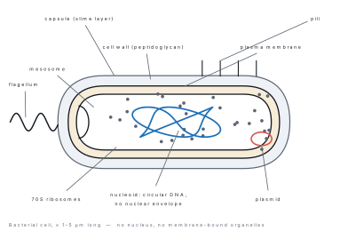
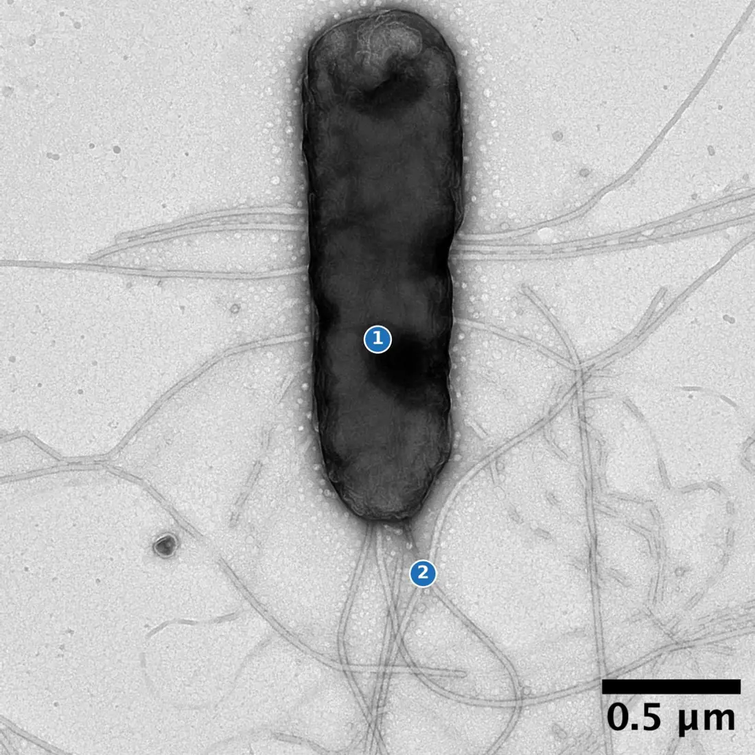
A prokaryotic cell has no nuclear envelope, so its DNA lies naked in the cytoplasm as a nucleoid. It has no membrane-bound organelles at all. Bacteria and cyanobacteria are the only prokaryotes.
A eukaryotic cell has a true nucleus bounded by a double envelope, and a whole set of membrane-bound organelles. Protists, fungi, plants and animals are eukaryotes.
| Feature | Prokaryotic cell | Eukaryotic cell |
|---|---|---|
| Size | 1–10 μm | 10–100 μm |
| Nuclear envelope | absent | present |
| Genetic material | single circular DNA, no histones | linear DNA bound to histones |
| Nucleolus | absent | present |
| Membrane-bound organelles | absent | mitochondria, plastids, ER, Golgi, lysosomes |
| Ribosomes | 70S | 80S in cytoplasm; 70S in mitochondria and plastids |
| Cell wall | peptidoglycan (murein) | cellulose in plants, chitin in fungi, absent in animals |
| Flagellum | one fibril of flagellin | 9 + 2 arrangement of microtubules |
| Division | binary fission, no spindle | mitosis or meiosis, with a spindle |
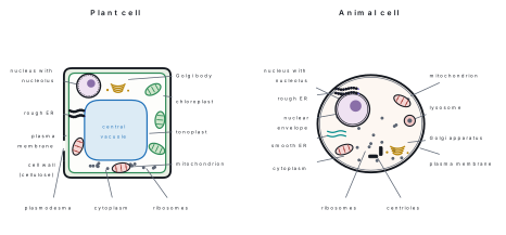
| Feature | Plant cell | Animal cell |
|---|---|---|
| Cell wall | present, cellulose | absent |
| Plastids | present | absent |
| Vacuole | one large central vacuole | small, few or none |
| Centriole | absent (except in some algae and lower plants) | present |
| Lysosome | rare | common |
| Food reserve | starch | glycogen |
| Shape | fixed and angular | flexible, often rounded |
| Cytokinesis | by a cell plate, growing outwards | by a cleavage furrow, growing inwards |
Problem. Treat two cells as cubes of side 10 μm and 30 μm. Calculate the surface area, volume and surface-area-to-volume ratio of each, and comment.
Solution. For a cube of side a, surface area = 6a^{2} and volume = a^{3}, so the ratio is
For a = 10\ \mu\text{m}: S = 600\ \mu\text{m}^{2}, V = 1000\ \mu\text{m}^{3}, S/V = 0.6\ \mu\text{m}^{-1}.
For a = 30\ \mu\text{m}: S = 5400\ \mu\text{m}^{2}, V = 27\,000\ \mu\text{m}^{3}, S/V = 0.2\ \mu\text{m}^{-1}.
Trebling the side divides the ratio by three. Materials enter through the surface but are used by the whole volume, so a large cell cannot supply its interior fast enough by diffusion. That is why cells stay small, and why cells that must absorb a lot — root hairs, intestinal cells — throw out extensions to raise the surface area instead of growing bigger.
Cell wall
The plant cell wall is a non-living, freely permeable layer outside the plasma membrane. It is laid down in three parts:
- the middle lamella, shared between neighbouring cells and made of calcium and magnesium pectate, which cements them together;
- the primary wall, thin and elastic, of cellulose microfibrils in a matrix of hemicellulose and pectin, laid down while the cell is still growing;
- the secondary wall, thick and rigid, deposited inside the primary wall when growth stops, often impregnated with lignin, suberin or cutin.
Thin unthickened spots called pits are crossed by fine cytoplasmic threads, the plasmodesmata, which link the protoplasm of neighbouring cells into one continuous system (the symplast). The wall gives shape, mechanical strength and protection, prevents bursting when the cell takes up water, and permits the turgor on which a herbaceous stem stands up. Fungal walls are chitin, bacterial walls peptidoglycan, diatom walls silica.
Cell membrane
Also called the plasma membrane or plasmalemma, it is about 7.5 nm thick and bounds every cell. The accepted account is the fluid-mosaic model of Singer and Nicolson (1972).
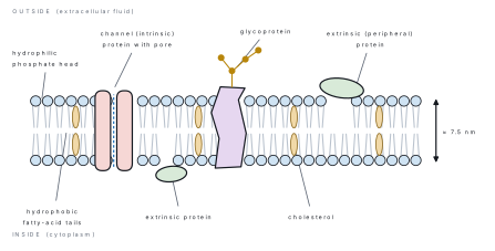
A bilayer of phospholipids forms the continuous framework. Each phospholipid has a hydrophilic phosphate "head" facing the watery surroundings and two hydrophobic fatty-acid "tails" facing inwards. Protein molecules are scattered through it like a mosaic: intrinsic (integral) proteins span the bilayer and act as channels, carriers and pumps, while extrinsic (peripheral) proteins sit on one surface. Cholesterol molecules wedged between the tails regulate fluidity, and short carbohydrate chains attached to lipids and proteins (glycolipids and glycoproteins) act as recognition markers on the outer face. Both layers can flow sideways — hence fluid.
The membrane is selectively permeable. Small non-polar molecules (O₂, CO₂) cross by simple diffusion; water by osmosis, largely through aquaporin channels; glucose and amino acids by facilitated diffusion through carrier proteins; and ions against their gradient by active transport, which needs ATP — the sodium–potassium pump is the standard example. Bulk material enters by endocytosis and leaves by exocytosis.
A semi-permeable membrane (like visking tubing) lets solvent through and holds solute back, purely by pore size. A living plasma membrane chooses: it can take in K⁺ while rejecting Na⁺ of the same size. Always write selectively permeable for a living membrane.
Mitochondria
Discovered by Altmann (1890) and named by Benda (1898), mitochondria are rod- or sausage-shaped bodies about 0.5–1.0 μm thick and 1–10 μm long, present in all aerobic eukaryotic cells.
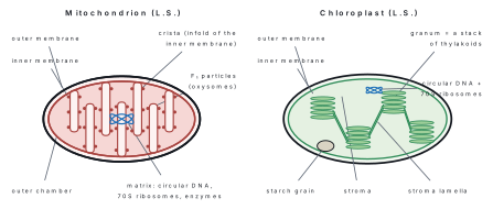
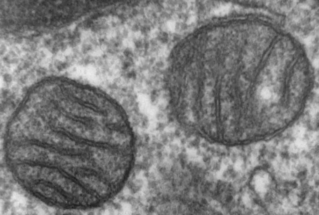
A mitochondrion has two membranes. The outer is smooth and permeable; the inner is folded inwards into shelves called cristae, which greatly increase the area available for the electron transport chain. The space between the two membranes is the outer chamber; the fluid inside the inner membrane is the matrix. Studding the inner membrane are stalked F₁ particles (oxysomes), which are the ATP synthase enzyme.
The matrix holds the enzymes of the Krebs cycle, and also circular DNA and 70S ribosomes of its own — so a mitochondrion can make some of its own proteins and divides by fission. It is therefore called a semi-autonomous organelle, and this is the evidence for the endosymbiotic theory. Because aerobic respiration and most ATP synthesis happen here, the mitochondrion is called the powerhouse of the cell.
Plastids
Plastids are double-membraned organelles found only in plants and some protists. Three types are recognised by their pigment:
- Chloroplasts — green, containing chlorophyll; the site of photosynthesis.
- Chromoplasts — yellow, orange or red, containing carotenoids; they colour petals, carrot roots and ripe tomatoes and attract pollinators and dispersers.
- Leucoplasts — colourless storage plastids of underground organs: amyloplasts store starch, elaioplasts oil and aleuroplasts protein.
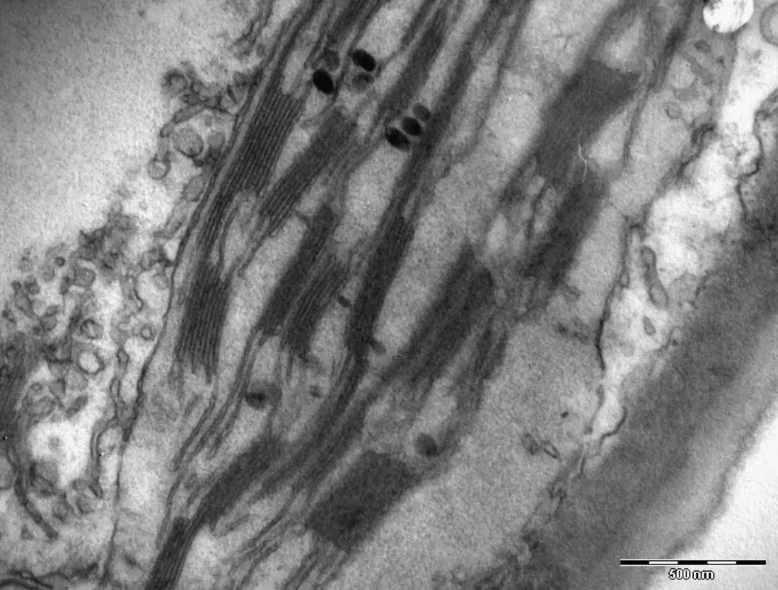
A chloroplast is a biconvex disc about 4–6 μm long. Inside the double envelope lies a colourless ground substance, the stroma, containing circular DNA, 70S ribosomes, starch grains and the enzymes of the Calvin cycle. Suspended in it is a system of flattened membranous sacs, the thylakoids. Where thylakoids are stacked like a pile of coins the stack is a granum (10–100 thylakoids, about 40–60 grana per chloroplast); the tubes connecting one granum to the next are the stroma lamellae. Chlorophyll sits in the thylakoid membrane, so the light reaction happens in the grana and the dark reaction in the stroma.
Endoplasmic reticulum
Discovered by Porter, Claude and Fullam (1945), the ER is a network of membranous tubules, vesicles and flattened sacs (cisternae) running through the cytoplasm and continuous with the outer membrane of the nuclear envelope. It is absent from prokaryotes and from mature red blood cells.
Rough ER carries ribosomes on its outer surface; it synthesises proteins for export and channels them to the Golgi body. Smooth ER has no ribosomes and makes lipids and steroids, stores calcium ions in muscle (as sarcoplasmic reticulum) and detoxifies drugs in liver cells. The ER also gives the cytoplasm mechanical support — it is often called the cell's skeletal framework — and its membranes supply material for the nuclear envelope after division.
Golgi apparatus
Described by Camillo Golgi in 1898, it consists of a stack of 4–8 flattened, slightly curved cisternae with tubules and vesicles at the edges. In plant cells the separate stacks are called dictyosomes. The convex face towards the ER is the cis (forming) face and the concave face is the trans (maturing) face.
Its work is packaging and dispatch. Proteins arrive from the rough ER in vesicles, are chemically modified — glycosylation, the addition of sugars is typical — sorted, concentrated and released at the trans face in secretory vesicles. The Golgi body also forms lysosomes, secretes the polysaccharides of the cell wall, and supplies the vesicles whose fusion builds the cell plate in plant cytokinesis.
Lysosomes
Isolated by Christian de Duve in 1955, lysosomes are simple spherical sacs bounded by a single membrane, 0.2–0.8 μm across, budded off the Golgi apparatus. They contain some fifty acid hydrolases — proteases, lipases, nucleases, phosphatases — which work best at about pH 5, maintained inside the sac by proton pumps.
Their functions are digestive:
- Heterophagy — digestion of food particles or bacteria taken in by endocytosis.
- Autophagy — controlled digestion of the cell's own worn-out organelles, the parts being recycled.
- Autolysis — if the membrane bursts, the enzymes digest the whole cell. For this reason lysosomes are called the suicide bags of the cell. Autolysis is a normal part of development: it removes the tadpole's tail.
Ribosomes
Ribosomes are the smallest organelles and the only ones with no membrane. First seen by George Palade (1953), each is a dense particle of roughly equal parts ribosomal RNA and protein, built of one large and one small subunit that join only in the presence of Mg²⁺ and only when protein synthesis begins.
Eukaryotic cytoplasmic ribosomes are 80S (60S + 40S). Prokaryotic ribosomes, and those inside mitochondria and chloroplasts, are 70S (50S + 30S). ("S" is the Svedberg unit of sedimentation rate, which is why 60 + 40 does not give 100.) Ribosomes occur free in the cytoplasm or attached to the rough ER; several translating the same mRNA form a chain called a polysome. The ribosome is where amino acids are joined in the order specified by mRNA, so it is called the protein factory of the cell.
Nucleus
Discovered by Robert Brown in 1831, the nucleus is the control centre. It is usually spherical, 5–25 μm across, and normally one per cell (though a Rhizopus hypha is multinucleate and a mature sieve tube has none).
It has four parts. The nuclear envelope is a double membrane, the outer one continuous with the ER, perforated by nuclear pores through which RNA and ribosomal subunits leave and proteins enter. The nucleoplasm is the ground fluid. The chromatin is a network of DNA-and-histone threads, staining darkly; loose, active regions are euchromatin and condensed, inactive regions heterochromatin. The nucleolus, a dense body without a membrane, is the site of rRNA synthesis and ribosome assembly — cells that make a lot of protein have large nucleoli.
Chromosomes
At division the chromatin condenses into distinct chromosomes, first named by Waldeyer (1888). Chemically a chromosome is about 35–40 % DNA, 50–55 % histone protein, plus non-histone protein and a little RNA. The DNA is wound about 1¾ turns round an octamer of histones to form a nucleosome, the bead-like repeating unit of chromatin.
A metaphase chromosome consists of two identical sister chromatids joined at a narrow primary constriction, the centromere, on which the disc-shaped kinetochore sits and to which spindle fibres attach. Some chromosomes have a secondary constriction cutting off a small knob, the satellite; these are SAT chromosomes and they organise the nucleolus. The ends are capped by telomeres, which stop chromosomes fusing.
| Type | Position of centromere | Shape at anaphase |
|---|---|---|
| Metacentric | exactly in the middle | V |
| Sub-metacentric | slightly off centre | L or J |
| Acrocentric | near one end | J, with one very short arm |
| Telocentric | at the very end | I (rod) |
The number of chromosomes is constant for a species — 46 in humans, 16 in onion (Allium cepa), 24 in pea, 20 in maize. Chromosomes that govern body characters are autosomes; those governing sex are sex chromosomes or allosomes.
Cilia and flagella
These are hair-like extensions of the cell surface used for movement, for creating a current, or as sensory structures. Cilia are short (5–10 μm) and very numerous; flagella are long (up to 150 μm) and few. Both have identical internal structure.
A cross-section shows the 9 + 2 arrangement: nine pairs (doublets) of microtubules arranged in a ring round two single central microtubules, the whole bundle called the axoneme and sheathed by an extension of the plasma membrane. The microtubules are made of the protein tubulin. Each outer doublet carries dynein arms, whose ATP-driven sliding against the neighbouring doublet bends the cilium. Beneath the surface each cilium arises from a basal body which has nine triplets and no central pair — a 9 + 0 arrangement, identical to a centriole. Bacterial flagella are quite different: a single solid fibril of the protein flagellin, with no 9 + 2 pattern.
Cell inclusions
Cell inclusions are the non-living substances lying free in the cytoplasm or vacuole — reserve food, secretions and waste. They are also called ergastic substances.
| Group | Examples | Where found |
|---|---|---|
| Reserve food | starch grains, glycogen granules, oil drops, aleurone (protein) grains | potato tuber, liver, castor seed, wheat grain |
| Secretory | enzymes, hormones, nectar, essential oils | glandular cells |
| Excretory / waste | calcium oxalate crystals (raphides, druses), cystoliths of calcium carbonate, tannins, resins, gums, alkaloids, latex | Colocasia petiole, Ficus leaf, Euphorbia and Calotropis stems |
| Pigments | anthocyanin in the cell sap | red and blue petals |
1.3 Cell division
The cell cycle
The cell cycle is the ordered sequence of events between the formation of a cell and its division into two. It has two unequal parts: a long interphase, and a short M phase in which division actually occurs.
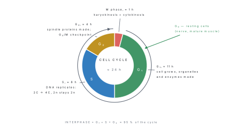
Interphase is not a resting stage — it is the busiest part of the cycle. It is divided into:
- G₁ (first gap) — the cell grows, makes RNA, proteins and organelles. The restriction point at the end of G₁ decides whether the cell divides at all.
- S (synthesis) — DNA replicates. Each chromosome becomes two chromatids, so the DNA content doubles from 2C to 4C while the chromosome number stays 2n.
- G₂ (second gap) — more growth; spindle proteins (tubulin) are made and the cell is checked for damaged DNA before mitosis is allowed to start.
Cells that stop dividing — mature nerve and skeletal muscle cells — leave the cycle at G₁ and enter a quiescent G₀ state. In a typical human cell the cycle takes about 24 hours, of which M phase is only about 1 hour.
After replication a cell has 2n chromosomes each of two chromatids — not 4n chromosomes. The chromosome number is counted by centromeres, and each chromosome still has exactly one.
Amitosis
Amitosis, or direct cell division, was first described by Remak (1841). The nucleus simply elongates, a constriction appears in the middle and it pinches into two, after which the cytoplasm divides. There is no spindle, no condensation of chromatin into visible chromosomes and no equal distribution of the genetic material, so the daughter cells may be genetically unequal.
Amitosis occurs in prokaryotes, in yeast, in Amoeba and other protozoa, in the endosperm of some seeds, in foetal membranes, and in old or diseased tissue — including cancerous cells. It is fast and cheap but unreliable, which is why almost all normal eukaryotic division is mitotic.
Mitosis
Mitosis (Flemming, 1882) is the division of a somatic cell that produces two daughter cells with exactly the same chromosome number as the parent. It is therefore called equational division. It occurs in the growing regions of a plant — root and shoot apical meristems, cambium — and throughout animal tissue during growth and repair. It has two steps: karyokinesis (division of the nucleus) followed by cytokinesis (division of the cytoplasm).
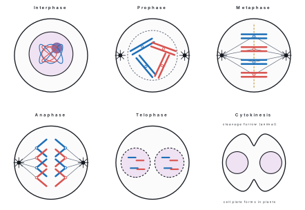
Prophase. Chromatin condenses into visible chromosomes, each already made of two sister chromatids joined at the centromere. The chromosomes thicken and shorten. In animals the two pairs of centrioles move to opposite poles and a spindle of microtubules begins to form between them; plants form the spindle without centrioles. By the end of prophase the nucleolus and the nuclear envelope have disappeared.
Metaphase. The spindle is complete. The chromosomes move to the equator and line up with their centromeres exactly on the equatorial plane, forming the metaphase plate, with the arms pointing outwards. Spindle fibres attach to the kinetochore of each chromatid from opposite poles. Chromosomes are at their shortest and thickest, so this is the best stage for counting and studying them.
Anaphase. Each centromere splits. The two sister chromatids — now daughter chromosomes — are pulled to opposite poles as the spindle fibres shorten. They trail their arms behind the centromere, giving the V, L, J and I shapes that reveal the centromere's position. This is the shortest stage.
Telophase. The daughter chromosomes reach the poles, uncoil and lengthen back into chromatin. The nuclear envelope re-forms round each group from ER membranes, the nucleolus reappears and the spindle breaks down. Two nuclei now exist in one cell.
Cytokinesis. In animal cells a ring of actin filaments pinches the cell in from outside — a cleavage furrow, growing centripetally. In plant cells the rigid wall prevents furrowing, so Golgi vesicles line up at the equator and fuse to form a cell plate that grows outwards (centrifugally) until it reaches the side walls and becomes the new middle lamella.
Mitosis matters because it keeps the chromosome number constant generation after generation of cells, provides growth and repair, replaces worn-out cells, and is the basis of asexual and vegetative reproduction — which is why a cutting of sugarcane or a potato tuber gives plants identical to the parent.
Meiosis
Meiosis (Farmer and Moore, 1905) is the division in which one diploid cell gives four haploid cells, the chromosome number being halved. It is therefore called reduction division. In animals it occurs in the gonads during gamete formation; in flowering plants it occurs in the anther (giving microspores) and in the ovule (giving the megaspore). It consists of two successive divisions, meiosis I and meiosis II, but the DNA replicates only once, before meiosis I.
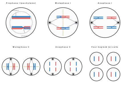
Meiosis I — the reduction division. Prophase I is long and is divided into five sub-stages:
| Sub-stage | What happens |
|---|---|
| Leptotene | chromosomes appear as long thin threads |
| Zygotene | homologous chromosomes pair lengthwise — synapsis — forming bivalents |
| Pachytene | each bivalent shows four chromatids (a tetrad); non-sister chromatids exchange segments — crossing over |
| Diplotene | homologues start to separate but stay joined at X-shaped chiasmata |
| Diakinesis | chiasmata slip to the ends (terminalisation); nucleolus and nuclear envelope disappear |
L Z P D D — Lazy Zebras Prefer Dry Deserts: Leptotene, Zygotene, Pachytene, Diplotene, Diakinesis. Remember that crossing over happens at pachytene but the chiasmata become visible at diplotene.
In metaphase I the bivalents line up at the equator with the two homologues of each pair on either side of the plane — a double metaphase plate. Which homologue faces which pole is decided independently for every pair; this is independent assortment and it is the second source of variation. In anaphase I whole chromosomes, each still made of two chromatids, move to opposite poles: the centromeres do not split. This is where the number is halved. Telophase I re-forms two haploid nuclei, and a short interkinesis follows with no S phase.
Meiosis II is essentially a mitotic division of each haploid cell. In prophase II the envelope breaks down again, in metaphase II the chromosomes line up singly, in anaphase II the centromeres split at last and sister chromatids separate, and in telophase II four haploid nuclei are formed. Cytokinesis gives four haploid cells, none of them genetically identical to any other.
| Feature | Mitosis | Meiosis |
|---|---|---|
| Where | somatic (body) cells | reproductive cells |
| Number of divisions | one | two |
| Daughter cells | 2 | 4 |
| Chromosome number | unchanged (2n → 2n) | halved (2n → n) |
| Pairing of homologues | absent | present (synapsis) |
| Crossing over | absent | present |
| Centromere splits in | anaphase | anaphase II only |
| Daughter cells are | genetically identical | genetically different |
| Purpose | growth, repair, asexual reproduction | gamete/spore formation, variation |
If gametes were made by mitosis, fertilisation would double the chromosome number every generation. Meiosis halves it first, so fusion restores exactly 2n and the species number stays constant. At the same time, crossing over and independent assortment shuffle the parental genes, producing the variation on which natural selection acts.
Problem. The onion Allium cepa has 2n = 16. For a root-tip cell, state (a) the number of chromosomes and of chromatids at mitotic metaphase, (b) the number of chromosomes moving to each pole at mitotic anaphase, and (c) the number of bivalents at metaphase I and the chromosome number of each cell at the end of meiosis in an anther.
Solution.
(a) DNA has replicated in S phase, so there are still 16 chromosomes, but each has 2 chromatids:
(b) At anaphase each centromere splits, so all 32 chromatids become separate chromosomes and half go to each pole:
— which is why each daughter cell is again 2n = 16.
(c) A bivalent is a pair of homologues, so
Meiosis halves the number, so each of the four microspores has n = 8 chromosomes.
Chapter summary
- Condensation joins monomers into polymers with the loss of water; hydrolysis reverses it. Carbohydrates \to glycosidic bonds, proteins \to peptide bonds, lipids \to ester bonds, nucleic acids \to phosphodiester bonds.
- Lipids yield about 39 kJ g⁻¹ against 17 kJ g⁻¹ for carbohydrate and protein.
- DNA is an antiparallel right-handed double helix: A=T (2 H-bonds), G≡C (3 H-bonds), 0.34 nm per base pair, 3.4 nm and 10 base pairs per turn, diameter 2 nm. Chargaff: A = T and G = C.
- Enzymes are protein catalysts that lower activation energy; they are specific and have an optimum temperature and pH. Induced fit, not lock and key, is the accepted model.
- Prokaryotes lack a nuclear envelope and membrane-bound organelles and have 70S ribosomes; eukaryotes have both and 80S cytoplasmic ribosomes.
- Mitochondria and chloroplasts are double-membraned and semi-autonomous, with circular DNA and 70S ribosomes; cristae and thylakoids increase membrane area.
- The plasma membrane is a fluid mosaic (Singer and Nicolson, 1972), about 7.5 nm thick and selectively permeable.
- Cell cycle = interphase (G₁, S, G₂) + M phase. DNA doubles 2C \to 4C in S phase but the chromosome number does not change.
- Mitosis is equational: 1 cell \to 2 identical cells, 2n \to 2n. Meiosis is reductional: 1 cell \to 4 different cells, 2n \to n, with crossing over at pachytene and reduction at anaphase I.
- Surface-area-to-volume ratio for a cube is 6/a; it falls as cells grow, which sets an upper limit on cell size.
Practice questions
Group A — Multiple choice (1 mark each)
- The bond that joins two amino acids in a protein is [1]aglycosidicbpeptidecesterdphosphodiester
- In a DNA molecule, adenine pairs with thymine by [1]aone hydrogen bondbtwo hydrogen bondscthree hydrogen bondsdan ionic bond
- Which organelle has no membrane at all? [1]alysosomebribosomecGolgi bodydchloroplast
- The stage of mitosis in which chromosomes are shortest and thickest and lie at the equator is [1]aprophasebmetaphasecanaphasedtelophase
- The chromosome number is actually halved during [1]aprophase Ibmetaphase Icanaphase Idanaphase II
Answers to Group A
1. (b) — condensation between the –COOH of one residue and the –NH₂ of the next.
2. (b) — A=T has two hydrogen bonds; G≡C has three.
3. (b) — the ribosome is the only non-membranous organelle.
4. (b) — at metaphase the chromosomes are maximally condensed on the metaphase plate.
5. (c) — homologues separate at anaphase I without the centromeres splitting.
Group B — Short answer (4 marks each)
- Draw a labelled diagram of a mitochondrion in longitudinal section and state two reasons for calling it a semi-autonomous organelle. [4]
- Differentiate between a prokaryotic and a eukaryotic cell (any four points). [4]
- A double-stranded DNA molecule contains 1200 base pairs, of which 30 % of the bases are guanine. Calculate the number of adenine, thymine, guanine and cytosine bases in it, and the length of the molecule in nanometres. [4]
- What is an enzyme? Describe the effect of temperature and of pH on enzyme activity. [4]
Answers to Group B
1. Diagram: outer membrane, inner membrane folded into cristae, outer chamber, matrix, F₁ particles. Semi-autonomous because (i) it contains its own circular DNA and 70S ribosomes and so makes some of its own proteins, and (ii) it multiplies by fission independently of nuclear division.
3. Total bases = 2 \times 1200 = 2400. Guanine is 30 % of all bases, so G = 0.30 \times 2400 = 720, and by Chargaff C = 720. The remaining bases are 2400 - 1440 = 960, shared equally, so A = T = 480. Length = number of base pairs \times\ 0.34\ \text{nm} = 1200 \times 0.34 = 408\ \text{nm}.
4. Definition: a protein catalyst that lowers activation energy and is unchanged at the end. Temperature: activity roughly doubles for each 10 °C rise up to the optimum (about 37 °C in mammals) as molecular collisions increase; above it the tertiary structure denatures and activity falls steeply and irreversibly. pH: each enzyme has a narrow optimum (pepsin 2, salivary amylase 6.8, trypsin 8) because extreme pH alters the charges that hold the active site in shape.
Group C — Long answer (8 marks each)
- Describe the various stages of meiosis I with the help of labelled diagrams, and explain how meiosis produces genetic variation. [8]
- (a) Draw a labelled diagram of a plant cell as seen under an electron microscope. [4](b) Give the structure and functions of the Golgi apparatus and of lysosomes. [4]
Answer outline to Group C question 1
Prophase I with the five sub-stages (leptotene — thin threads; zygotene — synapsis forming bivalents; pachytene — tetrads and crossing over; diplotene — chiasmata visible; diakinesis — terminalisation, envelope and nucleolus gone); metaphase I — bivalents on a double metaphase plate; anaphase I — whole homologous chromosomes separate, centromeres intact, number halved; telophase I — two haploid nuclei, then interkinesis with no DNA replication.
Variation arises from two independent sources: crossing over at pachytene, which exchanges segments between non-sister chromatids of homologues and creates new gene combinations on a single chromosome; and independent assortment at metaphase I, since each bivalent orients on the plate independently, giving 2^{n} possible gamete types from n pairs — 2^{23}, over 8 million, in humans. Random fertilisation then multiplies the variation further.
Practise more: Biology question bank