Unit 3 · Botany · 5 teaching hours · 21 min read
Introductory Microbiology
What the syllabus asks for 3
- Monera
- Virus
- Impacts of biotechnology in microbiology
Microbiology is the study of organisms too small to see with the naked eye. Two groups dominate this unit: the bacteria of kingdom Monera, which are true cells but have no nucleus, and the viruses, which are not cells at all. They matter out of all proportion to their size — they fix the nitrogen in a lentil field in Bara, set curd in a Kathmandu kitchen, rot the jute in a Jhapa retting pit, and cause cholera, typhoid and COVID-19. The unit ends with what happens when we deliberately re-engineer them: biotechnology.
Three items recur almost every year: a labelled diagram of a bacterial cell or of a bacteriophage; a difference table (Gram-positive vs Gram-negative, bacteria vs virus, virus living vs non-living); and a list-with-examples answer on economic importance or on applications of biotechnology. Learn the scientific names — an unnamed example earns no mark.
3.1 Monera
Monera is one of the five kingdoms of Whittaker's 1969 classification and holds all prokaryotes — organisms whose cells have no nuclear envelope and no membrane-bound organelles. In the modern three-domain scheme the kingdom splits into two domains, Archaea and Bacteria, while all other life forms one domain, Eukarya.
Monera is the kingdom of unicellular, prokaryotic organisms whose genetic material is a single naked circular DNA molecule lying free in the cytoplasm, whose cell wall (when present) contains peptidoglycan, and whose ribosomes are of the 70S type.
General characters
- Unicellular and prokaryotic; size usually 0.2–2 μm wide and 1–10 μm long. Mycoplasma (0.1–0.3 μm) is the smallest known living cell.
- No nuclear membrane, nucleolus, mitochondrion, plastid, ER, Golgi body, lysosome or true vacuole.
- Genetic material: one circular, double-stranded DNA molecule (the nucleoid) with no histone proteins, often plus small circular plasmids.
- Ribosomes are 70S (a 50S and a 30S subunit).
- Cell wall of peptidoglycan (murein) — a mesh of N-acetylglucosamine and N-acetylmuramic acid chains cross-linked by short peptides.
- Reproduction is asexual by binary fission; true meiosis and gametes are absent, but DNA can be exchanged by transformation, conjugation and transduction.
- They are the most widely distributed organisms on Earth — in soil, air, water, hot springs, snow, and inside other organisms.
Shape and arrangement
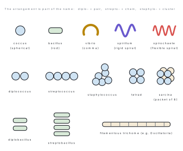
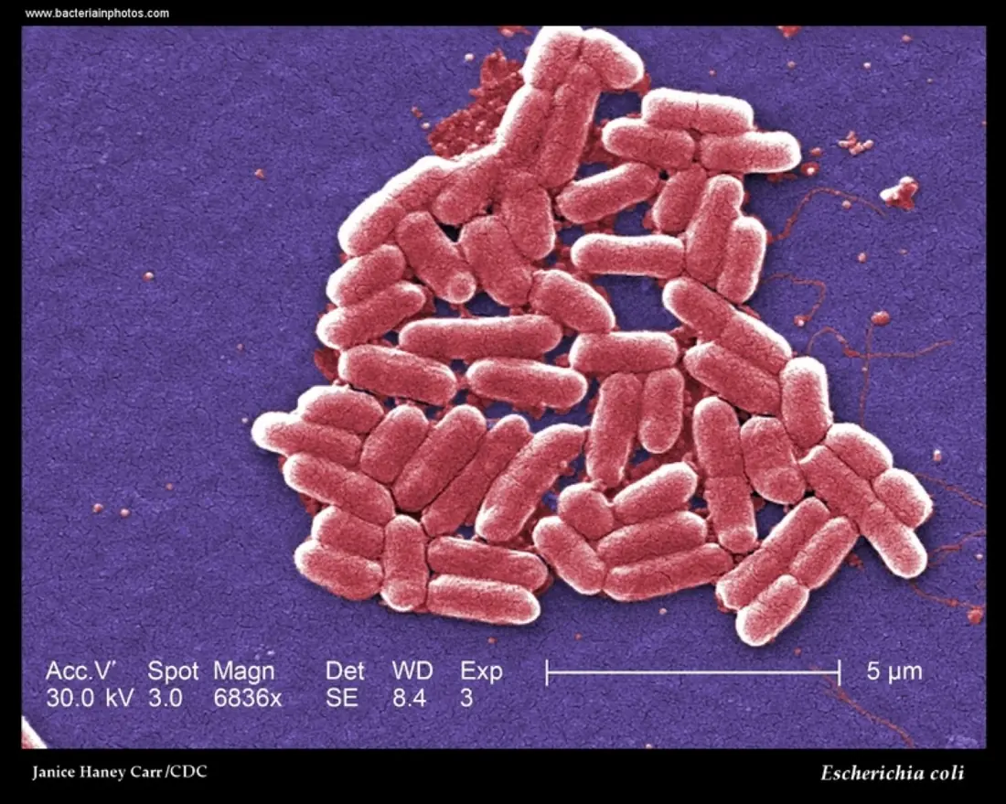
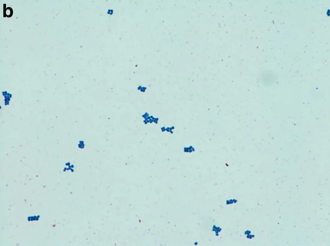
Bacteria are classified into four basic shapes. Because the daughter cells often stay stuck together after fission, each shape gives a set of characteristic arrangements, and the arrangement is part of the name.
Structure of a bacterial cell
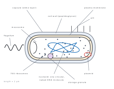
| Part | Structure | Function |
|---|---|---|
| Capsule / slime layer | outer mucilaginous polysaccharide sheath | protection, sticking to surfaces, resists drying and phagocytosis |
| Cell wall | rigid peptidoglycan mesh, 10–80 nm thick | shape, protection, resists osmotic bursting |
| Plasma membrane | lipoprotein bilayer, no cholesterol | selective transport, respiratory enzymes |
| Mesosome | infolding of the plasma membrane | increases surface area for respiration; helps in DNA replication |
| Nucleoid | one naked circular double-stranded DNA | carries all essential genes |
| Plasmid | small extra circular DNA | carries resistance, fertility and toxin genes; used as a vector in gene cloning |
| Ribosome | 70S (50S + 30S), free in cytoplasm | protein synthesis |
| Flagellum | flagellin fibre with basal body and hook | locomotion |
| Pili (fimbriae) | short hollow protein tubes | attachment; sex pili transfer DNA in conjugation |
| Inclusions | glycogen, PHB, volutin, gas vacuoles | food and gas storage |
A-M-A-L-C-P — Atrichous (none), Monotrichous (one, at one end), Amphitrichous (one at each end), Lophotrichous (a tuft at one end), Cephalotrichous (a tuft at both ends), Peritrichous (all over the body, as in Escherichia coli).
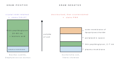
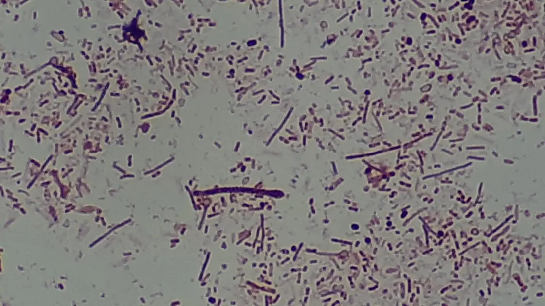
Gram's stain (Christian Gram, 1884) divides bacteria into two great groups, and the difference is a difference of wall chemistry.
| Feature | Gram-positive | Gram-negative |
|---|---|---|
| Colour after staining | violet / purple | pink / red |
| Peptidoglycan | thick, 20–80 nm, 40–90 % of wall | thin, 2–7 nm, 5–20 % of wall |
| Teichoic acid | present | absent |
| Outer membrane, lipopolysaccharide | absent | present |
| Periplasmic space | absent or very narrow | well developed |
| Sensitivity to penicillin | high | comparatively low |
| Examples | Bacillus subtilis, Staphylococcus aureus, Clostridium tetani | Escherichia coli, Vibrio cholerae, Salmonella typhi, Rhizobium leguminosarum |
Nutrition
| Mode | Energy / carbon source | Examples |
|---|---|---|
| Photoautotrophic | sunlight; CO₂ fixed | cyanobacteria (Nostoc), purple sulphur bacteria (Chromatium), green sulphur bacteria (Chlorobium) |
| Chemoautotrophic | oxidation of inorganic substances | Nitrosomonas (NH₃ → NO₂⁻), Nitrobacter (NO₂⁻ → NO₃⁻), Thiobacillus (S → SO₄²⁻) |
| Saprophytic | dead organic matter | Bacillus, most soil bacteria — the chief decomposers |
| Parasitic | living host, causes harm | Mycobacterium tuberculosis, Xanthomonas oryzae |
| Symbiotic | living host, mutual benefit | Rhizobium leguminosarum in the root nodules of legumes |
Reproduction
- Binary fission (amitosis) is the normal method. The DNA replicates, the cell elongates, a transverse septum grows inwards and the cell splits into two identical daughter cells. Under ideal conditions E. coli divides every 20 minutes.
- Endospore formation is not reproduction — it is survival. Bacillus and Clostridium lay down a thick, almost dehydrated spore inside the cell that can survive boiling, drought and disinfectants for years, then germinate into a single vegetative cell.
- Genetic recombination substitutes for sex. There are three routes: transformation — naked DNA is taken up from the medium (Griffith, 1928); conjugation — DNA passes through a sex pilus from a donor to a recipient (Lederberg and Tatum, 1946); transduction — DNA is carried from one cell to another by a bacteriophage (Zinder and Lederberg, 1952).
There is no spindle, no centromere and no nuclear envelope to break down, so binary fission is amitosis. Writing "prophase" or "metaphase" in a binary fission answer loses the mark.
The growth of a bacterial population
Because one cell becomes two at each division, a bacterial population grows geometrically. If N_0 cells grow for time t with a generation time g, the number of generations is n = t/g and
Problem. A culture starts with 1 \times 10^{3} cells of Escherichia coli whose generation time is 20 minutes. How many cells are present after 3 hours of exponential growth?
Solution. Time available t = 3 \times 60 = 180 minutes, so
Problem. A bacterial population rises from 5 \times 10^{3} to 3.2 \times 10^{5} cells in 2 hours. Find the number of generations and the generation time.
Solution. The growth factor is
so n = 6 generations. Then
Problem. A milk sample is diluted 10^{-5} times. 0.1\ \text{mL} of the dilution is spread on nutrient agar and gives 148 colonies. Calculate the number of colony-forming units (CFU) per millilitre of the original milk.
Solution. Each colony arose from one cell, so
The milk is heavily contaminated: the acceptable limit for pasteurised milk is about 10^{5}\ \text{CFU mL}^{-1}.
Classification of Monera
| Group | Wall | Distinguishing feature | Examples |
|---|---|---|---|
| Archaebacteria | no peptidoglycan; branched lipids with ether links | live in extreme habitats; oldest known prokaryotes | Methanobacterium (methanogen, in biogas and in cattle rumen), Halobacterium (salt lakes), Sulfolobus (hot acid springs) |
| Eubacteria — true bacteria | peptidoglycan | the ordinary heterotrophic and chemoautotrophic bacteria | Escherichia coli, Bacillus, Rhizobium, Vibrio cholerae |
| Eubacteria — cyanobacteria | peptidoglycan | oxygen-releasing photosynthesis with chlorophyll a | Nostoc, Anabaena, Oscillatoria, Spirulina |
| Mycoplasma (PPLO) | no cell wall | smallest living cells, pleomorphic; "jokers of the plant kingdom" | Mycoplasma gallisepticum; cause of little leaf of brinjal |
Cyanobacteria (blue-green algae) deserve special mention. They are prokaryotic but photosynthesise like plants, using chlorophyll a together with the blue pigment c-phycocyanin and the red c-phycoerythrin. Filamentous forms such as Nostoc and Anabaena have thick-walled colourless cells called heterocysts in which atmospheric nitrogen is fixed; this is why Anabaena azollae, living inside the water fern Azolla, is used as a biofertiliser in rice fields. When nutrient-rich water warms up, cyanobacteria multiply into a foul-smelling water bloom that deoxygenates the water and kills fish.
Economic importance of bacteria
| Useful role | Organism | Product or effect |
|---|---|---|
| Nitrogen fixation | Rhizobium leguminosarum, Azotobacter, Anabaena | converts N₂ into ammonia; raises soil fertility |
| Nitrification | Nitrosomonas, Nitrobacter | ammonia → nitrite → nitrate, the form roots absorb |
| Dairy | Lactobacillus, Streptococcus lactis | curd, cheese, dahi and mohi |
| Vinegar | Acetobacter aceti | ethanol → acetic acid |
| Antibiotics | Streptomyces griseus, S. venezuelae, Bacillus subtilis | streptomycin, chloramphenicol, subtilin |
| Retting | Clostridium, Pseudomonas | frees jute, flax and hemp fibres from the stem |
| Biogas | Methanobacterium | methane from cow dung — over 400,000 domestic plants in Nepal |
| Sewage treatment | mixed saprophytes | breaks organic waste into harmless minerals |
| Vitamins, amino acids | Pseudomonas denitrificans, Corynebacterium glutamicum | vitamin B₁₂, glutamic acid |
| Harmful effect | Disease | Causal bacterium |
|---|---|---|
| Human | cholera | Vibrio cholerae |
| Human | typhoid | Salmonella typhi |
| Human | tuberculosis | Mycobacterium tuberculosis |
| Human | tetanus | Clostridium tetani |
| Plant | bacterial blight of rice | Xanthomonas oryzae |
| Plant | citrus canker | Xanthomonas citri |
| Plant | crown gall | Agrobacterium tumefaciens |
| Other | food spoilage, denitrification, milk souring | many saprophytes |
3.2 Virus
The word virus is Latin for poison. In 1892 the Russian scientist D. J. Iwanowski showed that the sap of a tobacco plant with mosaic disease stayed infectious after being passed through a filter fine enough to stop all bacteria. In 1898 M. W. Beijerinck named the agent contagium vivum fluidum — a living infectious fluid. In 1935 W. M. Stanley crystallised tobacco mosaic virus and proved it was nucleoprotein, for which he received the Nobel Prize in 1946.
A virus is an ultramicroscopic, obligate intracellular parasite consisting of a single kind of nucleic acid (DNA or RNA, never both) enclosed in a protein coat called a capsid; it has no cytoplasm, no ribosomes and no metabolism of its own, and can multiply only inside a living host cell.
Living and non-living characters
| Living characters | Non-living characters |
|---|---|
| Contain nucleic acid, the genetic material | Can be crystallised and stored like a chemical |
| Multiply, giving thousands of copies | No respiration, no growth, no excretion |
| Show heredity and mutation | No cytoplasm, membrane, ribosome or enzyme system of their own |
| Are host- and tissue-specific | Inert outside the host — cannot multiply in a culture medium |
| Respond to heat, formalin and radiation | Obey the law of mass action, like a chemical |
Viruses are therefore said to be on the borderline between the living and the non-living: living inside the host, a chemical outside it.
Structure
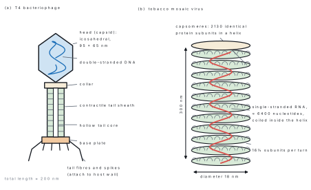
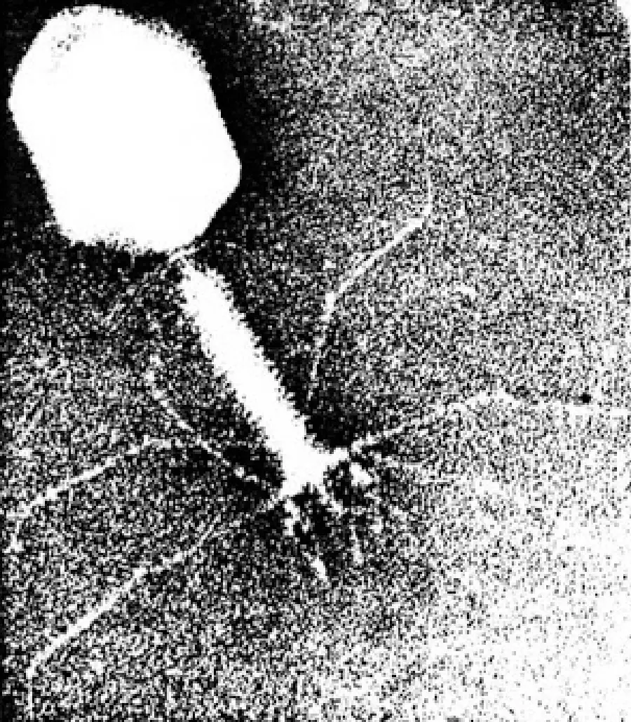
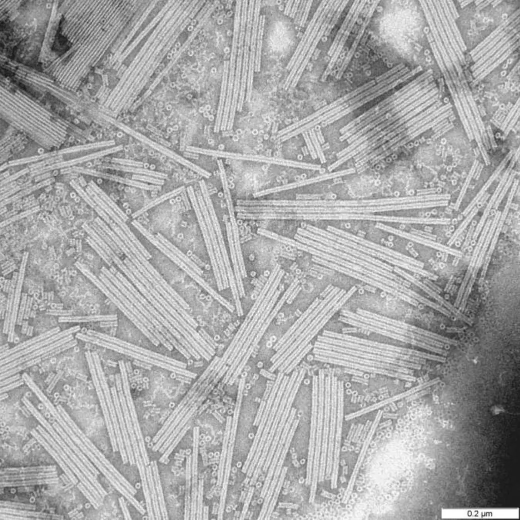
Every virus has two essential parts. The capsid is the protein coat, built of identical subunits called capsomeres; it protects the nucleic acid and decides which host cell the virus can attach to. The genome is one molecule (or a few pieces) of DNA or RNA, single- or double-stranded. Some animal viruses add an outer envelope of host membrane carrying viral spikes — influenza virus, HIV and SARS-CoV-2 are enveloped. Capsid plus genome is a nucleocapsid; one complete infective particle is a virion.
Notice a pleasing piece of arithmetic in TMV: three RNA nucleotides are bound by each protein subunit, so 6400/3 \approx 2133 — very close to the 2130 capsomeres actually counted.
| Basis | Types | Examples |
|---|---|---|
| Host | plant viruses, animal viruses, bacteriophages, mycophages | TMV; rabies virus; T4 phage |
| Nucleic acid | dsDNA, ssDNA, dsRNA, ssRNA | T4 phage; φX174; wound tumour virus; TMV, HIV |
| Shape | rod (TMV), spherical/polyhedral (poliovirus), tadpole (phage), brick (variola), bullet (rabies) | — |
| Envelope | naked (TMV, poliovirus) or enveloped (influenza, HIV) | — |
A virus carries one kind of nucleic acid. Most plant viruses are RNA viruses and most bacteriophages are DNA viruses. Retroviruses such as HIV are RNA viruses that copy their RNA into DNA using reverse transcriptase; this does not make them "DNA and RNA viruses".
Multiplication — the lytic cycle
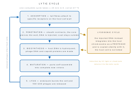
Problem. One T4 phage infects a single E. coli cell. Each lytic cycle takes 25 minutes and has a burst size of 200. Assuming every released phage at once finds a fresh host, how many free phages exist after 75 minutes?
Solution. In 75 minutes there are 75/25 = 3 complete cycles. Each phage becomes 200, so
from a single starting particle — which is why a phage-infected culture clears within an hour.
Two even simpler infectious agents exist. A viroid (T. O. Diener, 1971) is a short, naked circular RNA with no protein coat — potato spindle tuber viroid is the type example. A prion (S. Prusiner) is an infectious protein with no nucleic acid at all; prions cause scrapie in sheep, BSE ("mad cow disease") and Creutzfeldt–Jakob disease in humans.
| Group | Disease | Virus |
|---|---|---|
| Plant | tobacco mosaic | TMV |
| Plant | bunchy top of banana | banana bunchy top virus |
| Plant | yellow vein mosaic of lady's finger | bhindi yellow vein mosaic virus |
| Human | poliomyelitis | poliovirus |
| Human | rabies | rabies (lyssa) virus |
| Human | AIDS | HIV, a retrovirus |
| Human | COVID-19 | SARS-CoV-2 |
3.3 Impacts of biotechnology in microbiology
Biotechnology is the use of living organisms, their cells or their enzymes to manufacture products and provide services useful to humans. Modern biotechnology rests on recombinant DNA technology — transferring a chosen gene from one organism into another so that the new host makes the desired product.
Microorganisms are the ideal factories: they are small, they grow within hours on cheap media, their genetics is simple, and a single plasmid can carry a foreign gene into millions of cells. The key tools are:
- Restriction endonucleases — "molecular scissors" that cut DNA at a specific base sequence. EcoRI, from E. coli, leaves sticky ends.
- DNA ligase — the glue that seals the cut ends together.
- Vectors — carriers that take the gene into the host: plasmids (pBR322), the Ti plasmid of Agrobacterium tumefaciens for plants, and bacteriophage λ.
- Host cells — usually Escherichia coli or the yeast Saccharomyces cerevisiae.
- Bioreactor (fermenter) — a stirred, aerated, temperature-controlled vessel in which the engineered microbes are grown by the thousand litres.
I-C-I-T-S — Isolate the gene, Cut it out with a restriction enzyme, Insert it into a vector with ligase, Transform the host cell, then Select the recombinants and scale up in a bioreactor.
| Field | What microbial biotechnology delivers | Example |
|---|---|---|
| Medicine | human proteins made in bacteria | human insulin (Humulin, 1982) from E. coli; human growth hormone; interferon |
| Vaccines | safe subunit vaccines | hepatitis B vaccine made in yeast |
| Diagnostics | gene-based detection | PCR (K. Mullis, 1983) for tuberculosis and SARS-CoV-2; DNA fingerprinting |
| Agriculture | insect-resistant crops | cry genes from Bacillus thuringiensis in Bt cotton and Bt maize |
| Biofertiliser | living nitrogen fixers instead of urea | Rhizobium seed inoculum, Azotobacter, Azolla–Anabaena in paddy |
| Biopesticide | microbes that kill pests | Bt spore sprays; Trichoderma against soil fungi |
| Food | single-cell protein, fermented foods | Spirulina tablets; gundruk, kinema, curd |
| Energy | methane from waste | Methanobacterium in household biogas plants |
| Environment | bioremediation and bioleaching | Pseudomonas putida digests oil spills; Thiobacillus ferrooxidans extracts copper from low-grade ore |
| Industry | cheap enzymes | amylase and lipase in detergents; protease in leather tanning |
In Nepal the visible impacts are mainly agricultural and energy-related. Government and NGO programmes have installed more than 400,000 household biogas plants, each replacing firewood with methane from cattle dung. The Nepal Agricultural Research Council supplies Rhizobium inoculum for lentil, chickpea and soybean; tissue-culture laboratories multiply disease-free banana, potato and orchid plantlets; and the National Gene Bank at Khumaltar stores seed of indigenous crop landraces.
Against these gains stand real concerns: the escape of genes from genetically modified crops into wild relatives, the loss of local landraces when a single engineered variety is planted everywhere, the possibility of engineered pathogens being misused, patenting of genes taken from communities without their consent (biopiracy), and the cost of the technology, which keeps it in the hands of a few companies. Nepal regulates this work through its National Biosafety Framework, based on the Cartagena Protocol on Biosafety.
Answering "impacts of biotechnology"
Marks are awarded for named examples paired with the organism used. Write "human insulin produced in Escherichia coli", not "medicines are produced". If the question says impacts, give both benefits and hazards — two or three of each — or you forfeit half the marks.
Chapter summary
- Monera contains all prokaryotes: no nuclear envelope, no membrane-bound organelles, 70S ribosomes, a single naked circular DNA, and a peptidoglycan wall (absent in Mycoplasma, non-peptidoglycan in Archaebacteria).
- Bacterial shapes are coccus, bacillus, vibrio and spiral; the prefixes diplo-, strepto-, staphylo-, tetrad and sarcina name the arrangements.
- Gram-positive walls have thick peptidoglycan and teichoic acid and stain violet; Gram-negative walls have thin peptidoglycan plus an outer lipopolysaccharide membrane and stain pink.
- Reproduction is binary fission (amitosis); endospores are for survival, not reproduction; DNA is exchanged by transformation, conjugation and transduction.
- Population growth is geometric: N = N_0 \times 2^{n} with n = t/g.
- A virus is a nucleoprotein particle — one kind of nucleic acid in a capsid of capsomeres — that is inert outside its host and multiplies only inside it, by the lytic or the lysogenic cycle.
- T4 bacteriophage: icosahedral head of dsDNA, collar, contractile sheath, hollow core, base plate, tail fibres. TMV: helical rod 300 × 18 nm, 2130 capsomeres, ssRNA.
- Microbial biotechnology uses restriction enzymes, ligase, plasmid vectors and E. coli hosts to make insulin, vaccines, Bt crops, biofertilisers, biogas and bioremediation agents — with biosafety and biopiracy as the chief concerns.
Practice questions
Group A — Multiple choice (1 mark each)
- The ribosome of a bacterial cell is of the type [1]a80Sb70Sc60Sd55S
- Which structure allows a bacterium to transfer DNA during conjugation? [1]aflagellumbcapsulecsex pilusdmesosome
- Nitrogen fixation in Nostoc takes place in the [1]aakinetebheterocystchormogonedgas vacuole
- Tobacco mosaic virus contains [1]adouble-stranded DNAbsingle-stranded DNAcdouble-stranded RNAdsingle-stranded RNA
- An infectious agent made only of protein, with no nucleic acid, is a [1]aviroidbprioncprophagedmycoplasma
- The enzyme used as "molecular scissors" in gene cloning is [1]aDNA ligasebDNA polymerasecrestriction endonucleasedreverse transcriptase
Answers to Group A
1. (b) — prokaryotic ribosomes are 70S (50S + 30S); 80S is eukaryotic.
2. (c) — the F or sex pilus forms the conjugation tube.
3. (b) — the thick-walled colourless heterocyst holds the nitrogenase.
4. (d) — TMV has a single-stranded RNA genome of about 6400 nucleotides.
5. (b) — a prion is a proteinaceous infectious particle.
6. (c) — restriction endonucleases cut DNA at specific recognition sequences.
Group B — Short answer (4 marks each)
- Draw a labelled diagram of a bacterial cell and state the function of the mesosome and of the plasmid. [4]
- Differentiate between Gram-positive and Gram-negative bacteria (any four points, with one example of each). [4]
- A culture of Escherichia coli containing 2 \times 10^{4} cells is grown for 2 hours at 37 °C, where its generation time is 20 minutes. Calculate the final number of cells, and state how many generations occurred. [4]
- Why is a virus said to lie on the borderline between living and non-living? Give two characters of each kind. [4]
Answers to Group B
1. Diagram must show capsule, cell wall, plasma membrane, nucleoid, plasmid, 70S ribosomes, mesosome, flagellum and pili. The mesosome is an infolding of the plasma membrane that increases the surface area for respiratory enzymes and assists DNA replication and cell-wall formation. The plasmid is extra circular DNA carrying non-essential genes such as antibiotic resistance; it replicates independently and is used as a vector in genetic engineering.
3. Number of generations n = t/g = 120/20 = 6. Then
So six generations occur and the population reaches 1.28 \times 10^{6} cells.
4. Living: they possess nucleic acid and so show heredity and mutation, and they multiply to give thousands of progeny inside a host. Non-living: they can be crystallised and stored like a chemical, and they have no cytoplasm, no respiration and no enzyme system of their own, so they cannot grow on a nutrient medium.
Group C — Long answer (8 marks each)
- (a) Draw a well-labelled diagram of the T4 bacteriophage. [4](b) Describe the lytic cycle of a bacteriophage, and explain how it differs from the lysogenic cycle. [4]
- Describe the economic importance of bacteria under the headings agriculture, industry, medicine and disease, giving the scientific name of the organism in each case. [8]
Answer outline to Group C question 1
(a) Labels required: icosahedral head containing double-stranded DNA, collar, contractile tail sheath, hollow tail core, base plate, tail spikes and six long tail fibres; total length about 200 nm.
(b) Lytic cycle — (i) adsorption: tail fibres bind specific receptors on the wall of E. coli; (ii) penetration: the sheath contracts, the core pierces the wall and the DNA is injected while the empty coat (ghost) stays outside; (iii) biosynthesis (eclipse): host DNA is hydrolysed and the host machinery is diverted to make phage DNA and capsid proteins; (iv) maturation: parts assemble into complete virions; (v) lysis: endolysin dissolves the wall and 100–200 phages are released in about 25 minutes. The host always dies.
In the lysogenic cycle the injected DNA instead integrates into the bacterial chromosome as a prophage and is replicated silently with the host DNA for many generations; the host survives, and only when induced by ultraviolet light or chemicals does the prophage excise itself and enter the lytic cycle. Bacteriophage λ is the standard example.
Practise more: Biology question bank