Question bank · 85 min read
Biology: Unit-wise Subjective Question Bank
Groups B and C of the Biology paper carry 64 of the 75 marks: four 4-mark short answers and two 8-mark long answers in each of Botany and Zoology. The board's own model answers are never published, so this chapter does what a good teacher does: for every syllabus unit it lists the questions the board has actually set, adds further questions built on the same pattern, and gives a full-mark model answer in the shape a marker ticks — definition first, then structure or steps, then a labelled diagram wherever the question says "with diagram", then the significance. The marks in brackets such as [1+3] are the real splits, because the marker awards them part by part.
(NEB 2079), (NEB 2081) and (NEB 2082) mark a question the authors read in the printed board paper of that year (sets N, W and M). (NEB model 2079) is the board's official model question of that year. A question with OR in the paper is shown with the alternative named. (predicted) means the question has not been seen in the papers listed but follows from the pattern analysis in the next chapter; (original) means written for this book. The 2080 paper could not be obtained in full and is therefore not quoted in this chapter. Nothing here is a leak.
How to write a four-mark and an eight-mark answer
A 4-mark short answer is one idea, well built: a definition line, three or four points (a table if the question says "differentiate"), and a small labelled diagram if asked. An 8-mark long answer is five blocks: definition or introduction (1), main mechanism in steps (4), labelled diagram (2) and one closing line on significance or application (1). Draw the diagram first, in pencil, at full size and with straight label lines: examiners award the diagram marks separately, and it is the cheapest two marks in the paper.
9.1 Plant Anatomy
Most-repeated in this unit
| Topic | Seen in the papers | Shape of question |
|---|---|---|
| Meristem and its types | 2079 | short, 1 + 3 |
| Simple permanent tissue / parenchyma | 2081, 2082 | short, 4 |
| Vascular bundle, xylem and phloem | model 2079 | short, 1 + 1 + 2 |
| T.S. of root, stem, leaf; secondary growth | not yet asked as a long (gap = every paper so far) | long, predicted |
Every one of the four papers read for this book carried one Group B question on plant tissues. Learn the tissue chapter completely.
Short questions (4 marks)
- "Meristem is also known as juvenile tissue." Justify. Explain the types of meristem on the basis of position. [1+3] (NEB 2079)
- Define parenchyma tissue and explain its structure and function. [1+2+1] (NEB 2081)
- Differentiate between simple permanent tissue and complex permanent tissue on the basis of structure, types, cell composition and functions. [4] (NEB 2082)
- Define vascular bundle. What type of vascular bundle is present in a monocot root? Mention any two functions of a vascular bundle. [1+1+2] (NEB model 2079)
- Write four differences between a dicot stem and a monocot stem. [4] (original)
- Write four differences between a dicot root and a monocot root. [4] (original)
Model answers — short (1–6)
1. Justification [1]. A meristem is a group of cells that stay young: thin-walled, compact, with dense cytoplasm, a large nucleus and no or tiny vacuoles, dividing again and again throughout the life of the plant. Being always young, like a juvenile, it is called juvenile tissue.
Types by position [3]. - Apical meristem — at the tips of the root and the shoot; it lengthens the axis (primary growth). - Intercalary meristem — at the base of an internode or leaf (grasses, mint); it lets a grass regrow after grazing. - Lateral meristem — along the sides of the stem and root; vascular cambium and cork cambium; it increases the girth (secondary growth).
2. Definition [1]. Parenchyma is a living, simple permanent tissue made of thin-walled cells that can still divide. Structure [2]. The cells are round, oval or polygonal; the primary wall is thin cellulose; there is a large central vacuole, a peripheral nucleus and usually small intercellular spaces. Functions [1]. storage of food (cortex of potato tuber), photosynthesis (chlorenchyma in leaf), buoyancy (aerenchyma in aquatic plants), healing of wounds and regeneration, and secretion.
3.
| Basis | Simple permanent tissue | Complex permanent tissue |
|---|---|---|
| Structure | all cells alike in structure | cells of different structure working together |
| Types | parenchyma, collenchyma, sclerenchyma | xylem and phloem |
| Cell composition | one kind of cell | xylem: tracheids, vessels, fibres, parenchyma; phloem: sieve tubes, companion cells, fibres, parenchyma |
| Function | storage, photosynthesis, support | long-distance conduction of water and minerals (xylem) and food (phloem) |
4. Definition [1]. A vascular bundle is a strand of conducting tissue made up of xylem and phloem (with or without cambium) that runs through the plant body. Type in monocot root [1]. radial, with many (polyarch, more than six) separate patches of xylem and phloem on alternate radii; the xylem is exarch. Functions [2]. (i) Xylem conducts water and minerals upward; phloem conducts prepared food to all parts. (ii) The lignified xylem gives mechanical strength; the cambium, where present, produces secondary tissue.
5.
| Dicot stem | Monocot stem |
|---|---|
| Hypodermis collenchymatous | Hypodermis sclerenchymatous |
| Ground tissue divided into cortex, endodermis (starch sheath), pericycle, pith | Ground tissue not divided; one mass of parenchyma |
| Bundles in a ring, open (with cambium), wedge-shaped | Bundles scattered, closed (no cambium), oval, with sclerenchymatous sheath |
| Medullary rays present; secondary growth occurs | No medullary rays; no secondary growth |
6.
| Dicot root | Monocot root |
|---|---|
| Two to six xylem arms (diarch to hexarch) | More than six xylem arms (polyarch) |
| Pith small or absent | Pith large and well developed |
| Cambium appears between xylem and phloem; secondary growth possible | No cambium; no secondary growth |
| Conjunctive tissue parenchymatous; it gives the cambium | Conjunctive tissue sclerenchymatous in old roots |
Long question (8 marks)
- Describe the secondary growth in a dicot stem with the help of labelled diagrams. [1+5+2] (predicted: no long question on anatomy has appeared yet, which makes the unit overdue)
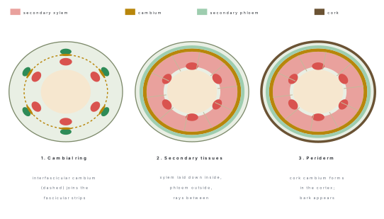
Model answer — long (7)
Definition [1]. Secondary growth is the increase in the girth of a dicot stem (and root) brought about by the lateral meristems: the vascular cambium and the cork cambium. It is absent in monocots.
Mechanism [5]. 1. Complete cambial ring. The cambium inside each bundle (fascicular cambium) is joined by interfascicular cambium, formed from the parenchyma of the medullary rays, to make a continuous ring. 2. Activity of the cambium. Cells of the ring divide tangentially. The inner daughter cells become secondary xylem and the outer ones secondary phloem. More xylem than phloem is formed, so the ring is pushed outward and the primary xylem stays at the centre, while the primary phloem is crushed. A few cambial cells form rays, bands of parenchyma that carry materials sideways. 3. Annual rings. In spring the cambium makes wide vessels (spring wood, light); in autumn, narrow vessels and fibres (autumn wood, dark). One pair is one annual ring; counting rings gives the age of the tree. The inner, dead, dark wood is heartwood; the outer, living, pale wood is sapwood, which conducts. 4. Cork cambium (phellogen). The stretched epidermis and cortex tear, so a new meristem arises in the outer cortex. It cuts phellem (cork) outward, which is suberised, dead and impermeable, and phelloderm (secondary cortex) inward. The three together are the periderm; all tissue outside it is bark. 5. Lenticels. Loose lens-shaped pores in the periderm let the inner cells exchange gases.
Diagram [2]. T.S. of the stem showing, from the outside: periderm (phellem, phellogen, phelloderm), cortex remnants, secondary phloem, vascular cambium, secondary xylem (with rings), primary xylem, pith and medullary rays.
9.2 Plant Physiology
Most-repeated in this unit
| Topic | Seen in the papers | Shape of question |
|---|---|---|
| Photosynthesis: light reaction, Hill reaction, cyclic and non-cyclic photophosphorylation, dark reaction | 2079 (Hill), 2081 (dark reaction), 2082 (photophosphorylation) | long, 8 marks, every year |
| Respiration: EMP pathway, Krebs cycle | model 2079 (EMP) | long |
| Water relations: transpiration vs guttation; diffusion vs osmosis | 2079 OR, 2082 OR | short, 4 |
| Germination and dormancy | model 2079 OR, 2081 OR | short, 4 |
Plant physiology supplied an 8-mark long question in all four papers read (Hill reaction, EMP, dark reaction, photophosphorylation). The board rotates through the sub-topics: light reaction, dark reaction and glycolysis have all had their turn; the Krebs cycle and the electron transport system, and C4 photosynthesis, have not.
Short questions (4 marks)
- Differentiate between transpiration and guttation. [4] (NEB 2079, as the first alternative)
- How does diffusion differ from osmosis on the basis of the types of molecules involved across a cell membrane? Explain how environmental factors influence the rate of diffusion. [2+2] (NEB 2082, as the alternative)
- Describe any four practical importances of seed dormancy in agriculture and horticulture. [2+2] (NEB 2081, as the alternative)
- Which influencing factors must be provided for the normal germination of seeds? What happens when seeds are kept at low temperature? [3+1] (NEB model 2079, as the alternative)
- Differentiate between C3 and C4 plants (any four points). [4] (original)
Model answers — short (1–5)
1.
| Transpiration | Guttation |
|---|---|
| Loss of water as vapour | Loss of water as liquid drops |
| Through stomata, cuticle and lenticels | Through hydathodes at the vein endings |
| Takes place mainly by day | Takes place at night or early morning, when transpiration is low |
| Caused by the sun's heat and the transpiration pull | Caused by root pressure |
| The water is pure | The water contains dissolved salts and sugars |
| Controlled by guard cells | Not controlled by any structure |
2. Difference [2]. Diffusion is the net movement of molecules of any substance — solid, liquid or gas, solute or solvent — from a region of higher to lower concentration. It needs no membrane. Osmosis is the movement of water (solvent) molecules only across a selectively permeable membrane from higher water potential to lower water potential. Environmental factors [2]. (i) Temperature: heat raises the kinetic energy of molecules and speeds diffusion. (ii) Concentration gradient: a steeper gradient makes it faster. (iii) Density of the medium and size of the molecule: small molecules move faster through a thinner medium (gases diffuse faster than liquids). (iv) Pressure (for gases), which raises the number of collisions.
3. Seed dormancy is useful because (any four): (i) it keeps seeds from germinating in an unfavourable season (winter, drought) and saves the seedling; (ii) it allows seeds to be stored and transported to distant places; (iii) it spreads germination over time, so a single bad spell cannot kill all seedlings; (iv) it prevents sprouting on the mother plant (vivipary) during a rainy harvest; (v) in horticulture, knowing the dormancy type lets growers break it by scarification or stratification, which gives uniform germination of apple, rose and many forest seeds; (vi) it lets seed banks keep germplasm for years.
4. Factors [3]. Water (imbibition softens the coat and activates enzymes), oxygen (for aerobic respiration that supplies energy), a suitable temperature (25–30 °C for most seeds; at best an optimum), plus a viable seed with a mature embryo and, for a few seeds (lettuce), light. Low temperature [1]. Enzymes work slowly, respiration and metabolism fall, so germination is delayed or stops and the seed stays dormant (and may be injured by freezing). Chilling for some weeks (stratification) breaks dormancy in apple and pear.
5.
| C3 plant | C4 plant |
|---|---|
| First stable product is 3-PGA (3C) | First stable product is oxaloacetate (4C) |
| Carboxylating enzyme RuBisCO only | PEP carboxylase in mesophyll, then RuBisCO in bundle sheath |
| No Kranz anatomy | Kranz anatomy: bundle sheath cells with large chloroplasts around the vein |
| Photorespiration high | Photorespiration negligible |
| Optimum temperature 20–25 °C | Optimum 30–45 °C; good in hot, dry climate |
| Wheat, rice, barley | Maize, sugarcane |
Long questions (8 marks)
- The source of oxygen in photosynthesis is water, not carbon dioxide. Explain with reasons. Briefly describe the Hill reaction. [2+6] (NEB 2079)
- Three German biochemists — Embden, Meyerhof and Parnas (EMP) — worked out a pathway that forms pyruvic acid from glucose. Identify the pathway and name the site. Construct the step-by-step pathway and list the net gain of ATP. [1+1+5+1] (NEB model 2079)
- The light-independent reactions of photosynthesis convert carbon dioxide into glucose. Explain the steps involved and how ATP and NADPH from the light reaction are used. [5+3] (NEB 2081)
- How do the different photosynthetic pigments help in capturing light energy? Explain how cyclic and non-cyclic photophosphorylation generate ATP and NADPH, and illustrate them with suitable charts. [2+3+3] (NEB 2082)
- Describe the link reaction and the Krebs cycle. Calculate the ATP, NADH and FADH₂ formed from one molecule of glucose in these stages. [2+4+2] (predicted: the Krebs cycle and electron transport have not yet been the long question)
Model answers — long (6–10)
6. Reasons [2]. (i) Van Niel's comparison. Purple sulphur bacteria photosynthesise with H₂S and release sulphur, not oxygen: CO₂ + 2H₂S → (CH₂O) + H₂O + 2S. So in green plants the hydrogen donor must be water, and the gas released must come from it: CO₂ + 2H₂O → (CH₂O) + H₂O + O₂. (ii) Ruben and Kamen (1941) used heavy oxygen. Chlorella given water labelled with ¹⁸O released ¹⁸O₂; when the CO₂ was labelled and the water was ordinary, the oxygen released was not labelled. So O₂ comes from H₂O. Hill reaction [6]. Robert Hill (1937) showed that isolated chloroplasts can release oxygen in the light, without any CO₂, if a suitable hydrogen acceptor is provided. Experiment. Chloroplasts are isolated from crushed spinach leaves in cold buffered sucrose. A blue dye, DCPIP (or ferricyanide), is added as the Hill oxidant. Tubes: (a) chloroplasts + dye in light; (b) chloroplasts + dye in dark; (c) boiled chloroplasts + dye in light; (d) dye alone in light. Observation. Only tube (a) turns from blue to colourless (the dye is reduced) and gives off O₂. Equation. 2H₂O + 2A → 2AH₂ + O₂ (chloroplast, light), where A is the Hill oxidant (in the living leaf A = NADP⁺). Conclusions. (1) Oxygen is released by photolysis of water, in the light reaction, in the grana. (2) The light reaction can occur without CO₂ fixation. (3) Light and intact chloroplast are needed (tubes b and c fail). (4) The electrons taken from water reduce the acceptor; in plants they finally reduce NADP⁺ to NADPH.
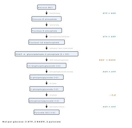
7. Pathway [1]: glycolysis (EMP pathway). Site [1]: the cytoplasm (cytosol) of the cell; no oxygen is needed. Steps [5]: see the figure above. Preparatory phase (energy investment). (1) Glucose + ATP → glucose-6-phosphate (hexokinase). (2) → fructose-6-phosphate (isomerase). (3) + ATP → fructose-1,6-bisphosphate (phosphofructokinase). (4) → dihydroxyacetone phosphate + glyceraldehyde-3-phosphate (aldolase); (5) the two trioses are interconverted, so 2 G3P continue. Pay-off phase (energy yield), twice per glucose. (6) G3P + NAD⁺ + Pᵢ → 1,3-bisphosphoglycerate + NADH. (7) → 3-phosphoglycerate + ATP (substrate-level phosphorylation). (8) → 2-phosphoglycerate. (9) → phosphoenolpyruvate + H₂O (enolase). (10) → pyruvate + ATP. Net gain [1]. 4 ATP made − 2 ATP used = 2 ATP, plus 2 NADH and 2 pyruvate: glucose + 2NAD⁺ + 2ADP + 2Pᵢ → 2 pyruvate + 2NADH + 2ATP + 2H₂O.
8. The light-independent (dark) reaction is the Calvin–Benson cycle, in the stroma. Steps [5]. 1. Carboxylation. CO₂ joins ribulose-1,5-bisphosphate (RuBP, 5C); RuBisCO forms an unstable 6-carbon compound that splits into two molecules of 3-phosphoglycerate (3-PGA), the first stable product. 2. Reduction. ATP phosphorylates 3-PGA to 1,3-bisphosphoglycerate; NADPH then reduces it to glyceraldehyde-3-phosphate (G3P), a triose sugar. 3. Regeneration. Of every six G3P, five are rearranged using ATP to regenerate three RuBP; one G3P leaves the cycle and becomes glucose, sucrose or starch. Three turns fix three CO₂ and yield one G3P; six turns give one glucose. Use of ATP and NADPH [3]. ATP is the energy that drives steps 2 and 3; NADPH is the reducing power (hydrogen and electrons) that reduces 3-PGA to sugar. For each CO₂: 3 ATP + 2 NADPH; for one glucose (6 CO₂): 18 ATP + 12 NADPH. Without the light reaction supplying them, the cycle stops within seconds, so the "dark" reaction depends on light indirectly.
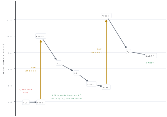
9. Pigments [2]. Chlorophyll a (blue-green) is the main pigment and the reaction-centre molecule (P680, P700). Chlorophyll b and the carotenoids (carotene, xanthophyll) are accessory pigments: they absorb other wavelengths (blue and green-blue) and pass the energy on to chlorophyll a, so the usable range of the spectrum widens; the carotenoids also shield chlorophyll from photo-oxidation. Non-cyclic photophosphorylation [3]. Light excites P680 of photosystem II; its electron travels through phaeophytin, plastoquinone, cytochrome b₆f and plastocyanin to photosystem I, releasing energy that pumps H⁺ into the thylakoid lumen; ATP synthase uses the gradient to make ATP (chemiosmosis). Photosystem I is excited by light; its electron goes through ferredoxin and NADP reductase to reduce NADP⁺ to NADPH. The electron lost by P680 is replaced by photolysis of water: 2H₂O → 4H⁺ + 4e⁻ + O₂. Products: ATP + NADPH + O₂. Cyclic photophosphorylation. Only photosystem I works. The excited electron goes to ferredoxin, then back through cytochrome b₆f and plastocyanin to P700, making ATP on the way. No water is split, no NADPH and no O₂ are formed; it tops up ATP when the Calvin cycle needs more ATP than NADPH. Charts [3]. Non-cyclic: H₂O → PS II (P680) → plastoquinone → cyt b₆f → plastocyanin → PS I (P700) → ferredoxin → NADP⁺ → NADPH (ATP made at cyt b₆f). Cyclic: PS I (P700) → ferredoxin → plastoquinone → cyt b₆f → plastocyanin → back to PS I (ATP made at cyt b₆f).
10. Link reaction [2]. In the mitochondrial matrix each pyruvate is decarboxylated and oxidised and joined to coenzyme A: pyruvate + NAD⁺ + CoA → acetyl CoA + CO₂ + NADH (pyruvate dehydrogenase). Krebs cycle [4]. Acetyl CoA (2C) + oxaloacetate (4C) → citrate (6C) → isocitrate → α-ketoglutarate (5C) (CO₂, NADH) → succinyl CoA (4C) (CO₂, NADH) → succinate (GTP, which makes ATP) → fumarate (FADH₂) → malate → oxaloacetate (NADH), ready for the next turn. Accounting [2].
| Stage (per glucose) | ATP | NADH | FADH₂ | CO₂ |
|---|---|---|---|---|
| Link reaction (2 pyruvate) | — | 2 | — | 2 |
| Krebs cycle (2 turns) | 2 | 6 | 2 | 4 |
| Total | 2 | 8 | 2 | 6 |
In the electron transport system each NADH gives about 2.5 and each FADH₂ about 1.5 ATP (total 30–32 ATP per glucose; older books say 36–38).
9.3 Genetics
Most-repeated in this unit
| Topic | Seen in the papers | Shape of question |
|---|---|---|
| DNA structure, replication (semi-conservative, enzymes) | model 2079, 2081, 2082 OR | long (and short) |
| Crossing over / linkage | model 2079 OR, 2082 short OR | long and short |
| Mutation: gene and chromosomal | 2079, 2081 OR, MCQ 2079 model, 2081, 2082 | long |
| Sex-linked inheritance: Drosophila, colour blindness, criss-cross | 2079 OR, 2082, MCQ 2080 | long |
| Incomplete dominance; polyploidy | 2081 OR, 2079 OR | short |
Genetics is the most generous unit for the 8-mark question: a genetics long question appeared in every paper read (2079, model 2079, 2081, 2082). The three favourites are replication, mutation and sex-linkage; crossing over is the fourth and is always paired with a labelled diagram.
Short questions (4 marks)
- If you were assigned to make a Watson and Crick model of DNA, what essential components would you use to make the exact structure? [2+2] (NEB model 2079, as the alternative)
- Explain the mechanism of incomplete dominance up to the F₂ generation with a suitable example. How does it differ from complete dominance? [2+2] (NEB 2081, as the alternative)
- In which process are linked genes inherited together from one generation to another with parental and non-parental combinations? Explain the process with a suitable example. [4] (NEB 2082)
- Explain in brief the significance of polyploidy with examples. [4] (NEB 2079, as the alternative)
- Write four reasons why Drosophila melanogaster is chosen for genetic studies. [4] (original)
- Distinguish between a point mutation and a frame-shift mutation, with an example of each. [4] (original)
Model answers — short (1–6)
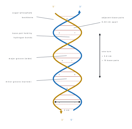
1. Components [2]. A nucleotide = one deoxyribose sugar + one phosphate + one nitrogenous base. Four kinds of base are needed: the purines adenine (A) and guanine (G), drawn as double rings, and the pyrimidines thymine (T) and cytosine (C), as single rings. Building the model [2]. (i) Join the nucleotides by phosphodiester bonds (3′ carbon of one sugar to the 5′ phosphate of the next) into two long chains of sugar–phosphate backbone. (ii) Pair the bases across the two chains by hydrogen bonds: A with T (two bonds) and G with C (three bonds), so that a purine always faces a pyrimidine and the ladder keeps a uniform width of 2 nm. (iii) Run the two chains antiparallel (one 5′→3′, the other 3′→5′) and twist them into a right-handed helix with 10 base pairs per turn, a pitch of 3.4 nm and 0.34 nm between pairs.
2. Mechanism [2]. In incomplete dominance neither allele masks the other, so the heterozygote is intermediate. In the four o'clock plant (Mirabilis jalapa), RR (red) × rr (white) gives an F₁ that is all Rr, pink. On selfing the F₁:
| R | r | |
|---|---|---|
| R | RR red | Rr pink |
| r | Rr pink | rr white |
F₂: 1 red : 2 pink : 1 white — the phenotypic ratio equals the genotypic ratio, 1 : 2 : 1. Difference [2]. In complete dominance the F₁ looks like the dominant parent, the heterozygote cannot be told from the dominant homozygote, and the F₂ phenotypic ratio is 3 : 1 (genotypic 1 : 2 : 1). In incomplete dominance the heterozygote is a distinct intermediate and the F₂ ratio is 1 : 2 : 1 for both.
3. The process is linkage. Definition. Genes that lie on the same chromosome tend to be inherited together; they do not assort independently. Because crossing over separates some of them, the offspring show parental combinations more often than recombinants. Example (Morgan, Drosophila). Grey body and long wings (b⁺b⁺ vg⁺vg⁺) × black body and vestigial wings (bb vgvg) gives F₁ grey long. A test cross of the F₁ female with a black-vestigial male gave 965 grey-long and 944 black-vestigial (parental, 83 %) but only 206 grey-vestigial and 185 black-long (recombinant, 17 %). If the genes had assorted independently the four classes would have been 25 % each. The recombination frequency (391/2300 = 17 %) is a measure of the distance between the genes: 1 % = 1 map unit.
4. Polyploidy is the presence of more than two complete sets of chromosomes. Significance: (i) it makes cells and organs larger — bigger fruits, flowers and seeds (gigas effect), as in polyploid tomato and sugar beet; (ii) autotriploids are sterile and so give seedless fruit: banana (3n) and watermelon; (iii) it restores the fertility of hybrids: triticale, an allopolyploid of wheat × rye, combines the yield of wheat and the hardiness of rye; (iv) it is a source of new species in nature — bread wheat is hexaploid (6n = 42) and more than a third of flowering plants are polyploid; (v) colchicine treatment lets breeders produce polyploids on purpose.
5. (i) Small and cheap to keep in bottles on banana or cornmeal medium; (ii) a short life cycle of about 10–14 days, so many generations can be followed in a few months; (iii) one mating gives hundreds of offspring, so ratios are reliable; (iv) only four pairs of chromosomes (2n = 8), and the giant salivary-gland chromosomes can be seen with banding; (v) males and females are easily told apart, and there are many visible mutant traits (white eye, vestigial wing, yellow body).
6.
| Point mutation | Frame-shift mutation |
|---|---|
| One base is replaced by another (substitution): transition (A↔G, C↔T) or transversion (purine ↔ pyrimidine) | One or two bases are added (insertion) or lost (deletion) |
| Only one codon changes; effect may be silent, missense or nonsense | Every codon after the change is read differently, so the whole downstream protein is altered |
| Example: sickle-cell anaemia, GAG → GTG, glutamic acid → valine | Example: deletion of one base in the β-globin gene that leads to a truncated β-thalassaemia protein |
Long questions (8 marks)
- Mutation is important from the evolutionary point of view; give reasons. How does gene mutation occur? Explain its types. [2+2+4] (NEB 2079)
- Why is Drosophila melanogaster selected for genetical study? "The inheritance of a sex-linked trait follows the criss-cross pattern." Justify this statement with special reference to the eye colour of Drosophila. [3+5] (NEB 2079, as the alternative)
- Explain how colour blindness is inherited as a sex-linked trait in humans. Illustrate it by suitable genetic crosses and analyse the phenotypic outcomes. On that basis justify why the pattern is called criss-cross inheritance. [2+4+2] (NEB 2082)
- Semi-conservative replication is the most common mode of DNA replication and gives two identical daughter molecules. Explain the enzymes and the mechanism involved, with necessary diagrams. [2+4+2] (NEB model 2079; the 2082 alternative asked the same topic as [3+5])
- "Genes are inherited in new combinations due to crossing over in the gametes." Interpret this statement with its mechanism and necessary diagrams. Assess its significance also. [4+2+2] (NEB model 2079, as the alternative)
- In what ways does DNA serve as the genetic material? Discuss its double-helical structure with a labelled diagram. Analyse the similarities and differences between DNA and RNA and evaluate their significance. [1+4+2+1] (NEB 2081)
- How can chromosomal mutations alter the number and structure of chromosomes? Discuss the types of chromosomal mutation and analyse their significance. [1+4+3] (NEB 2081, as the alternative)
Model answers — long (7–13)
7. Why mutation matters in evolution [2]. Mutation is the ultimate source of new alleles. Natural selection can only choose among variants that already exist, so mutation supplies the raw material; useful mutations spread, and over many generations they lead to adaptation and new species. How gene mutation occurs [2]. A gene mutation is a change in the base sequence of a gene. It arises spontaneously by a mistake in DNA replication (a base pairs with its rare tautomer, or the polymerase slips) that escapes repair, or it is induced by mutagens: physical (UV light, X-rays, γ-rays), chemical (nitrous acid, ethyl methane sulphonate, 5-bromouracil, mustard gas) or biological (transposons, some viruses). Types [4]. (a) Substitution (point) mutation — one base replaced by another. Transition: purine ↔ purine or pyrimidine ↔ pyrimidine (A→G, C→T). Transversion: purine ↔ pyrimidine (A→T). Result may be silent (same amino acid), missense (different amino acid; sickle-cell anaemia GAG→GTG, Glu→Val) or nonsense (a stop codon). (b) Frame-shift mutation — insertion or deletion of one or two bases; the reading frame moves and all later codons change; example: β-thalassaemia from a single-base deletion.
8. Reasons [3]. Small, cheap, short life cycle (10–14 days), many progeny, only 2n = 8, giant salivary chromosomes, many visible mutants, sexes easily separated. Criss-cross inheritance [5]. The gene for eye colour (w) lies on the X chromosome; the Y carries no allele. Red (W) is dominant, white (w) is recessive. A male has one X (hemizygous), so one w makes him white-eyed. Cross 1: red-eyed female (X^W X^W) × white-eyed male (X^w Y). F₁: all red-eyed; females X^W X^w (carriers), males X^W Y. F₁ × F₁: F₂ females X^W X^W and X^W X^w (all red), males X^W Y (red) and X^w Y (white): 3 red : 1 white, and every white-eyed fly is a male. Cross 2 (reciprocal): white-eyed female (X^w X^w) × red-eyed male (X^W Y). F₁: all daughters X^W X^w red, all sons X^w Y white. F₂: 1 red female : 1 white female : 1 red male : 1 white male. Justification. In cross 2 the white-eye trait passes from the mother to her sons, and in the next generation, through the carrier daughters, to the grandsons. The trait therefore crosses from mother to son and from father (via daughter) to grandson: the two sexes exchange it, like a crisscross. The reciprocal crosses give different results, which never happens for an autosomal gene.
9. Sex-linked trait [2]. The gene for red–green colour vision lies on the X chromosome, in the region that has no partner on the Y. The allele for colour blindness (X^c) is recessive. Females (XX) must be X^cX^c to be colour-blind; males (XY) are hemizygous, so a single X^c makes a male colour-blind. Hence the disorder is commoner in men. Crosses [4]. (C = normal, c = colour-blind)
| Cross | Daughters | Sons |
|---|---|---|
| Carrier female X^CX^c × normal male X^CY | all normal (½ carrier) | ½ normal, ½ colour-blind |
| Normal female X^CX^C × colour-blind male X^cY | all normal, all carriers | all normal |
| Colour-blind female X^cX^c × normal male X^CY | all normal, all carriers | all colour-blind |
| Carrier female X^CX^c × colour-blind male X^cY | ½ carrier, ½ colour-blind | ½ normal, ½ colour-blind |
Why criss-cross [2]. In the third cross the colour-blind mother passes the allele to all her sons, and the normal father passes his X to his daughters, who become carriers; those carrier daughters can give it to their sons. The gene moves from mother to son and from father through the daughter to the grandson, never from father to son. That diagonal flow is criss-cross inheritance.
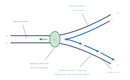
10. Enzymes [2]. Helicase unwinds the double helix; single-strand binding proteins keep the strands apart; topoisomerase relieves the coiling ahead of the fork; primase makes a short RNA primer; DNA polymerase III adds nucleotides in the 5′→3′ direction; DNA polymerase I removes the primers and fills the gaps; DNA ligase seals the nicks. Mechanism [4]. (1) Replication starts at the origin of replication, where helicase opens a bubble with two replication forks. (2) Each parental strand serves as a template. (3) Primase lays an RNA primer; DNA polymerase III extends it. (4) On the leading strand (template 3′→5′) synthesis is continuous towards the fork. (5) On the lagging strand (template 5′→3′) synthesis is discontinuous: short Okazaki fragments are made away from the fork, each from its own primer. (6) Polymerase I replaces the primers with DNA and ligase joins the fragments. (7) Each daughter molecule consists of one old strand and one new strand: the mode is semi-conservative. Proof: Meselson and Stahl (1958) grew E. coli in ¹⁵N and transferred it to ¹⁴N; after one generation all DNA was hybrid, after two, half hybrid and half light. Diagrams [2]. The replication fork above.
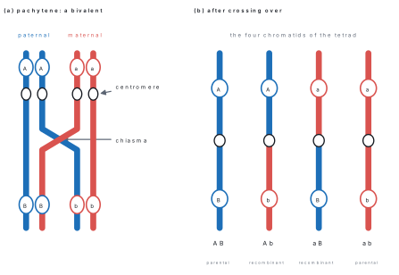
11. Interpretation and mechanism [4]. Crossing over is the exchange of corresponding segments between non-sister chromatids of homologous chromosomes during pachytene of prophase I. Homologues pair (synapsis) at zygotene to form a bivalent; at pachytene an enzyme (endonuclease) breaks one chromatid of each homologue at the same level; the broken pieces are exchanged and joined by ligase; at diplotene the crossing points are seen as chiasmata, and the homologues separate. Of the four chromatids two remain parental (AB, ab) and two become recombinant (Ab, aB), so the gametes carry new combinations of alleles that neither parent had. Significance [2]. (i) It produces genetic variation, the raw material for natural selection and evolution; (ii) the frequency of crossing over allows genetic maps to be drawn (1 % = 1 map unit); (iii) breeders use it to combine useful genes from different parents; (iv) it separates linked genes, including a harmful allele from a useful one. Diagram [2]. Crossing over in a bivalent, as above.
12. Why DNA is the genetic material [1]. It stores information in the sequence of bases, replicates exactly, passes from cell to cell, is expressed through RNA into protein, and is stable but can mutate. Evidence: Griffith's transformation, Avery–MacLeod–McCarty's identification of DNA as the transforming principle and Hershey–Chase's experiment, in which ³²P-labelled DNA entered the bacterium and ³⁵S-labelled protein did not. Double helix [4]. Watson and Crick (1953): two polynucleotide chains wound right-handed round a common axis, antiparallel; backbone of deoxyribose and phosphate on the outside joined by phosphodiester bonds; bases inside, paired A=T and G≡C by hydrogen bonds (Chargaff: A = T, G = C); diameter 2 nm, 10 base pairs per turn, pitch 3.4 nm, 0.34 nm between pairs; major and minor grooves. The labelled diagram is given above. DNA vs RNA [2]. Similarities: both are polymers of nucleotides, have a sugar–phosphate backbone and four bases and carry genetic information (RNA in some viruses).
| DNA | RNA |
|---|---|
| Deoxyribose sugar | Ribose sugar |
| Bases A, T, G, C | Bases A, U, G, C |
| Double-stranded helix | Mostly single-stranded |
| Mostly in the nucleus; stores information | Nucleus and cytoplasm; mRNA, tRNA, rRNA carry out expression |
| Replicates itself | Made from DNA by transcription |
Significance [1]. DNA keeps and passes on the genetic information; RNA carries it out, so genes become proteins and traits.
13. Definition [1]. A chromosomal mutation is a change in the structure or number of whole chromosomes. Types [4]. Structural: deletion (loss of a piece: cri-du-chat syndrome); duplication (a piece repeated: Bar eye of Drosophila); inversion (a piece turned through 180°, paracentric or pericentric); translocation (a piece moves to a non-homologous chromosome, as in the Philadelphia chromosome). Numerical: aneuploidy — one chromosome more or less: trisomy 21 (Down's syndrome, 47), monosomy X (Turner's, 45, X), XXY (Klinefelter's, 47); euploidy/polyploidy — whole extra sets (3n, 4n), induced by colchicine; caused by non-disjunction at meiosis. Significance [3]. (i) Polyploidy and translocations have created new species, so they drive evolution; (ii) breeders use them: seedless watermelon (3n), triticale (allopolyploid), larger crop varieties; (iii) in humans they cause syndromes, recognisable in a karyotype and offered in genetic counselling; (iv) they change the gene order and so affect linkage.
9.4 Embryology
Most-repeated in this unit
| Topic | Seen in the papers | Shape of question |
|---|---|---|
| Dicot embryo | 2079 | short with diagram, 2 + 2 |
| Pollen grain / male gametophyte; why two male gametes | 2081 | short with diagram |
| Vegetative propagation (grafting) | 2082 | short with diagram |
| MCQs: porogamy (2080, 2082), parthenogenesis/parthenocarpy (model 2079, 2081), degenerating cells of the embryo sac (2079) | all papers | MCQ |
Embryology has never supplied an 8-mark question in the papers read; it is worth about 5 marks per paper in the short and MCQ groups. Female gametophyte, male gametophyte and fertilisation are the long questions the board has not yet set from this unit.
Short questions (4 marks)
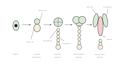
- Write the salient features of a dicot embryo in reference to its development pattern, with labelled diagrams. [2+2] (NEB 2079)
- Study the diagram of a pollen grain. (i) When is the pollen grain produced in a flower? (ii) Identify the parts A and C. (iii) In angiosperms the pollen tube carries two male gametes; what is the purpose when a single gamete could fertilise the egg? [1+1+2] (NEB 2081)
- The diagram shows a type of artificial vegetative propagation in an angiosperm, with parts P and Q. (i) Identify it and name P and Q. (ii) Explain it briefly with an example. (iii) Mention any two applications. [1.5+1.5+1] (NEB 2082)
- What is double fertilisation? Explain it and state its significance. [1+2+1] (original)
- Distinguish between wind pollination (anemophily) and insect pollination (entomophily). [4] (original)
Model answers — short (1–5)
1. Development pattern [2]. The zygote divides (after a rest) to give a two-celled proembryo: the upper embryonal cell and the lower suspensor cell. The suspensor cell forms the suspensor, which pushes the embryo into the endosperm; the embryonal cell passes through the globular, heart-shaped (two cotyledons begin) and torpedo stages to the mature embryo. Features of the mature embryo [2]. It has an embryonal axis with the radicle at the lower end (covered by a root cap), the hypocotyl, the epicotyl and the plumule (shoot tip), and two cotyledons attached to the axis; the suspensor lies at the radicle end. Draw the stages figure below.
2. (i) The pollen grain is produced in the anther (microsporangium) when the flower is forming, from the microspore mother cells by meiosis. (ii) A standard pollen grain shows: the thick outer exine of sporopollenin with a germ pore, the thin inner intine, the large vegetative (tube) cell and the small generative cell. The letters A and C in the paper's figure mark two of these; name whichever structures the arrows point to from this list. (iii) The two male gametes are needed for double fertilisation. One fuses with the egg to give the diploid zygote (syngamy); the other fuses with the two polar nuclei (triple fusion) to give the triploid primary endosperm nucleus, which becomes the food tissue of the seed. With only one gamete the embryo would have no nourishment.
3. (i) Grafting. P is the stock (the rooted lower part) and Q the scion (the cut twig of the desired variety). (ii) A scion from a plant with desirable fruit is cut obliquely, fitted on a stock of a related plant so that the cambium layers touch, tied and sealed with wax or tape. The cambia unite and the scion grows with the stock's root system; used for mango, apple, citrus and rose. (iii) Applications: it propagates varieties that do not come true from seed and gives earlier fruiting; it combines the stock's disease resistance and hardiness with the scion's fruit quality; it repairs damaged trees; it speeds multiplication of superior clones.
4. Definition [1]. The fusion of one male gamete with the egg and the other with the two polar nuclei, both in the same embryo sac, is double fertilisation; it is found only in angiosperms. Explanation [2]. The pollen tube enters the embryo sac (usually through the micropyle, porogamy) and releases two male gametes. Syngamy: gamete 1 + egg → diploid zygote (2n) → embryo. Triple fusion: gamete 2 + two polar nuclei → triploid primary endosperm nucleus (3n) → endosperm. The synergids and antipodals then degenerate. Significance [1]. The endosperm starts to form only when fertilisation has occurred, so no food is wasted on an unfertilised ovule; it also gives hybrid vigour to the endosperm (xenia). It was discovered by Nawaschin (1898).
5.
| Anemophily | Entomophily |
|---|---|
| Pollen carried by wind | Pollen carried by insects |
| Flowers small, dull, scentless, no nectar | Flowers large, coloured, scented, with nectar |
| Pollen light, dry, smooth and produced in huge amounts | Pollen sticky, spiny, produced in smaller amounts |
| Stigma large, feathery or branched; anthers exposed | Stigma small and sticky |
| Examples: maize, wheat, grasses | Examples: mustard, sunflower, rose |
Long question (8 marks)
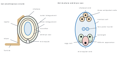
- Describe the development of the female gametophyte (embryo sac) in an angiosperm. Draw a labelled diagram of the mature embryo sac. [1+5+2] (predicted)
Model answer — long (6)
Definition [1]. The female gametophyte is the embryo sac, a haploid seven-celled structure that develops inside the ovule from one functional megaspore. Development [5] (Polygonum type, monosporic). 1. Megasporogenesis. A single cell of the nucellus near the micropyle becomes the megaspore mother cell (2n). It divides by meiosis into four haploid megaspores, in a row. 2. Three megaspores, on the micropylar side, degenerate; the one nearest the chalaza is the functional megaspore. 3. Megagametogenesis. The functional megaspore enlarges and its nucleus divides by mitosis: two nuclei move to opposite poles (2-nucleate stage). 4. Each divides twice more, giving four nuclei at each pole (8-nucleate stage). 5. One nucleus from each pole, a polar nucleus, migrates to the centre; the remaining three at the micropylar end form the egg apparatus (an egg and two synergids) and the three at the chalazal end become the antipodal cells. The two polar nuclei lie in the large central cell. The result is 7 cells and 8 nuclei. Diagram [2]. Mature embryo sac: egg and two synergids at the micropylar end (synergids with filiform apparatus), central cell with two polar nuclei, three antipodals at the chalazal end; see the figure above. After fertilisation the synergids and antipodals degenerate.
9.5 Biotechnology
Most-repeated in this unit
| Topic | Seen in the papers | Shape of question |
|---|---|---|
| Biotechnology and food scarcity / GMOs and food security | model 2079, 2082 | short, 4 (paired with "justify your opinion") |
| Hybridisation as a breeding method | 2079 | short, 1 + 3 |
| Micropropagation and conservation | 2081 | short, 2 + 2 |
| MCQ: embryo culture (2079) | 2079 | MCQ |
This unit is worth exactly one Group B question in every paper read, and the board likes an opinion stem ("would you think…", "justify", "what are the concerns").
Short questions (4 marks)
- Biotechnology is an emerging discipline. Do you think it can be used to mitigate the problem of food scarcity in the context of Nepal? Justify your opinion. [4] (NEB model 2079)
- "Hybridisation is a modern breeding method for the development of crop plants with desired characters as well as hybrid vigour." Justify the statement by describing the steps of the hybridisation procedure. [1+3] (NEB 2079)
- Describe the process of micropropagation to rapidly produce a large number of identical plantlets and how it can be used in the conservation of endangered plant species. [2+2] (NEB 2081)
- How can GMOs be used to address food-security challenges, and what are the possible environmental concerns and health risks associated with them? [2+2] (NEB 2082)
- Distinguish between a biofertiliser and a biopesticide, giving two examples of each. [4] (original)
Model answers — short (1–5)
1. Yes, with limits. Arguments for [3]. (i) Tissue culture multiplies disease-free banana, potato, orchid and cardamom plantlets quickly, raising yield per hectare on small hill farms. (ii) Genetic engineering can give crops that resist pests, drought and saline soil, or contain more nutrients (Golden Rice with β-carotene), so less is lost. (iii) Biofertilisers (Rhizobium, Azotobacter, Azolla) and biopesticides (Bt, Trichoderma) cut the cost of chemicals that a landlocked country imports. (iv) Marker-assisted breeding shortens the time to develop varieties for Nepal's altitude zones. Limits [1]. Seed and gene technology must be affordable, regulated and safe, and scarcity also comes from poor roads and storage, so biotechnology is one part of the answer, not all of it.
2. Justification [1]. Hybridisation crosses two genetically different parents so that desirable characters are combined in one line, and the F₁ often shows hybrid vigour (heterosis): higher yield, vigour, disease resistance. Steps [3]. (i) Selection of parents with the desired traits (e.g. high yield and disease resistance). (ii) Emasculation: removal of the anthers from the flowers of the female parent, before they open, to prevent self-pollination. (iii) Bagging the emasculated flower with a fine paper bag. (iv) Cross-pollination: when the stigma is receptive, pollen of the male parent is dusted on it, and the flower is re-bagged and tagged. (v) Raising and selecting the F₁, and selfing or backcrossing to fix the desired combination; the best lines are tested and released as a variety.
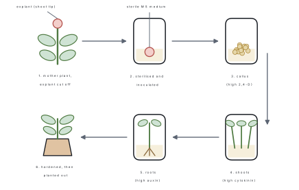
3. Process [2]. Micropropagation is the rapid clonal multiplication of a plant from a small piece (explant, e.g. shoot tip or node) in sterile conditions. Steps: (1) choose a healthy mother plant and surface-sterilise the explant; (2) place it on sterile MS medium with sugar and growth regulators for shoot multiplication (high cytokinin); (3) transfer the shoots to a rooting medium (high auxin); (4) harden the plantlets in a greenhouse and move them to soil. Thousands of genetically identical plantlets come from one explant. Conservation [2]. Endangered plants, such as the orchid Dendrobium or the medicinal Taxus and Rauvolfia, can be multiplied without harvesting wild plants, the cultures can be stored at low temperature (in vitro gene bank), and the plantlets can be used to re-stock forests.
4. Use for food security [2]. GM crops can carry genes for insect resistance (Bt cotton, Bt maize), herbicide tolerance, drought or salt tolerance and extra nutrients (Golden Rice: vitamin A), raising yield, cutting losses and improving nutrition. Concerns [2]. Environmental: gene flow to wild relatives, loss of biodiversity, evolution of resistant pests and superweeds, harm to non-target insects. Health: possible allergens, unknown long-term effects of new proteins, antibiotic-resistance marker genes. Social: patent control by seed companies and dependence of farmers.
5.
| Biofertiliser | Biopesticide |
|---|---|
| Living microbes that add nutrients to soil or plants | Living organisms or their products that kill pests and pathogens |
| Raise soil fertility (nitrogen fixation, phosphate solubilisation) | Protect crops from insects, fungi and weeds |
| Rhizobium (legumes), Azotobacter (free-living), Azolla + Anabaena (rice fields) | Bacillus thuringiensis (Bt toxin), Trichoderma (fungal diseases), nuclear polyhedrosis virus |
| Replace chemical fertiliser | Replace chemical pesticide |
Long question (8 marks)
- What is plant tissue culture? Describe the steps of tissue culture technique and mention its applications. [1+4+3] (predicted)
Model answer — long (6)
Definition [1]. Plant tissue culture is the growing of plant cells, tissues or organs on an artificial, sterile nutrient medium under controlled conditions; it depends on totipotency — every living plant cell can regenerate a whole plant. Steps [4]. (1) Selection and sterilisation of the explant (shoot tip, leaf, node) with ethanol and sodium hypochlorite. (2) Culture medium: Murashige–Skoog (MS) medium with mineral salts, sucrose, vitamins and the hormones auxin and cytokinin, gelled with agar, sterilised in an autoclave. (3) Inoculation of the explant into the vessel in a laminar-flow chamber. (4) Callus formation: the explant makes an undifferentiated mass of cells. (5) Differentiation: a high cytokinin : auxin ratio gives shoots, a high auxin : cytokinin ratio gives roots. (6) Hardening and transfer of the plantlets to soil. Applications [3]. Mass multiplication (banana, orchids, potato); virus-free plants from meristem culture; haploid plants from anther culture; embryo culture to save aborting hybrid embryos; somatic hybrids by protoplast fusion; production of secondary metabolites such as shikonin; conservation of endangered plants and germplasm storage.
9.6 Animal Tissues
Most-repeated in this unit
| Topic | Seen in the papers | Shape of question |
|---|---|---|
| Epithelial tissue: simple epithelia, squamous vs cuboidal | 2079 (with diagrams), 2082 | short, 3 + 1 and 1 + 3 |
| Connective tissue: areolar vs adipose | model 2079 | short, 2 + 2 |
| Muscle: voluntary vs involuntary | 2081 | short, 2 + 2 |
| MCQs: adipose, cartilage lacks blood supply, phagocytic white cells, vaginal epithelium, mast cells | all papers | MCQ |
Every paper read had exactly one Group B question on animal tissues (Q18 in 2079, 2081 and 2082; Q20 in the model paper), and it has always been a "differentiate/compare" or "types with diagram" stem. Expect it again; the nervous tissue (neuron) and cartilage/bone have not been asked yet.
Short questions (4 marks)
- How would you elaborate the different types of simple epithelial tissues in animals? Support with diagrams. [3+1] (NEB 2079)
- Compare and contrast areolar and adipose connective tissues. [2+2] (NEB model 2079)
- Compare and contrast voluntary and involuntary muscles. [2+2] (NEB 2081)
- "Squamous epithelium is called pavement epithelium." Give the reason. Write any three differences between squamous and cuboidal epithelium. [1+3] (NEB 2082)
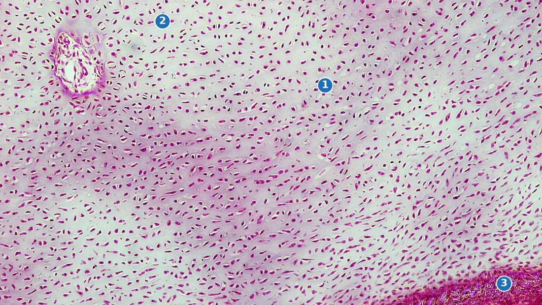
- Name the three types of cartilage and state where each is found; why does cartilage heal slowly? [3+1] (original)
Model answers — short (1–5)
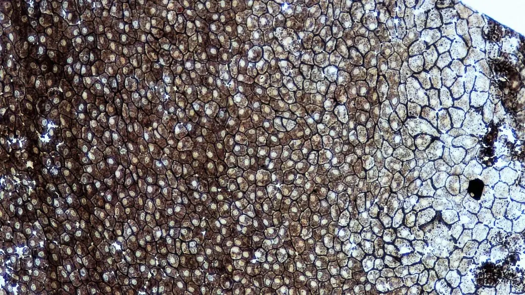
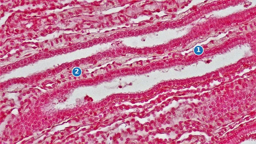
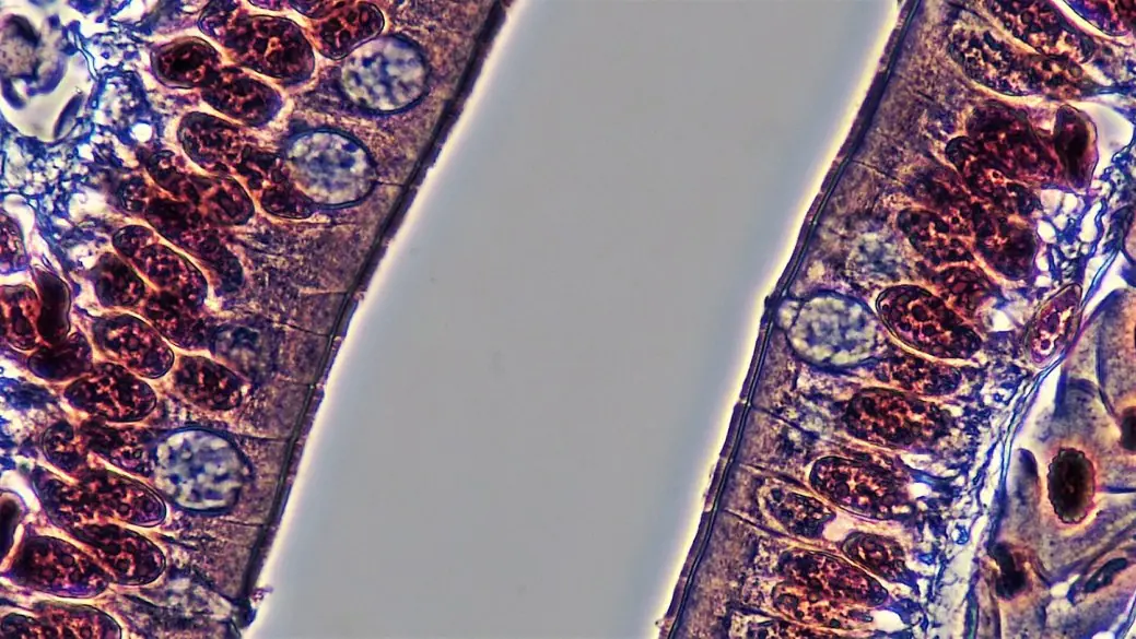
1. Simple epithelium is a single layer of cells resting on a basement membrane. Types [3]: - Squamous — flat, plate-like cells with a central nucleus; lining of alveoli, blood vessels (endothelium), Bowman's capsule; diffusion and filtration. - Cuboidal — cube-shaped cells with a round central nucleus; thyroid follicles, kidney tubules (brush border in the PCT), ducts of glands; secretion and absorption. - Columnar — tall, narrow cells with a basal nucleus; stomach and intestine (goblet cells secrete mucus; microvilli absorb); secretion and absorption. A ciliated form lines the trachea and oviduct and moves mucus or eggs. - Glandular — single cells (goblet cells) or sheets that secrete. Diagram [1]: as in the figure.
2.
| Areolar tissue | Adipose tissue |
|---|---|
| Loose connective tissue with a semi-fluid matrix | A type of loose connective tissue packed with fat cells |
| Cells: fibroblasts, macrophages, mast cells, plasma cells | Cells: adipocytes, each with one large fat droplet that pushes the nucleus to the side |
| White collagen and yellow elastic fibres present, in a network | Few fibres; matrix scanty |
| Packing tissue between organs and under skin; binds skin to muscle | Stores fat, insulates, cushions kidney and heart |
3.
| Voluntary (skeletal) muscle | Involuntary muscle |
|---|---|
| Under control of the will | Not under the will; controlled by the autonomic nervous system |
| Striated; cylindrical, unbranched, multinucleate fibres | Smooth muscle: unstriated, spindle-shaped, uninucleate; cardiac muscle: striated, branched, with intercalated discs |
| Attached to bones by tendons; found in limbs, tongue, face | In the walls of the gut, blood vessels, bladder (smooth) and in the heart (cardiac) |
| Contract quickly, tire quickly | Contract slowly and rhythmically, do not tire (cardiac never does) |
4. Reason [1]. The cells are thin, flat and polygonal and fit edge to edge in a single layer, like the slabs of a paved floor, so it is called pavement epithelium. Differences [3].
| Squamous epithelium | Cuboidal epithelium |
|---|---|
| Cells flat, plate-like | Cells cube-shaped |
| Nucleus flat, central | Nucleus round, central |
| Lines alveoli, blood vessels, Bowman's capsule | Lines kidney tubules, thyroid follicles, gland ducts |
| Function: diffusion and filtration | Function: secretion and absorption |
5. Hyaline cartilage — the commonest: ends of long bones (articular), nose, trachea, larynx, ribs; glassy matrix. Elastic cartilage — pinna of the ear, epiglottis; yellow elastic fibres. Fibrocartilage — intervertebral discs, pubic symphysis; dense collagen, strongest. Slow healing [1]. Cartilage is avascular; its cells are fed by diffusion from the perichondrium, so repair is slow.
Long question (8 marks)
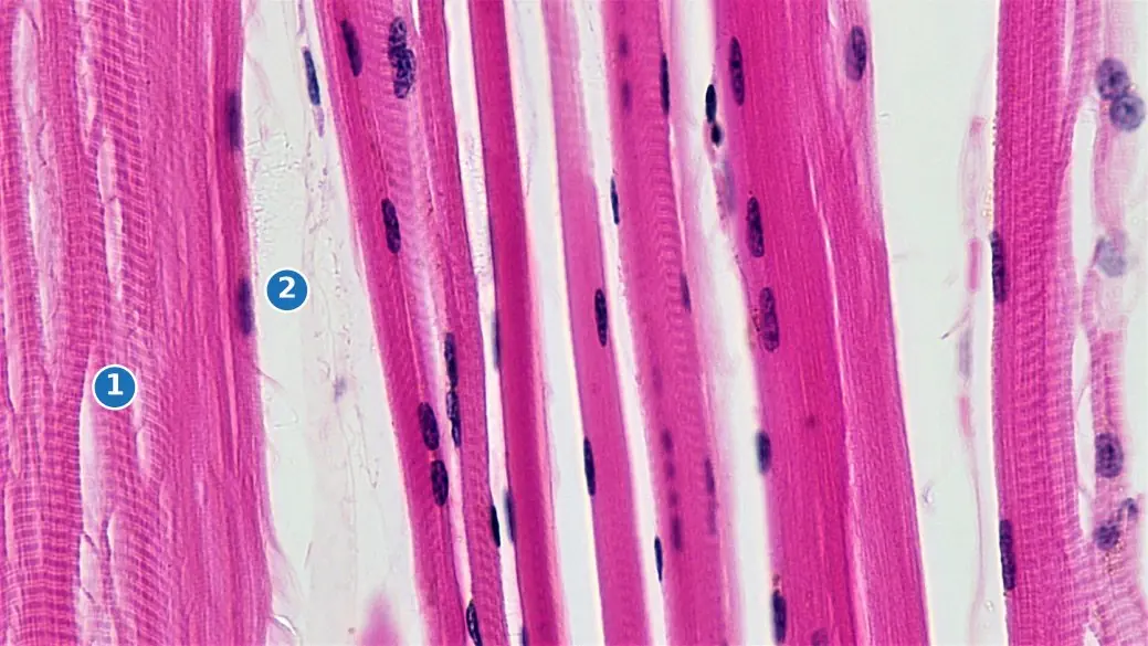
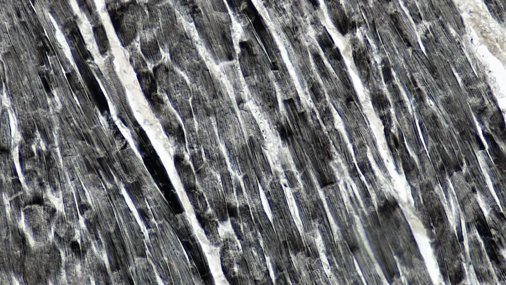
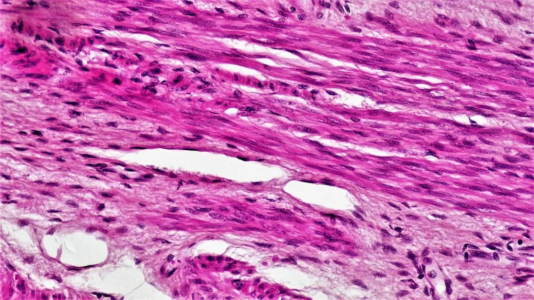
- Describe the three types of muscular tissue with labelled diagrams. [6+2] (predicted)
Model answer — long (6)
1. Skeletal (striated, voluntary) muscle [2]. Long, cylindrical, unbranched fibres up to several centimetres long; multinucleate, with nuclei at the periphery just under the sarcolemma; cross-striations from alternating dark A bands and light I bands (actin and myosin); attached to bones by tendons; contracts quickly and under the will; in limbs, tongue, face, body wall.
2. Smooth (unstriated, involuntary) muscle [2]. Spindle-shaped cells with tapering ends, one central nucleus, no striations; in the walls of the stomach, intestine, bladder, blood vessels and uterus; contracts slowly and rhythmically (peristalsis), controlled by the autonomic nervous system.
3. Cardiac muscle [2]. Found only in the heart; cylindrical, branched fibres, striated, uninucleate (nucleus central); fibres joined end to end by intercalated discs; involuntary and never fatigues; beats rhythmically, starting at the SA node. Diagrams [2]. Draw the three fibre types side by side as in the figure and label: nucleus, striations, sarcolemma, intercalated disc. Summary. A table of the three types (voluntary/involuntary, striated or not, nuclei, shape, location) makes this answer complete.
9.7 Developmental Biology
Most-repeated in this unit
| Topic | Seen in the papers | Shape of question |
|---|---|---|
| Stages of frog development: blastulation, coelom, neurulation | model 2079, 2081, 2082 | short, 4 |
| Oogenesis / spermatogenesis / IVF | 2079 (oogenesis with diagram; IVF as OR), 2081 MCQ | short, 3 + 1 |
| MCQs: gametogenesis sequence, spermiogenesis, coelom from mesoderm, sequence of frog stages | 2079, model 2079, 2081, 2082 | MCQ |
Developmental biology shows up as a four-mark question: on one stage of frog development in three of the four papers (blastulation in model 2079, coelom in 2081, neurulation in 2082) and on oogenesis in 2079. Gastrulation and cleavage are the stages that have not had their turn; prepare them with the same sequence.
Short questions (4 marks)
- Define blastulation. Write the process of blastulation during the development of the frog. [1+3] (NEB model 2079)
- What is coelom? How is it formed in the development of the frog? [1+3] (NEB 2081)
- Describe the process of oogenesis in animals with a diagram. [3+1] (NEB 2079)
- Write about the process of neurulation in the development of the frog. [4] (NEB 2082)
- Define the term in-vitro fertilisation and describe its process. [1+3] (NEB 2079, as the alternative)
- Describe gastrulation in the frog. [4] (predicted)
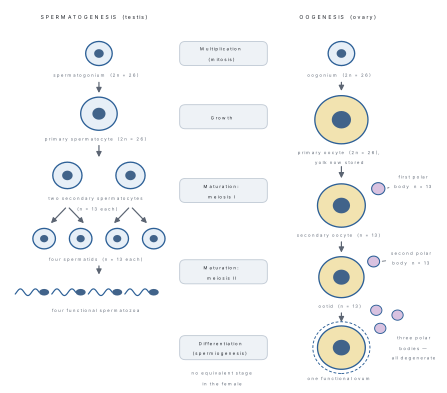
Model answers — short (1–6)
1. Definition [1]. Blastulation is the stage of development in which the morula is converted into a hollow blastula with a central cavity, the blastocoel. Process [3]. After the 32–64-cell morula forms, the cells rearrange and fluid accumulates among the smaller animal-pole cells (micromeres). The fluid-filled blastocoel appears and lies towards the animal pole, pushed off-centre by the bulky yolky vegetal cells (macromeres). The wall of the blastula is formed of several layers of cells: a roof of small micromeres, a floor of large macromeres and a marginal zone between them; such a blastula is a coeloblastula. The embryo, still the size of the egg, is ready for gastrulation.
2. Coelom [1]. The coelom is the body cavity lying between the body wall and the gut, lined with mesoderm. Formation [3]. In the frog the coelom is schizocoelic. During gastrulation the mesoderm arises from the roof and sides of the archenteron (and from cells rolling in at the lips of the blastopore) and spreads between ectoderm and endoderm. The mesoderm splits into two layers: an outer somatic (parietal) layer against the ectoderm and an inner splanchnic (visceral) layer against the endoderm. The space between the two layers is the coelom; it later holds the heart, gut and other viscera.
3. Oogenesis [3]. It takes place in the ovary in three phases. Multiplication: germinal epithelium cells form oogonia (2n) by mitosis. Growth: an oogonium becomes a primary oocyte, which grows in size by storing yolk and stops in prophase I. Maturation: the primary oocyte finishes meiosis I and forms a large secondary oocyte (n) and a tiny first polar body; the secondary oocyte starts meiosis II but stops at metaphase II; it completes meiosis only if a sperm enters, giving a large ovum and a second polar body. The first polar body may also divide. One primary oocyte therefore gives one ovum and three polar bodies, which degenerate. Diagram [1]. As in the figure.
4. Neurulation is the formation of the nervous system that follows gastrulation. The notochord (mesoderm) induces the overlying ectoderm to thicken into the neural plate. The edges of the plate rise as neural folds, with the neural groove between them. The folds meet and fuse in the mid-line, turning the groove into a hollow neural tube, which sinks below the ectoderm; its front end swells into the brain, the rest becomes the spinal cord. The remaining ectoderm closes over as the epidermis.
5. Definition [1]. In-vitro fertilisation (IVF) is the fertilisation of an egg by a sperm outside the body, in a laboratory dish, followed by return of the embryo to the uterus. Process [3]. (i) The woman receives hormones (FSH-like drugs) to produce many eggs; the eggs are collected from the ovary. (ii) The man gives semen, from which active sperm are selected. (iii) Eggs and sperm are mixed in a culture dish, where fertilisation takes place. (iv) The zygotes are grown for 2–5 days to form embryos at the 8-cell or blastocyst stage. (v) One or two healthy embryos are placed in the uterus (embryo transfer); the pregnancy is confirmed by test. Babies so born are called test-tube babies (Louise Brown, 1978); a surrogate mother may carry the pregnancy.
6. Gastrulation converts the blastula into a gastrula with the three germ layers. It begins when a crescent-shaped groove, the dorsal lip of the blastopore, appears near the grey crescent. Cells of the animal hemisphere spread (epiboly) and roll in (invagination, involution) over the dorsal lip, forming the roof of a new cavity, the archenteron (future gut); the blastocoel is squeezed out. The lip extends round as a circle enclosing the large yolky cells as the yolk plug. By the end, ectoderm covers the outside, endoderm lines the archenteron, and mesoderm lies between them.
Long question (8 marks)
- Describe the development of the frog from fertilisation to the formation of the neurula. [1+6+1] (predicted)
Model answer — long (7)
Fertilisation [1]. Sperm enter the egg in water at the animal pole; the egg cortex rotates and the grey crescent appears opposite the point of entry, marking the future dorsal side. Cleavage [2]. Rapid mitoses without growth: the first two furrows are vertical, the third is horizontal and above the equator, giving four small micromeres above and four large macromeres below (8 cells); the cleavage is holoblastic but unequal, slowed by yolk in the vegetal half. The embryo becomes a solid ball of 32–64 cells, the morula. Blastulation [1]. Fluid fills the blastocoel in the animal half; the hollow blastula results. Gastrulation [2]. Invagination at the dorsal lip of the blastopore, involution, and epiboly form the archenteron, the yolk plug and three germ layers: ectoderm, mesoderm, endoderm. Neurulation [1]. The notochord induces the neural plate, folds and neural tube. Germ layers [1]. Ectoderm: epidermis, nervous system. Mesoderm: notochord, muscles, heart, kidney, coelom. Endoderm: gut lining, liver, lungs. Diagram. Draw the blastula, gastrula and neurula in section as in the figures of this unit.
9.8 Human Biology
Most-repeated in this unit (28 of the 64 Zoology teaching hours, and it supplied Zoology Q23, the second 8-mark question, in every paper read)
| Topic | Seen in the papers | Shape of question |
|---|---|---|
| Digestion, alimentary canal with diagram | 2079, model 2079 | long, 8 |
| Circulation: SA node, heart-beat, blood group | 2079 (OR) | long, 3 + 2 + 3 |
| Respiration: transport of CO₂ | model 2079 (OR) | long |
| Reproductive systems: male, female, ovulation | 2081, 2082 | long |
| Excretion: nephron, ADH; renal disorders | 2082 (OR), 2079, 2082 | long and short |
| Sense organs: eye, ear | 2079 (eye), 2081 (OR, eye), 2082 (ear) | short and long |
| Endocrine: pituitary and its hormones | 2081, model 2079 | short, 1 + 1 + 2 |
The pattern for Zoology Q23 is very regular: a system with a labelled diagram plus a one-mark or two-mark "why/what if". Q22 is a communicable disease (9.10). Learn, for each of digestion, circulation, excretion, reproduction and the eye and ear, (a) the labelled diagram, (b) the function of each part, (c) one "what happens if" consequence.
Short questions (4 marks)
- Study the diagram of the pituitary gland. (i) Label parts A and B. (ii) Name two hormones released from part B. (iii) Which hormone is called the birth hormone and why? [1+1+2] (NEB 2081)
- Study the given diagram of the vertical section of the human eye.(a) Label A, B, C and D.(b) Write one function of each labelled part. [2+2] (NEB 2079)
- Study the given figure of the human internal ear. (i) Label parts A, B, C and D. (ii) Write the function of A and D. [2+2] (NEB 2082)
- Highlight any two common renal disorders in human beings. [4] (NEB 2079)
- Renal disorder is the malfunctioning of the kidneys. Identify any two risk factors responsible for kidney disorders and suggest any two healthy habits to protect the kidneys. [2+2] (NEB 2082)
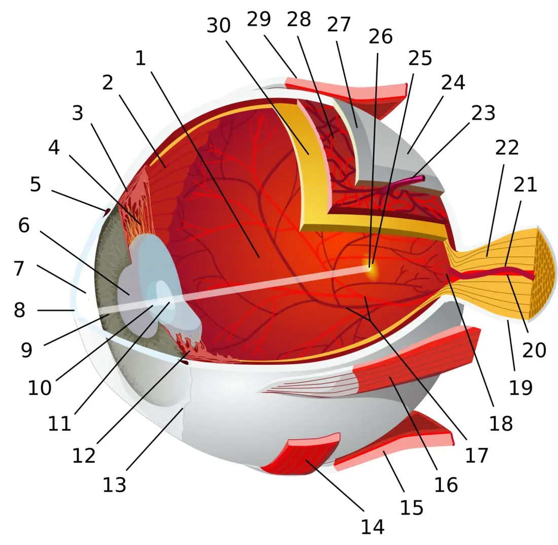
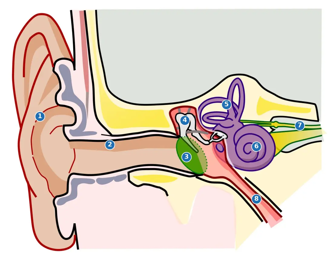
Model answers — short (1–5)
1. (i) The figure shows the two lobes of the pituitary: A = anterior lobe (adenohypophysis) and B = posterior lobe (neurohypophysis). (ii) The posterior lobe releases oxytocin and antidiuretic hormone (ADH, vasopressin); both are made in the hypothalamus and stored and released here. (iii) Oxytocin is the birth hormone: at the end of pregnancy it makes the smooth muscle of the uterus contract strongly, pushing the baby out; contractions stimulate more oxytocin (positive feedback) until delivery; it also causes milk ejection during suckling.
2. The printed diagram marks four parts of the eye; the four that are asked most often are below (match the letters to the arrows in your paper). (a)/(b) - Cornea — the transparent front of the sclera; refracts most of the light and protects the eye. - Lens — transparent, elastic, biconvex; focuses the image on the retina (accommodation by the ciliary muscles). - Retina — inner layer with rods (dim light) and cones (colour); the image forms here and is converted to nerve impulses. - Optic nerve — carries impulses from the retina to the visual cortex. (Other labelled parts may be the iris, which controls the size of the pupil, or the sclera, the tough white outer coat that protects and maintains the shape.)
3. On the usual reading of the printed figure: A = semicircular canals, B = vestibule (utricle and saccule), C = Eustachian tube (the tube running towards the throat, at the bottom of the figure) and D = cochlea (the coiled tube). Function of A: the three canals, in three planes, sense rotational movement of the head; fluid (endolymph) moves and bends hair cells, giving dynamic balance. Function of D: the cochlea contains the organ of Corti; its hair cells convert the sound vibrations that arrive through the oval window into nerve impulses — the organ of hearing. (If your paper labels the Eustachian tube D, its function is to equalise the air pressure on the two sides of the eardrum.)
4. (i) Kidney stones (renal calculi). Crystals of calcium oxalate or phosphate form in the pelvis when urine is concentrated; causes severe pain, blood in the urine; treated by drinking water, lithotripsy or surgery. (ii) Glomerulonephritis. Inflammation of the glomeruli, often after a streptococcal infection; proteins and blood leak into urine, face and legs swell, blood pressure rises. (iii) Renal failure (uraemia). The kidneys stop filtering, so urea builds up in the blood; treated by dialysis or kidney transplant. (Any two, each with cause and effect.)
5. Risk factors [2]. (i) Chronic high blood pressure damages the blood vessels of the nephron; (ii) uncontrolled diabetes mellitus damages the glomeruli over the years; also frequent urinary infections, kidney stones, pain-killers and smoking. Healthy habits [2]. (i) Drink plenty of water (about 2–3 litres a day) so that the urine is dilute and does not crystallise; (ii) eat a balanced, low-salt diet, do not overuse painkillers, keep blood pressure and sugar under control, exercise, avoid smoking.
Long questions (8 marks)
- Draw a labelled diagram of the alimentary canal of a human being. How would you explain the digestion of proteins in the gut? Justify the relationship between amylase and lipase enzymes in the digestion of food. [3+4+1] (NEB 2079)
- A human takes complex foods in the diet but the body needs their simpler forms. Describe how complex foods are converted into simple forms within the alimentary canal, with a well-labelled diagram of the alimentary canal. What would happen in digestion if the gall bladder were removed surgically? [4+2+2] (NEB model 2079)
- On oxidation of glucose in the cells CO₂ is released. Explain how CO₂ is transported in the human body. What is the role of the enzyme carbonic anhydrase? Mention the significance of the diaphragm in breathing. [5+1+2] (NEB model 2079, as the alternative)
- Justify the statement "the sino-auricular node is a natural pacemaker". What would you suggest if the natural pacemaker does not function normally? How do you test the blood group in human beings? [3+2+3] (NEB 2079, as the alternative)
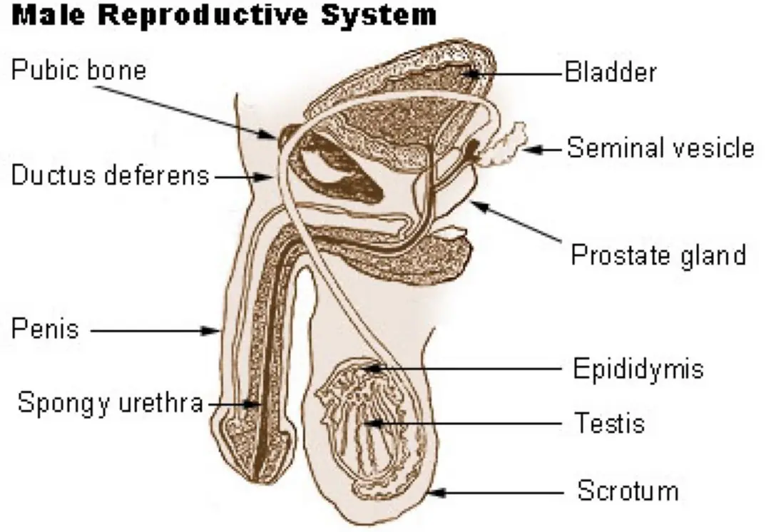
- Elaborate the organs of the male reproductive system with the help of a labelled diagram. Why are the testes located outside the abdominal cavity? [5+2+1] (NEB 2081)
- Eyes facilitate binocular vision. Draw a V.S. of the human eye and explain its structure. "The blind spot of the eye is devoid of the ability of vision." Justify. [2+5+1] (NEB 2081, as the alternative)
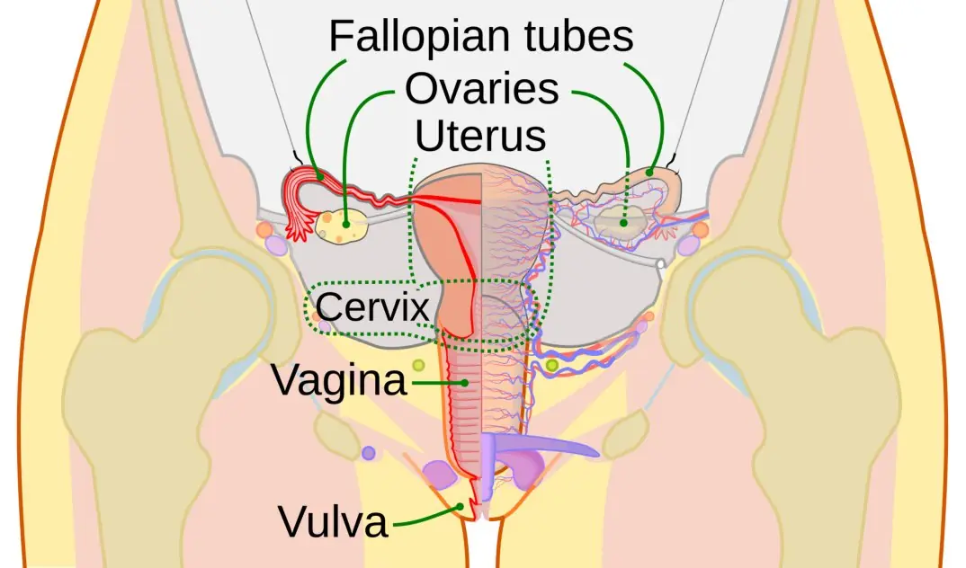
- Reproduction is a process of continuation of the human race. Describe the female reproductive system with a well-labelled diagram. How would you explain ovulation in the human female? [4+3+1] (NEB 2082)
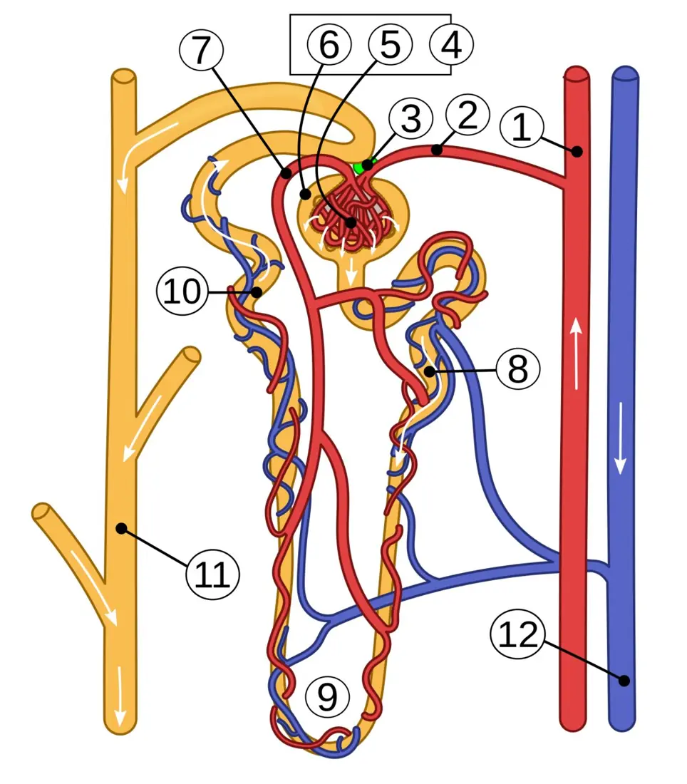
- Nephrons are the structural and functional units of the kidney. Illustrate the structure of a nephron with a labelled diagram. What would happen if ADH is secreted insufficiently? [4+3+1] (NEB 2082, as the alternative)
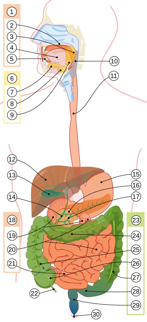
Model answers — long (6–13)
6. Diagram [3]. Mouth (teeth, tongue, salivary glands) → pharynx → oesophagus → stomach → small intestine (duodenum, jejunum, ileum) → large intestine (caecum with appendix, colon, rectum) → anus; with liver, gall bladder and pancreas (figure above). Digestion of protein [4]. Mouth and oesophagus: no protein digestion. Stomach: the oxyntic cells release HCl (pH 1.5–2), which kills microbes and activates pepsinogen from the chief cells into pepsin; pepsin splits proteins into proteoses and peptones; in infants rennin curdles milk casein. Duodenum: pancreatic juice brings trypsinogen, activated by enterokinase into trypsin, plus chymotrypsin and carboxypeptidase; they break peptones into peptides and dipeptides in the alkaline medium made by bile and bicarbonate. Ileum: the intestinal juice contains aminopeptidase and dipeptidase, which finish the job: amino acids, absorbed into the villus capillaries. Amylase and lipase [1]. Both are hydrolases of pancreatic juice working in the alkaline duodenum, but on different substrates: amylase on starch (→ maltose), lipase on emulsified fat (→ fatty acids and glycerol). They are complementary, not dependent: each digests a separate food class, so absence of one leaves the other intact, and together with proteases they complete the digestion of a mixed meal.
7. Digestion [4].
| Region | Juice / enzyme | Action |
|---|---|---|
| Mouth | saliva: salivary amylase (ptyalin) | starch → maltose |
| Stomach | gastric juice: HCl, pepsin, rennin | protein → proteoses and peptones |
| Duodenum | bile (no enzyme), pancreatic amylase, trypsin, lipase | bile emulsifies fat; starch → maltose; peptones → peptides; fat → fatty acids + glycerol |
| Ileum | intestinal juice: maltase, sucrase, lactase, peptidases, lipase | disaccharides → monosaccharides; peptides → amino acids; fats finished |
The end products are glucose, amino acids, fatty acids and glycerol, which the villi absorb. Diagram [2]. As in question 6. Gall bladder removal [2]. The gall bladder stores and concentrates bile. After removal, the liver still makes bile and it drips into the duodenum, but it cannot be released in a surge after a fatty meal. Emulsification of fat becomes incomplete, so lipase digests less fat, the stools become fatty, and the absorption of the fat-soluble vitamins (A, D, E, K) falls. The person must take small, low-fat meals.
8. Transport of CO₂ [5]. Tissue cells release CO₂, which diffuses into the blood and is carried in three ways: (1) As bicarbonate (about 70 %). CO₂ enters the red cell, where carbonic anhydrase combines it with water: CO₂ + H₂O ⇌ H₂CO₃ ⇌ H⁺ + HCO₃⁻. The HCO₃⁻ leaves the cell into the plasma, and Cl⁻ enters to keep the charge balanced (chloride shift); the H⁺ is taken up by haemoglobin, which acts as a buffer. (2) As carbaminohaemoglobin (20–25 %). CO₂ binds to the amino groups of the globin part of haemoglobin. (3) Dissolved in plasma (about 7 %). In the lungs the reactions reverse: CO₂ is released from these forms, diffuses into the alveolar air and is exhaled. Carbonic anhydrase [1]. This zinc-containing enzyme of the red cell speeds up the hydration and dehydration of CO₂ thousands of times, so the CO₂ is moved quickly in both tissue and lung. Diaphragm [2]. It is the dome-shaped muscle between thorax and abdomen. Contraction flattens it, increases the volume of the thoracic cavity, and lowers the lung pressure, so air enters (inspiration); relaxation returns it to the dome, and air leaves (expiration). Together with the intercostal muscles it does most of quiet breathing.
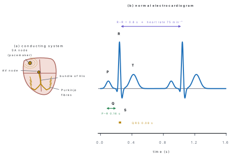
9. SA node [3]. The sino-atrial node is a small mass of modified cardiac muscle in the wall of the right atrium near the opening of the superior vena cava. It is myogenic: it fires on its own about 70–75 times a minute, faster than any other part, so it sets the rate of the whole heart. Each impulse spreads over the atria (atrial systole), reaches the AV node (delayed 0.1 s), and passes by the bundle of His, its branches and the Purkinje fibres to the ventricles (ventricular systole). Hence the SA node is the natural pacemaker. If it fails [2]. The AV node takes over at a slower 40–60 beats a minute, but if the rate is too low or irregular, an artificial pacemaker, a battery-driven device implanted under the skin, sends regular impulses through wires to the heart; drugs and ECG monitoring follow. Blood grouping test [3]. Take three drops of blood on a clean glass slide. Add one drop each of anti-A, anti-B and anti-D (Rh) antiserum, mix with separate sticks, and watch for agglutination (clumping) after one to two minutes.
| Clumping with | Blood group |
|---|---|
| anti-A only | A |
| anti-B only | B |
| anti-A and anti-B | AB |
| neither | O |
| anti-D | Rh-positive; none = Rh-negative |
10. Organs [5]. Testes — a pair of oval glands in the scrotum; each has coiled seminiferous tubules where sperm are made (spermatogenesis, with Sertoli cells) and interstitial (Leydig) cells that make testosterone. Epididymis — coiled tube on each testis that stores and matures sperm. Vas deferens — carries sperm from the epididymis up into the abdomen to the ejaculatory duct. Seminal vesicles — secrete fructose-rich fluid for sperm energy. Prostate gland — secretes alkaline fluid that neutralises vaginal acidity. Bulbourethral (Cowper's) glands — mucus lubricant. Urethra and penis — common passage for urine and semen; the penis is the organ of copulation. Diagram: as in the figure (a). Why outside [2+1]. Spermatogenesis needs a temperature about 2–3 °C lower than the body's core temperature (37 °C). The scrotum hangs outside the abdomen, and the cremaster and dartos muscles raise or lower the testes to regulate it. If a testis stays inside the abdomen (cryptorchidism) the sperm count falls and the man can be infertile.
11. Diagram [2]. As in the figure. Structure [5]. The wall has three layers. (1) Sclera — tough white outer coat that protects and keeps the shape; in front it is the transparent cornea, which refracts light. (2) Choroid — middle, dark and vascular layer that nourishes the eye and absorbs stray light; in front it forms the ciliary body (with the ciliary muscles that change the lens shape) and the iris, the coloured diaphragm with a central opening, the pupil, which controls the amount of light. (3) Retina — inner layer, containing the photoreceptors: rods (dim light, rhodopsin) and cones (bright light, colour); the fovea centralis has only cones and gives the sharpest vision. The lens is a transparent biconvex structure held by the suspensory ligaments; aqueous humour fills the space in front of it and vitreous humour the large chamber behind it. The optic nerve leaves at the back. Blind spot [1]. At the optic disc, where the optic nerve and the retinal vessels leave the eye, there are no rods or cones. An image falling there produces no impulse and is not seen; hence it is the blind spot. Binocular vision hides it, since each eye sees the part the other misses.
12. Description [4]. The female reproductive system consists of a pair of ovaries in the lower abdomen that produce ova (oogenesis) and the hormones oestrogen and progesterone; a pair of fallopian tubes (oviducts) — funnel-shaped infundibulum with finger-like fimbriae that catch the ovum, a wide ampulla where fertilisation occurs, and the narrow isthmus; the pear-shaped uterus, with an outer perimetrium, a muscular myometrium and an inner endometrium where the embryo implants; the cervix and the vagina, the birth canal and the receptacle of semen; externally the vulva. Diagram [3]. As in figure (b), labelled: ovary, fallopian tube with fimbriae, uterus, endometrium, cervix, vagina. Ovulation [1]. About day 14 of the 28-day cycle a surge of LH (with FSH) makes the mature Graafian follicle rupture and release the secondary oocyte, which is swept into the fallopian tube by the fimbriae; the empty follicle becomes the corpus luteum, which secretes progesterone.
13. Structure [4]. A nephron is a long tube about 3 cm long, with about a million in each kidney. Its parts in order: (1) Malpighian body — the cup-shaped Bowman's capsule holding a tuft of capillaries, the glomerulus, fed by a wide afferent and drained by a narrow efferent arteriole, which gives high pressure for ultrafiltration; (2) proximal convoluted tubule (PCT) with a brush border, which reabsorbs most of the glucose, amino acids, salts and two-thirds of the water; (3) loop of Henle — a U-shaped tube with a descending limb permeable to water and an ascending limb permeable to salt, which creates the concentration gradient of the medulla; (4) distal convoluted tubule (DCT) for secretion and ADH-controlled reabsorption; (5) collecting duct, which carries urine to the renal pelvis. Diagram [3]. As in the figure. ADH [1]. ADH (vasopressin) makes the DCT and the collecting duct permeable to water. If too little is secreted, water is not reabsorbed, and the person passes a large volume (up to 10–20 litres a day) of dilute urine: diabetes insipidus; thirst and dehydration follow.
9.9 Human Population and Health Disorders
Most-repeated in this unit
| Topic | Seen in the papers | Shape of question |
|---|---|---|
| Smoking, drug abuse, peer pressure: causes, prevention, awareness plan | model 2079 (peer pressure and smoking), 2081 (drugs) | short, 4 (Q21) |
| Renal disorders and healthy habits | 2079, 2082 | short, 4 (Q21) |
| Immunity and its types; transplants | model 2079 (OR), MCQ 2079, 2082 | short / MCQ |
| Population: growth, family planning, immigration | MCQ 2079, 2082 | MCQ |
Zoology Q21 is always a health-and-society question: peer pressure and smoking (model 2079), kidney disorders (2079), drug addiction (2081), kidney risks (2082). It asks for your view and a plan, not a definition. Answers with two concrete, realistic actions score full marks.
Short questions (4 marks)
- Peer pressure plays a negative role in provoking smoking habits among adolescents. As the school captain, plan any two activities you would organise with the help of senior students and any two you would like the school authorities to organise to tackle the problem. [2+2] (NEB model 2079)
- Taking drugs for a non-medical purpose causes addiction. In your view, what motivates youngsters to take drugs and how can this be prevented? [4] (NEB 2081)
- What is immunity? Explain its types. [1+3] (NEB model 2079, as the alternative)
- Define population growth. Explain the factors that influence the size of a human population. [1+3] (original)
- What is AIDS? State how it is transmitted and how it can be prevented. [1+1.5+1.5] (original)
Model answers — short (1–5)
1. Activities with senior students [2]. (i) A peer-education circle: trained senior students talk to Grade 8–10 students in small groups about what tobacco does to the lungs and heart, and teach refusal skills ("How to say no"). (ii) An awareness campaign: posters, street drama and a rally to the neighbouring shops that sell cigarettes to children. Activities by the school [2]. (iii) Declare the campus and its surroundings tobacco-free, display warnings and put a rule in the code of conduct. (iv) Set up a counselling desk with a teacher or a health worker, organise talks with doctors and former smokers, and involve parents and the local police. (Any two and two, each with the means.)
2. Motives [2]. Curiosity and thrill-seeking; peer pressure; stress, exam pressure or family problems; loneliness or broken homes; the false belief that drugs give energy or confidence; easy availability and lack of awareness. Prevention [2]. Parental guidance and open talk at home; drug-awareness programmes and life-skills education in schools; healthy activities (sports, music, clubs); strict control of sale; early counselling; treatment and rehabilitation for those addicted instead of punishment.
3. Immunity [1]. The power of the body to resist and fight pathogens and their toxins. Types [3]. - Innate (natural) immunity — present from birth, non-specific: skin and mucus, stomach acid, phagocytes, inflammation. - Acquired immunity — developed in life, specific, with memory: - Active: the body makes its own antibodies — natural after an infection, artificial after a vaccine. - Passive: ready-made antibodies received — natural from the mother through the placenta and breast milk, artificial by antiserum injection (anti-snake-venom, tetanus antitoxin).
4. Definition [1]. Population growth is the change in the number of individuals in a population over a period. Factors [3]. growth = (natality − mortality) + (immigration − emigration). Natality (birth rate) rises with early marriage, desire for sons, poor use of contraception; mortality (death rate) falls with better health care, vaccination and nutrition; immigration and emigration (Nepal has large labour migration abroad). Education of girls, age at marriage, economic conditions and government policy (family planning) set these rates; the sum is shown in the demographic transition model.
5. AIDS [1]. Acquired Immuno-Deficiency Syndrome, caused by HIV, an RNA retrovirus that destroys CD4 helper T cells, so the body cannot fight infections. Transmission [1.5]. Unprotected sexual contact; contaminated blood or blood products; shared needles and syringes; from an infected mother to her child (pregnancy, birth, breast-feeding). It is not spread by handshake, mosquito bite or sharing food. Prevention [1.5]. Safe sex and condom use; screening of donated blood; use of a new syringe for each injection; antiretroviral drugs for pregnant women to protect the baby; AIDS education and counselling.
Long question (8 marks)
- Explain malaria: the causative agent and its life cycle in man and mosquito, symptoms, and control measures. [1+3+2+2] (predicted: a communicable-disease long question appears in every paper — the three seen so far covered typhoid, cholera and TB; malaria, hepatitis and AIDS are the others on the syllabus)
Model answer — long (6)
Causative agent [1]. Malaria is a protozoan disease caused by Plasmodium — P. vivax (the commonest in Nepal's Terai), P. falciparum (the most dangerous), P. malariae and P. ovale — carried by the female Anopheles mosquito. Life cycle [3]. In man (asexual). When an infected mosquito bites, sporozoites enter the blood and go to the liver, where they multiply (schizogony) into merozoites. These enter the red blood cells and multiply again; the cells burst every 48 hours (vivax) and release merozoites and toxin. Some merozoites become gametocytes. In the mosquito (sexual). The female Anopheles sucks the blood; the gametocytes form male and female gametes in its gut and fuse, and the zygote becomes an ookinete, then an oocyst on the gut wall, which produces thousands of sporozoites that move to the salivary glands. Symptoms [2]. Chills, high fever and sweating in cycles (every 48 hours for vivax), headache, vomiting, anaemia and enlarged spleen; cerebral malaria in falciparum. Control [2]. Kill the vector: drain stagnant water, spray indoor residual insecticide, use larvicides and larvivorous fish (Gambusia); avoid bites: insecticide-treated bed nets, repellents, screens; early diagnosis (rapid test, blood smear) and treatment with chloroquine/artemisinin-based drugs; community awareness.
9.10 Applied Biology
Most-repeated in this unit
| Topic | Seen in the papers | Shape of question |
|---|---|---|
| Typhoid | model 2079, 2081 | long, 8 marks |
| Cholera | 2079 | long, 8 |
| Tuberculosis and "disease of poverty" | 2082 | long, 8 |
| Poultry (pullets, scope), fish culture (indigenous fish, integrated fish farming) | 2081 OR, 2082 OR | short, 1 + 3 |
| MCQs: bacterial disease sets, waterborne diseases | model 2079, 2082 | MCQ |
Zoology Q22 is always a communicable bacterial disease — cholera (2079), typhoid (model 2079 and 2081), tuberculosis (2082) — and always asks for the same five boxes: causative agent, mode of transmission, symptoms, prevention/control, and one "why" or "which organ" line. Typhoid is the only disease that has been asked twice; the three not yet asked as a long question are malaria, hepatitis and AIDS (see 9.9), which are the likely next ones.
Short questions (4 marks)
- What are pullets? How would you express the scope of poultry farming in Nepal? [1+3] (NEB 2081, as the alternative)
- Name any two indigenous fish species of Nepal. Discuss how integrated fish farming is advantageous over traditional fish farming. [1+3] (NEB 2082, as the alternative)
- Describe the three castes of the honeybee and their functions. [4] (original)
- Describe the life cycle of the silkworm Bombyx mori and state the stage at which silk is obtained. [4] (original)
- What is vermicomposting? Explain its method and advantages. [1+2+1] (original)
Model answers — short (1–5)
1. Pullets [1]. Young female chickens (fowls) that have not yet started laying eggs, usually from about 8 weeks to the start of laying. Scope in Nepal [3]. (i) Eggs and meat are an affordable source of protein, and demand is rising with urban growth and tourism; (ii) poultry needs a small land area and capital and can be kept in backyards, so it gives self-employment, especially for women and in the hills; (iii) poultry waste is a good manure and a feed for fish; (iv) farms, hatcheries, feed mills and marketing provide jobs and reduce the import of eggs and chicken; (v) improved breeds (White Leghorn for eggs; Cobb and Ross for broiler meat) and vaccination against Ranikhet disease make it profitable.
2. Indigenous fish [1]. Asala (snow trout, Schizothorax) and Sahar (golden mahseer, Tor putitora); also Katle. Advantages of integrated fish farming [3]. It combines fish culture with another farm enterprise, such as poultry, duck, pig or paddy and vegetables. (i) The waste of the animals falls in the pond as manure and feed, cutting the cost of fish feed and fertiliser; (ii) the nutrient-rich pond water and mud can irrigate and fertilise the crop; (iii) the same land gives more than one product and more income; (iv) it makes use of waste and saves money, which a traditional single-crop pond cannot do.
3. Queen — one per colony, large fertile female with long abdomen; she lays up to 1500–2000 eggs a day and produces the pheromone that holds the colony together; she eats royal jelly throughout life. Workers — sterile females (2n), the smallest and most numerous; they collect nectar and pollen, make honey and wax, build comb, feed the brood, guard and clean the hive, and ventilate it. Drones — males, haploid, hatched from unfertilised eggs, larger eyes; their only function is to mate with the queen (during the nuptial flight), and they are driven out or killed after the season.
4. The silkworm shows complete metamorphosis: egg (laid by the moth, about 400–500 eggs) → larva (caterpillar) that feeds greedily on mulberry leaves and moults four times → pupa inside a cocoon spun from a single continuous thread of fibroin and sericin from its salivary glands → adult moth. Silk is obtained from the cocoon: the pupa is killed by hot air or steam before the moth emerges (otherwise it cuts the thread), and the cocoons are boiled to soften the sericin and the thread is reeled off.
5. Vermicomposting [1]. Preparation of compost from organic waste using earthworms. Method [2]. A shaded pit, box or bed is filled with a bedding of partly decomposed dry leaves and cow dung; kitchen and farm waste is added in layers; about 1000 worms (Eisenia fetida) are released per square metre; the bed is kept moist (60–70 %) and covered with jute; after 45–60 days the dark, crumbly vermicast is sieved out. Advantages [1]. Cheap, rich in NPK and humus, improves soil structure, recycles waste, and gives extra income from worms.
Long questions (8 marks)
- Why is typhoid called enteric fever? Illustrate its causative agent, mode of transmission, symptoms and preventive measures. [1+1+2+2+2] (NEB model 2079; asked again in NEB 2081 as [1+2+2+2+1]: name the causative agent, transmission, symptoms, prevention, and the organ affected in severe cases)
- Explain cholera as an acute diarrhoeal disease. Discuss its causative agent, mode of transmission and symptoms. Mention its control measures. [1+1+2+2+2] (NEB 2079)
- Mention the causative agent, mode of transmission and symptoms of pulmonary tuberculosis. Highlight the strategies for its prevention. Why is it called a "disease of poverty"? [1+2+2+2+1] (NEB 2082)
Model answers — long (7–9)
7. Why enteric [1]. The disease is caused by an organism that lives in the intestine (enteron), and the fever starts when bacteria invade the intestinal wall and the blood; hence enteric fever. Causative agent [1]. Salmonella Typhi (written Salmonella typhi in older books), a motile, rod-shaped Gram-negative bacterium. Transmission [2]. The faeco-oral route: food and water contaminated with the faeces or urine of patients or carriers (Typhoid Mary was a famous carrier); flies carry bacteria to food; poor sanitation and unwashed hands; street food and untreated water. Symptoms [2]. Stepwise rise of fever for a week to 40 °C, headache, weakness and loss of appetite, abdominal pain with constipation or diarrhoea, pink rose spots on the trunk, a coated tongue and enlarged spleen. The incubation period is 10–14 days. Organ affected in severe cases [1 in the 2081 version]. The small intestine: ulcers in the Peyer's patches may bleed or perforate. Prevention [2]. Drink boiled or chlorinated water; wash hands with soap after the toilet and before food; eat hot, freshly cooked food and avoid open street food; control flies; proper sewage disposal; typhoid vaccine (Vi polysaccharide or conjugate); identify and treat carriers; diagnosis by Widal test or blood culture and treatment with antibiotics (azithromycin, ceftriaxone).
8. Cholera [1]. An acute diarrhoeal disease with sudden, profuse, watery diarrhoea that can kill through dehydration within hours. Causative agent [1]. Vibrio cholerae, a comma-shaped Gram-negative bacterium with a single polar flagellum; its enterotoxin makes the intestinal cells pump out water and salts. Transmission [2]. Faeco-oral: water contaminated with faeces (wells, rivers after floods, the monsoon), uncooked food and shellfish, flies and unwashed hands; epidemics in crowded places. Symptoms [2]. Sudden rice-water stools, vomiting, severe dehydration (sunken eyes, dry mouth, loss of skin turgor), muscle cramps, low blood pressure, rapid pulse and collapse; incubation a few hours to 5 days. Control [2]. Clean, chlorinated drinking water and safe sewage disposal; hand-washing and food hygiene; oral cholera vaccine in outbreaks; immediate oral rehydration therapy (ORS, and intravenous fluid in severe cases), zinc and antibiotics (doxycycline); notify and isolate cases.
9. Causative agent [1]. Mycobacterium tuberculosis, a rod-shaped acid-fast bacillus. Transmission [2]. Airborne droplets when a person with active lung TB coughs, sneezes or talks; sometimes infected milk (M. bovis); the risk rises in crowded, badly ventilated rooms. Symptoms [2]. Cough for more than two weeks, sometimes with blood in the sputum, evening fever, night sweats, weight loss, loss of appetite, chest pain and fatigue. Prevention [2]. BCG vaccination at birth; finding and treating cases early with DOTS (directly observed treatment, short course: rifampicin, isoniazid, pyrazinamide and ethambutol for two months, then rifampicin and isoniazid, six months in all) to cut the source; cover the mouth when coughing; good ventilation and sunlight; nutrition; screening of contacts; avoid spitting. Disease of poverty [1]. It spreads where people live in overcrowded, poorly ventilated houses, are malnourished and have little access to health care, and poverty keeps them from completing treatment; the disease in turn makes the earner ill and deepens poverty.
Chapter summary
- Write to the marks. A 4-mark answer is one built idea (definition + 3 points or a table + a small diagram); an 8-mark answer is definition, steps, labelled diagram and significance. The bracket splits [1+3], [2+2+4] are how the marker awards marks.
- Botany: tissues are asked every year (Q6); one 8-mark photosynthesis or respiration question appears every year; one genetics question (replication, mutation, sex-linkage, crossing over) appears every year and always wants a diagram.
- Zoology: Q18 is a tissue or frog-stage short; Q21 is a health-and-society short; Q22 is a bacterial disease (typhoid, cholera, TB) with the same five boxes; Q23 is a system with a labelled diagram plus a "what if".
- Diagrams: epithelia, dicot embryo, alimentary canal, eye, ear, nephron, reproductive systems, heart conduction, replication fork and crossing over have all been demanded; practise drawing them with straight leader lines.
- Honesty about the data: only four full papers (2079, model 2079, 2081, 2082) could be read; the 2080 paper could not be obtained, so the repeat counts are a guide to the board's habits, not a prediction.