Chapter 3 · Evolution and Diversity · 4 questions in the paper · 31 min read
Animal Tissues and Histology
What the syllabus asks for 1
- Epithelial, connective, muscular and nervous tissues: structure, location and function
Animal Tissues and Histology carries 4 of the 40 Zoology questions. The syllabus asks for the four primary tissues — epithelial, connective, muscular and nervous — with their structure, location and function, so almost every question is "which tissue lines/supports/forms this organ" or "which cell or fibre does this job". The marks sit in the epithelium table, the three cartilages and bone, the blood cells with their numbers, the three kinds of muscle with the sarcomere, and the parts of a neuron.
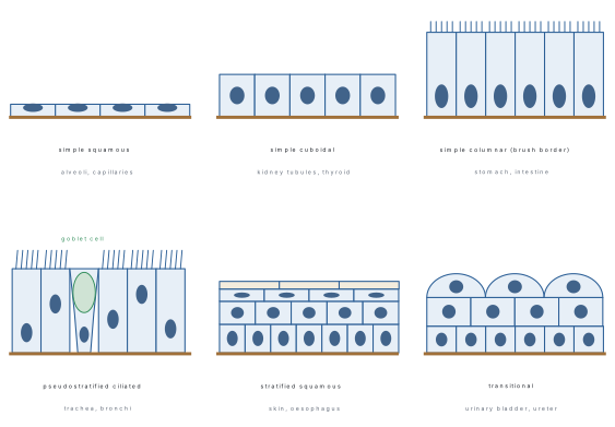
- Epithelia: name and location of each type — squamous (alveoli, blood vessels), cuboidal (kidney tubules, thyroid), columnar (stomach, intestine), ciliated (oviduct, bronchioles), pseudostratified (trachea), stratified squamous (skin, oesophagus), transitional (urinary bladder).
- Glands: exocrine, endocrine and heterocrine; merocrine, apocrine and holocrine secretion.
- Connective tissue cells (fibroblast, macrophage, mast cell, plasma cell, adipocyte) and fibres (collagen, elastic, reticular); tendon versus ligament.
- Cartilage (hyaline, elastic, fibrocartilage) versus bone (ossein, hydroxyapatite, Haversian system, osteoblast/osteoclast).
- Blood as fluid connective tissue: plasma, RBC, the five WBCs in their percentages, platelets.
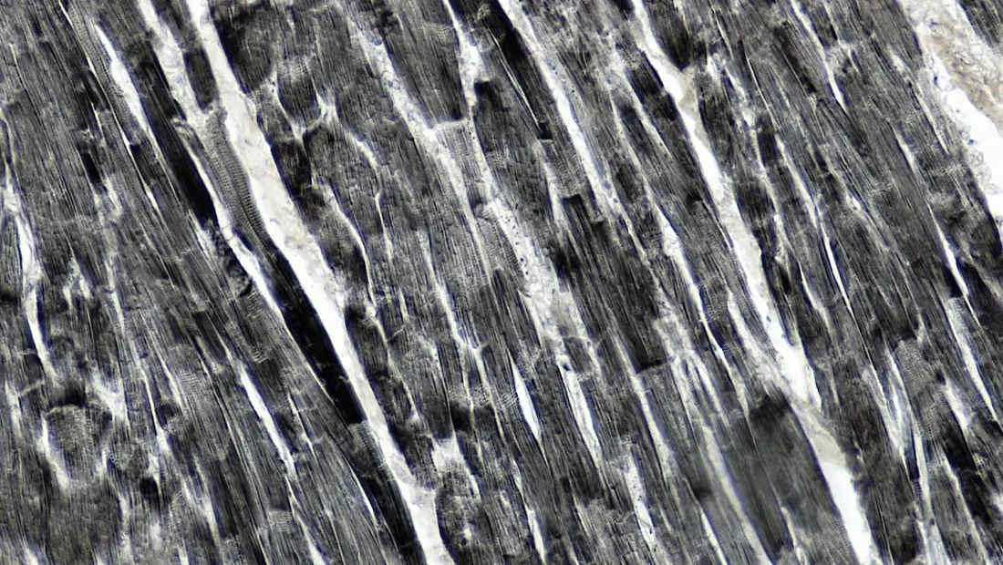
- Skeletal, smooth and cardiac muscle: shape, nuclei, striations, intercalated discs, control; the sarcomere bands in contraction.
- Neuron structure (Nissl granules, axon, myelin, nodes of Ranvier), neuron types and neuroglia.
3.1 Epithelial, connective, muscular and nervous tissues: structure, location and function
A tissue is a group of similar cells, with their intercellular substance, that has a common origin and performs a common function. Histology is the study of tissues. Animals have four primary tissues.
| Tissue | Germ layer | Cells and matrix | Main job | Examples |
|---|---|---|---|---|
| Epithelial | all three | cells packed close, almost no matrix, on a basement membrane | covering, lining, protection, secretion, absorption | epidermis, lining of gut, glands |
| Connective | mesoderm | few cells scattered in abundant matrix with fibres | binding, support, transport, storage, defence | areolar tissue, tendon, cartilage, bone, blood |
| Muscular | mesoderm (mostly) | elongated contractile muscle fibres | movement, contraction | skeletal, smooth, cardiac muscle |
| Nervous | ectoderm | neurons and neuroglia | receive, conduct and transmit impulses; coordination | brain, spinal cord, nerves |
Epithelial tissue
Epithelium is a sheet of closely packed cells with negligible intercellular matrix, resting on a non-cellular basement membrane, that covers the body surface and lines its cavities, ducts and tubes.
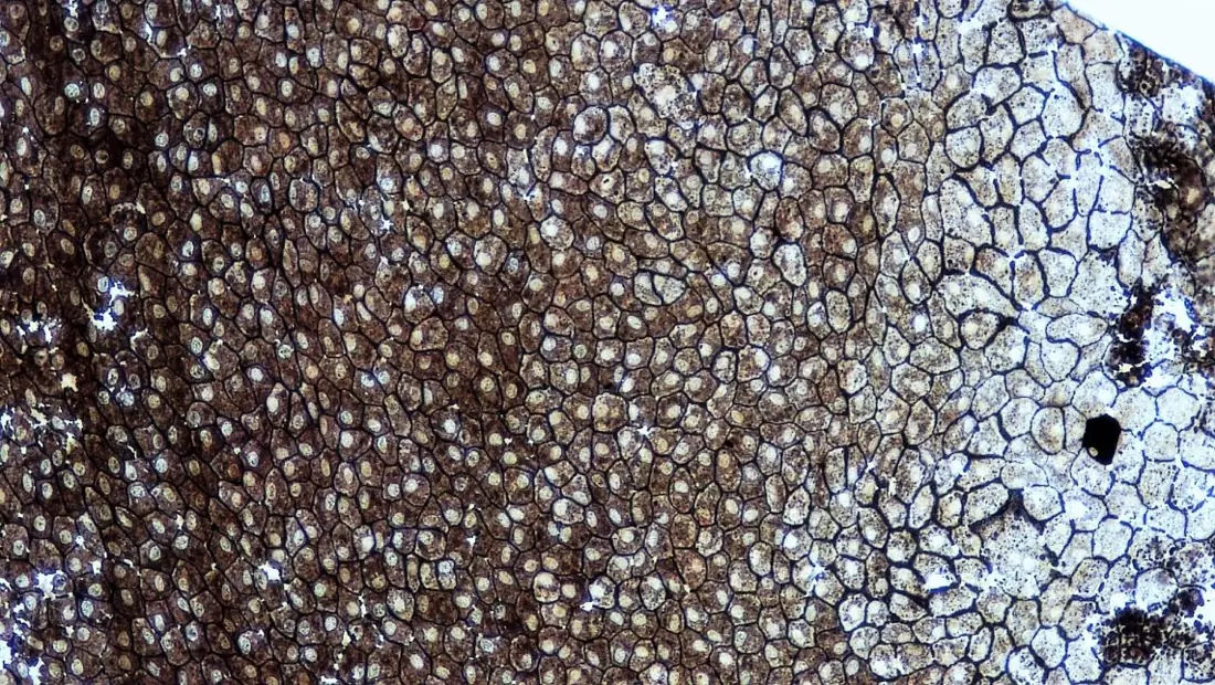
- It arises from all three germ layers — epidermis (ectoderm), gut lining (endoderm), lining of blood vessels and kidney tubules (mesoderm).
- Avascular: no blood vessels enter it; nutrients diffuse from capillaries of the connective tissue below.
- Cells are polar (free apical and basal surfaces) and joined by cell junctions: tight junctions (stop leakage between cells), adhering junctions / desmosomes (cement neighbouring cells), gap junctions (cytoplasmic channels for ions and small molecules).
- High power of regeneration; free surfaces may bear microvilli (absorption), cilia (movement) or stereocilia (long immotile microvilli, as in the epididymis).
- The lining of blood vessels and the heart is called endothelium; of the body cavities (pleura, peritoneum, pericardium) mesothelium.
| Epithelium | Structure | Location | Function |
|---|---|---|---|
| Simple squamous (pavement) | one layer of thin, flat, polygonal cells with a central flattened nucleus | alveoli of lungs, walls of blood capillaries and vessels (endothelium), Bowman's capsule, thin loop of Henle, serous membranes | diffusion, filtration |
| Simple cuboidal | one layer of cube-shaped cells, round central nucleus | kidney tubules (PCT with a brush border of microvilli), thyroid follicles, ducts of glands, surface of the ovary (germinal epithelium) | secretion, absorption |
| Simple columnar | tall cells, oval nucleus near the base; often with microvilli and goblet cells | lining of stomach, small intestine (brush border), gall bladder | secretion, absorption |
| Ciliated columnar | columnar cells with cilia | fallopian tubes (oviducts), small bronchioles, uterus, central canal of spinal cord | moves ova, mucus or fluid in one direction |
| Pseudostratified ciliated columnar | one layer, but cells of different heights with nuclei at different levels, so it looks layered; all cells touch the basement membrane; goblet cells | trachea, bronchi, nasal passages | traps dust in mucus, cilia sweep it up |
| Stratified squamous keratinised | many layers; only the basal (germinative) layer divides; surface cells dead, filled with keratin | epidermis of skin | waterproof, resists wear |
| Stratified squamous non-keratinised | many layers, surface cells living and moist | buccal cavity, pharynx, oesophagus, vagina, cornea | resists abrasion |
| Stratified cuboidal | two or more layers of cuboidal cells | ducts of sweat glands, salivary and mammary ducts | protection, secretion |
| Stratified columnar | several layers, columnar at the surface | part of the male urethra, conjunctiva, large ducts | protection |
| Transitional (urothelium) | 4–6 layers; large dome-shaped (umbrella) surface cells that flatten when the organ is stretched | urinary bladder, ureters, renal pelvis | stretches without tearing |
Glandular epithelium. Epithelial cells specialised for secretion form glands. A unicellular gland is the mucus-secreting goblet cell of the intestine and respiratory tract. Multicellular glands are classified three ways.
| Basis | Type | Example |
|---|---|---|
| Where the secretion goes | exocrine — through a duct | salivary, sweat, sebaceous, gastric, mammary glands |
| endocrine — ductless, into the blood (hormones) | thyroid, pituitary, adrenal | |
| heterocrine (mixed) — both | pancreas (enzymes + insulin), gonads | |
| Mode of secretion | merocrine (eccrine) — secretion leaves by exocytosis, cell intact | salivary glands, pancreas, most sweat glands |
| apocrine — the apical part of the cell pinches off with the secretion | mammary glands, sweat glands of the armpit | |
| holocrine — the whole cell breaks down to form the secretion | sebaceous glands | |
| Shape | simple tubular / coiled tubular / branched tubular | intestinal crypts / sweat glands / gastric glands |
| simple branched alveolar | sebaceous glands | |
| compound tubulo-alveolar | salivary glands, pancreas, mammary glands |
Connective tissue
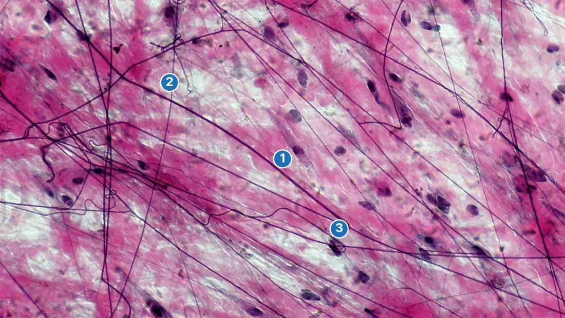
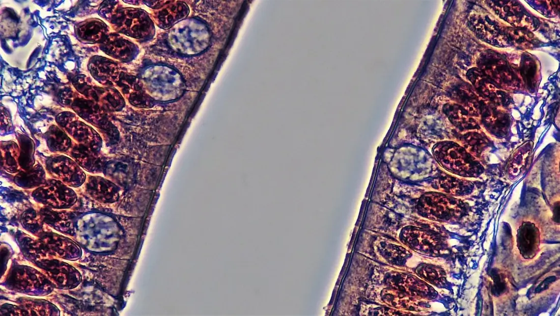
Connective tissues are the most abundant and widespread tissues. They arise from the mesoderm, have few cells in a large amount of matrix, are usually vascular (cartilage is the exception) and bind, support, protect, store and transport.
The fibres
| Fibre | Protein | Appearance | Property | Where |
|---|---|---|---|---|
| White (collagen) | collagen — the commonest protein of the body | wavy unbranched bundles | great tensile strength, little stretch; boiled → gelatin | tendons, dermis, bone, cartilage |
| Yellow (elastic) | elastin | single, branched fibres | stretch and recoil | ligaments, walls of arteries, lungs, pinna |
| Reticular | reticulin (a fine collagen) | delicate branched network | supporting framework (stroma) | lymph nodes, spleen, liver, bone marrow |
The cells of loose connective tissue
| Cell | Job |
|---|---|
| Fibroblast | the commonest cell; secretes fibres and matrix |
| Macrophage (histiocyte) | phagocytosis — engulfs microbes and debris |
| Mast cell | secretes heparin (anticoagulant), histamine (dilates vessels; allergy) and serotonin |
| Plasma cell | derived from B lymphocytes; secretes antibodies |
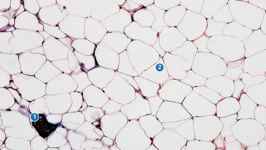
| Adipocyte (fat cell) | stores fat in one large droplet; nucleus pushed to the edge (signet-ring cell) |
| Melanocyte, lymphocyte | pigment; immunity |
Classification of connective tissue
| Group | Tissue | Structure | Location |
|---|---|---|---|
| Loose (proper) | Areolar | loose mesh of collagen and elastic fibres, all the cell types, semi-fluid matrix | under the skin, round blood vessels, nerves and muscles, in mesenteries — "packing tissue" |
| Adipose | fat cells packed together; white fat (one droplet) and brown fat (many droplets, many mitochondria — makes heat) | subcutaneous layer, round kidneys, heart, mesentery, yellow bone marrow; brown fat in newborns and hibernating mammals | |
| Reticular | reticular fibres with reticular cells | lymph nodes, spleen, bone marrow | |
| Dense | Dense regular | fibres in parallel bundles | tendons (white collagen; join muscle to bone, inelastic); ligaments (with yellow elastic fibres; join bone to bone, slightly elastic) |
| Dense irregular | collagen fibres in all directions | dermis of skin, capsules of organs, periosteum, perichondrium | |
| Supporting (skeletal) | Cartilage | chondrocytes in lacunae in a firm matrix of chondrin (chondroitin sulphate); covered by perichondrium; avascular | see the cartilage table |
| Bone | osteocytes in lacunae in a hard matrix of ossein (collagen) and calcium salts | skeleton | |
| Fluid | Blood | cells in fluid plasma | blood vessels |
| Lymph | plasma-like fluid with lymphocytes, no RBCs or platelets | lymph vessels |
Cartilage
| Type | Matrix | Location |
|---|---|---|
| Hyaline — the commonest, glassy, bluish-white | homogeneous; fine collagen fibres invisible | articular ends of long bones, costal cartilages of ribs, rings of the trachea and bronchi, larynx, nasal septum; the embryonic skeleton |
| Elastic (yellow) | many yellow elastic fibres | pinna (external ear), epiglottis, Eustachian tube |
| White fibrocartilage | dense bundles of collagen fibres — the strongest | intervertebral discs, pubic symphysis, menisci of the knee |
| Calcified | hyaline cartilage hardened by calcium salts | suprascapula of the frog; heads of the humerus and femur of the frog |
Bone is the hardest connective tissue. Its matrix is about two-thirds inorganic — mainly calcium phosphate as hydroxyapatite with some calcium carbonate — and one-third organic, chiefly the protein ossein. Acid dissolves the salts and leaves a flexible bone; burning removes the ossein and leaves a brittle one.
- Osteoblasts form bone; when trapped in their own matrix they become osteocytes, which sit in lacunae and reach each other through fine canaliculi. Osteoclasts resorb bone.
- Compact bone (shafts of long bones) of mammals is built of Haversian systems (osteons): a central Haversian canal carrying blood vessels and nerves, surrounded by concentric lamellae of matrix with lacunae between them. Transverse Volkmann's canals link neighbouring Haversian canals. Outside is the periosteum, inside the endosteum.
- Spongy (cancellous) bone (ends of long bones, flat bones) is a lattice of trabeculae with red bone marrow (makes blood cells) in the spaces; the marrow cavity of the shaft holds yellow (fatty) marrow in adults.
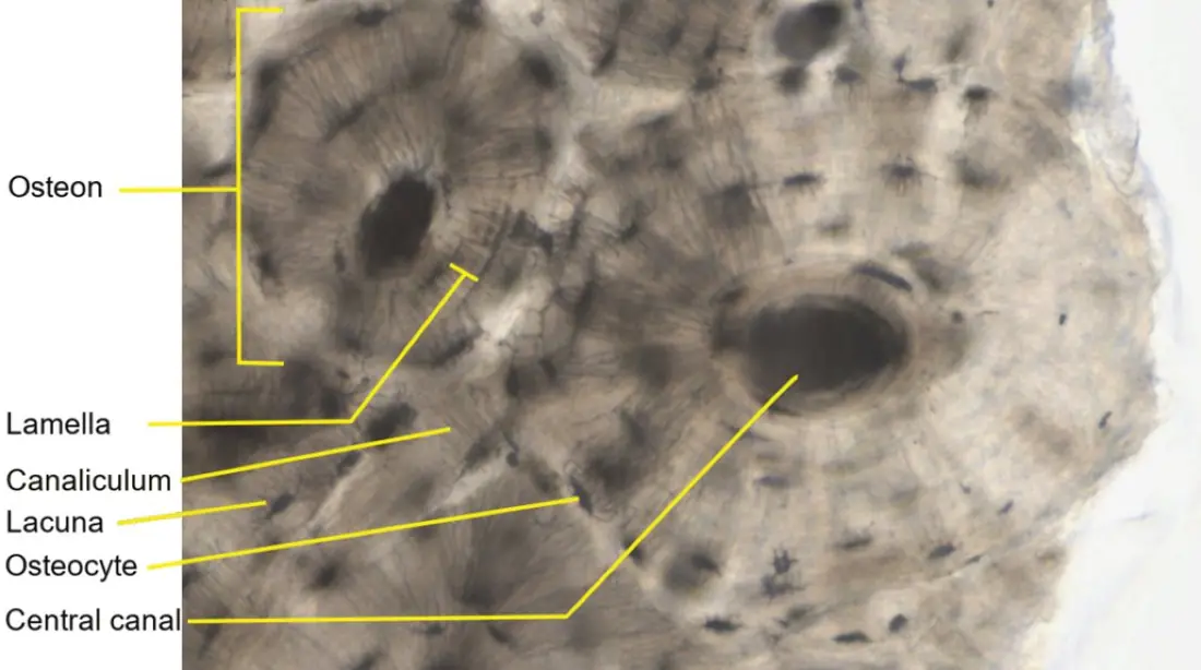
| Feature | Cartilage | Bone |
|---|---|---|
| Matrix | chondrin, soft and flexible | ossein + calcium salts, hard and rigid |
| Blood vessels | absent (nourished by diffusion from the perichondrium) | present (Haversian and Volkmann's canals) |
| Cells | chondrocytes, 1–4 per lacuna | osteocytes, 1 per lacuna |
| Canaliculi | absent | present |
| Growth | from inside and from the surface | from the surface (and at growth plates) |
| Marrow | absent | present |
Blood is a fluid connective tissue: about 55 % plasma and 45 % formed elements. Plasma is 90–92 % water and 6–8 % proteins — albumin (osmotic pressure), globulins (antibodies, transport) and fibrinogen (clotting) — with glucose, amino acids, salts, hormones and urea.
| Element | Number per mm³ | Structure | Life span | Job |
|---|---|---|---|---|
| RBC (erythrocyte) | 5–5.5 million (men), about 4.5 million (women) | biconcave, no nucleus in mammals, about 7.5 μm; full of haemoglobin (12–16 g per 100 mL blood); made in red bone marrow | 120 days; destroyed in the spleen (the "graveyard of RBCs") and liver | carry O₂ and some CO₂ |
| WBC (leucocyte) | 6000–8000 | nucleated, colourless, amoeboid; can squeeze out of capillaries (diapedesis) | days | defence |
| Platelets (thrombocytes) | 1.5–3.5 lakh | non-nucleated fragments of megakaryocytes of bone marrow | about a week | blood clotting |
| WBC | Type | % of WBCs | Nucleus | Job |
|---|---|---|---|---|
| Neutrophil | granulocyte | 60–65 (most abundant) | 3–5 lobes | phagocytosis of bacteria; first to arrive; pus is dead neutrophils |
| Eosinophil (acidophil) | granulocyte | 2–3 | bilobed | allergy and parasitic worm infections (rises in both) |
| Basophil | granulocyte | 0.5–1 (fewest) | S-shaped or lobed | releases histamine, heparin and serotonin (inflammation) |
| Lymphocyte | agranulocyte | 20–25 | large round nucleus filling the cell | immunity: B cells (antibodies), T cells |
| Monocyte | agranulocyte | 6–8 | kidney- (bean-) shaped; the largest WBC | phagocytosis; becomes a macrophage in tissues |
"Never Let Monkeys Eat Bananas" — Neutrophils (60–65 %) > Lymphocytes (20–25 %) > Monocytes (6–8 %) > Eosinophils (2–3 %) > Basophils (0.5–1 %).
Muscular tissue
Muscle is made of long, contractile muscle fibres (cells) whose cytoplasm (sarcoplasm) is packed with myofibrils of the proteins actin (thin filaments) and myosin (thick filaments). The cell membrane is the sarcolemma; the endoplasmic reticulum, which stores Ca²⁺, is the sarcoplasmic reticulum. Muscles are mesodermal, except the muscles of the iris and the myoepithelial cells of glands, which are ectodermal.
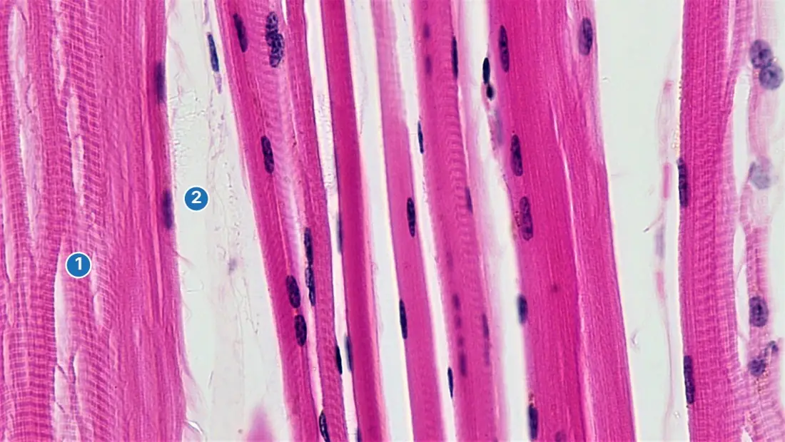
| Feature | Skeletal (striated, voluntary) | Smooth (unstriated, involuntary, visceral) | Cardiac |
|---|---|---|---|
| Shape | long, cylindrical, unbranched | spindle-shaped, unbranched | cylindrical, branched |
| Nuclei | many, at the periphery (a syncytium) | one, central | one (sometimes two), central |
| Striations | prominent light and dark bands | absent | present, fainter |
| Intercalated discs | absent | absent | present (gap junctions and desmosomes) |
| Control | voluntary (somatic nerves) | involuntary (autonomic) | involuntary; myogenic — beats on its own |
| Contraction | fast, powerful, fatigues quickly | slow, sustained, no fatigue | rhythmic, never fatigues |
| Location | limbs, body wall, tongue, pharynx, diaphragm, attached to bones by tendons | walls of alimentary canal, blood vessels, urinary bladder, uterus, iris, ciliary body, arrector pili | wall of the heart (myocardium) |
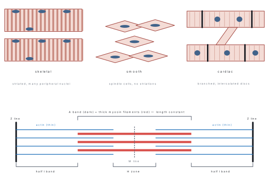
The sarcomere is the functional unit of a myofibril — the segment between two Z lines (Krause's membranes).
| Band | Contains | Name | In contraction |
|---|---|---|---|
| A band | myosin (with overlapping actin at its ends) | anisotropic, dark | length unchanged |
| I band | actin only | isotropic, light; bisected by the Z line | shortens |
| H zone | myosin only, centre of the A band | Hensen's zone; bisected by the M line | shortens / disappears |
| Sarcomere | Z line to Z line (about 2–3 μm at rest) | — | shortens |
Sliding filament theory (Huxley and Hanson, 1954). A nerve impulse releases Ca²⁺ from the sarcoplasmic reticulum; Ca²⁺ binds troponin, which shifts tropomyosin off the binding sites on actin; the myosin heads (ATPase) form cross-bridges, swing and pull the actin towards the M line. ATP is needed to detach each cross-bridge — after death ATP runs out and the muscles lock (rigor mortis). The filaments themselves do not shorten.
| Red (slow, type I) fibres | White (fast, type II) fibres |
|---|---|
| much myoglobin, many mitochondria, rich blood supply | little myoglobin, few mitochondria |
| aerobic, slow, sustained contraction, resist fatigue | anaerobic (lactic acid), fast, brief, fatigue quickly |
| back muscles, flight muscles of birds | eye muscles, breast muscles of the domestic fowl |
Nervous tissue
Nervous tissue is ectodermal and consists of neurons, the structural and functional units that conduct impulses, and neuroglia, which support them.
The neuron
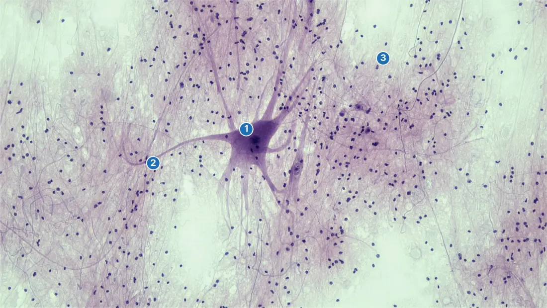
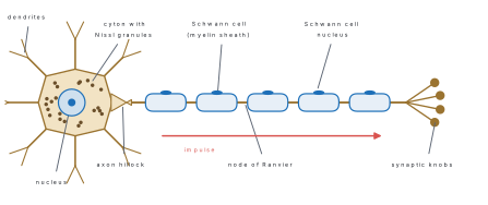
- Cyton (cell body, perikaryon) — a large nucleus with a prominent nucleolus; cytoplasm with Nissl granules (clumps of rough ER with ribosomes: protein synthesis) and neurofibrils. Mature neurons have no functional centrioles and do not divide.
- Dendrites — short, branched processes that carry impulses towards the cyton; they contain Nissl granules.
- Axon — usually one long process carrying impulses away from the cyton; it arises from the axon hillock (no Nissl granules), and ends in branches with synaptic knobs full of neurotransmitter vesicles.
- Myelin sheath — a fatty (lipoprotein) wrapping formed by Schwann cells in the PNS and oligodendrocytes in the CNS; the outer sheath of the Schwann cell is the neurilemma. Gaps in the myelin are the nodes of Ranvier, where the impulse jumps (saltatory conduction, up to about 120 m s⁻¹).
| Neuron type | Processes | Where |
|---|---|---|
| Unipolar | one | early embryo; invertebrates |
| Pseudounipolar | one process that splits into two | sensory neurons of the dorsal root ganglia |
| Bipolar | one axon + one dendrite | retina, olfactory epithelium, inner ear |
| Multipolar | one axon + many dendrites (the commonest) | motor neurons, brain and spinal cord |
By function neurons are sensory (afferent), motor (efferent) or interneurons (association neurons). Myelinated fibres form the white matter and the cranial and spinal nerves; non-myelinated fibres occur in the grey matter and the autonomic nervous system. A nerve is a bundle of nerve fibres wrapped in connective tissue (endoneurium, perineurium, epineurium).
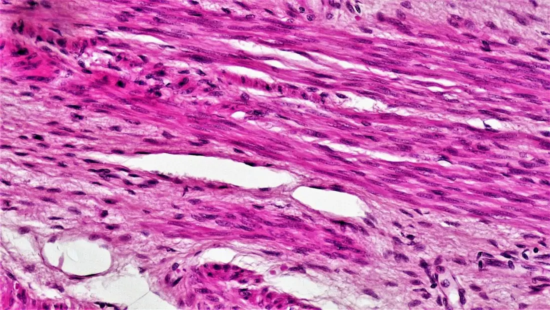
Neuroglia — non-conducting supporting cells that make up about half the volume of nervous tissue and, unlike neurons, can still divide.
| Glial cell | Job |
|---|---|
| Astrocytes | star-shaped; support neurons, form the blood–brain barrier, regulate ions |
| Oligodendrocytes | form myelin in the CNS |
| Schwann cells | form myelin in the PNS and the neurilemma |
| Microglia | phagocytic scavengers of the CNS (of mesodermal origin) |
| Ependymal cells | line the brain ventricles and central canal; help make CSF |
- The trachea is lined by pseudostratified ciliated columnar epithelium, not stratified.
- The oesophagus and vagina have non-keratinised stratified squamous epithelium; only the skin is keratinised.
- Tendon = muscle to bone (white, collagen, inelastic); ligament = bone to bone (yellow, elastic).
- Pinna and epiglottis are elastic cartilage; intervertebral discs are fibrocartilage; the ends of long bones are hyaline.
- In contraction the A band does not change; the I band and H zone shorten.
- Monocytes are the largest WBCs, neutrophils the most numerous, basophils the fewest; eosinophils rise in allergy and worm infection.
Answering "which tissue" questions
Decide first whether the organ needs to diffuse (squamous), secrete or absorb (cuboidal/columnar), move something along (ciliated), resist wear (stratified) or stretch (transitional). The location follows from the job.
Problem. Which specific epithelium lines (a) Bowman's capsule and pulmonary alveoli, (b) the proximal convoluted tubule (PCT) of the nephron, (c) the fallopian tube and bronchioles, and (d) the urinary bladder?
Solution. (a) Simple squamous (thin diffusion barrier); (b) brush-bordered simple cuboidal (microvilli maximise reabsorption); (c) ciliated columnar/cuboidal (propels ovum or mucus); (d) transitional epithelium / urothelium (stretches without tearing).
Problem. When a skeletal muscle fibre contracts under the sliding-filament mechanism, state what happens to the length of (a) the A-band, (b) the I-band, (c) the H-zone, and (d) the sarcomere (Z-to-Z distance).
Solution. Thick (myosin) and thin (actin) filaments do not shorten; actin slides into the A-band. Therefore (a) the A-band stays constant, while (b) the I-band, (c) the H-zone, and (d) the sarcomere all shorten.
Problem. Histological slide X shows concentric lamellae around Haversian canals connected by Volkmann's canals; slide Y shows chondrocytes in fluid-filled lacunae (2–4 per capsule) in an avascular basophilic matrix without Haversian systems. Identify X and Y.
Solution. Haversian systems (osteons) are the hallmark of mammalian compact bone (X); avascular matrix with chondrocyte cell nests (isogenous groups) lacking Haversian canals is hyaline cartilage (Y).
Formula and fact sheet
| Item | Value or name |
|---|---|
| Basement membrane, no matrix, avascular | epithelium |
| Endothelium / mesothelium | lining of blood vessels / of body cavities |
| Brush border | PCT of kidney (cuboidal), small intestine (columnar) |
| Stereocilia | epididymis, vas deferens |
| Goblet cell | unicellular mucous gland |
| Heterocrine gland | pancreas, gonads |
| Holocrine / apocrine / merocrine | sebaceous / mammary / salivary, pancreas |
| Commonest cell of areolar tissue | fibroblast |
| Heparin and histamine | mast cells (and basophils) |
| Antibodies | plasma cells (from B lymphocytes) |
| Brown fat | thermogenesis — newborns, hibernating animals |
| Collagen / elastin / reticulin | white / yellow / reticular fibres |
| Tendon / ligament | muscle to bone / bone to bone |
| Cartilage matrix / bone matrix protein | chondrin / ossein |
| Bone salt | calcium phosphate as hydroxyapatite (about 2/3 of the matrix) |
| Bone-forming / bone-resorbing cells | osteoblasts / osteoclasts |
| Haversian and Volkmann's canals | compact bone of mammals |
| Blood | 55 % plasma, 45 % cells; plasma 90–92 % water, 6–8 % protein |
| RBC | 5–5.5 million/mm³; biconcave, no nucleus; 120 days; spleen = graveyard |
| WBC | 6000–8000/mm³ — N 60–65, L 20–25, M 6–8, E 2–3, B 0.5–1 % |
| Platelets | 1.5–3.5 lakh/mm³; megakaryocyte fragments; clotting |
| Haemoglobin | 12–16 g per 100 mL |
| Skeletal muscle | striated, voluntary, multinucleate, unbranched |
| Smooth muscle | unstriated, involuntary, uninucleate, spindle-shaped |
| Cardiac muscle | striated, involuntary, branched, intercalated discs, myogenic |
| Sarcomere | Z to Z; A band constant; I band and H zone shorten |
| Ca²⁺ store / Ca²⁺ receptor | sarcoplasmic reticulum / troponin |
| Nissl granules | rough ER + ribosomes in cyton and dendrites; absent from the axon hillock |
| Myelin | Schwann cells (PNS), oligodendrocytes (CNS) |
| Saltatory conduction | nodes of Ranvier |
| Bipolar / pseudounipolar / multipolar | retina / dorsal root ganglion / motor neurons |
| Blood–brain barrier / phagocytic glia | astrocytes / microglia |
Chapter summary
- A tissue is a group of similar cells with a common origin and function; the four primary tissues are epithelial, connective, muscular and nervous.
- Epithelium rests on a basement membrane, has almost no matrix, is avascular and arises from all three germ layers; it is named by layers and cell shape — squamous (alveoli, capillaries), cuboidal (kidney tubules, thyroid), columnar (stomach, intestine), ciliated (oviduct), pseudostratified (trachea), stratified squamous (skin, oesophagus), transitional (urinary bladder).
- Glands are exocrine, endocrine or heterocrine (pancreas), and secrete in merocrine (salivary), apocrine (mammary) or holocrine (sebaceous) fashion.
- Connective tissue has few cells (fibroblasts, macrophages, mast cells, plasma cells, fat cells) in abundant matrix with collagen, elastic and reticular fibres: areolar, adipose, tendon, ligament, cartilage, bone, blood and lymph.
- Cartilage is avascular with chondrin matrix — hyaline (joints, trachea), elastic (pinna, epiglottis), fibrocartilage (intervertebral discs); bone has ossein and hydroxyapatite, osteocytes in lacunae and Haversian systems.
- Blood: 55 % plasma; RBCs (5 million/mm³, 120 days, no nucleus), WBCs (6000–8000/mm³; neutrophils most, basophils fewest, monocytes largest) and platelets (clotting).
- Muscle: skeletal (striated, voluntary, multinucleate), smooth (spindle, involuntary) and cardiac (branched, intercalated discs, myogenic); in contraction actin slides over myosin, the A band stays constant and the I band and H zone shorten.
- Nervous tissue: neurons (cyton with Nissl granules, dendrites, axon, myelin from Schwann cells or oligodendrocytes, nodes of Ranvier) and neuroglia (astrocytes, oligodendrocytes, Schwann cells, microglia, ependymal cells).
Practice questions
Set A — Recall (25 questions)
- The inner lining of blood vessels (endothelium) is made ofasimple cuboidal epitheliumbsimple columnar epitheliumcsimple squamous epitheliumdtransitional epithelium
- The urinary bladder is lined byatransitional epitheliumbkeratinised stratified squamous epitheliumcsimple columnar epitheliumdpseudostratified epithelium
- The trachea is lined byasimple squamous epitheliumbstratified squamous epitheliumcsimple cuboidal epitheliumdpseudostratified ciliated columnar epithelium
- Cuboidal epithelium with a brush border of microvilli lines theatracheabproximal convoluted tubulecurinary bladderdepidermis
- Sebaceous glands secrete by theamerocrine modebapocrine modecholocrine modedendocrine mode
- Mammary glands are an example ofaapocrine glandsbholocrine glandscendocrine glandsdunicellular glands
- The most abundant cell of areolar connective tissue is theamast cellbmacrophagecplasma celldfibroblast
- Heparin and histamine are secreted in connective tissue byafibroblastsbmast cellscplasma cellsdadipocytes
- A tendon connectsamuscle to bonebbone to bonecmuscle to muscledbone to cartilage
- The pinna and the epiglottis are supported byahyaline cartilagebwhite fibrocartilagecelastic cartilagedcalcified cartilage
- The intervertebral discs are made ofahyaline cartilagebwhite fibrocartilagecelastic cartilagedspongy bone
- Haversian canals are found inahyaline cartilagebelastic cartilagecred bone marrowdcompact bone of mammals
- Bone-forming cells are calledaosteoblastsbosteoclastscchondroblastsdfibroblasts
- The protein of the bone matrix isachondrinbkeratincosseindelastin
- The most numerous white blood cells in human blood arealymphocytesbmonocytesceosinophilsdneutrophils
- The largest white blood cell is thealymphocytebmonocytecbasophildneutrophil
- Antibodies are secreted byamast cellsbmacrophagescplasma cellsderythrocytes
- The average life span of a human red blood cell isa120 daysb12 daysc60 daysd7 days
- Intercalated discs are found inaskeletal musclebsmooth musclectendonsdcardiac muscle
- Smooth muscle is found in theabicepsbwall of the intestinecheartdtongue
- The functional unit of a myofibril is theasarcomerebsarcolemmacsarcoplasmdsarcoplasmic reticulum
- The myelin sheath of peripheral nerve fibres is formed byaoligodendrocytesbastrocytescSchwann cellsdmicroglia
- Nissl granules of a neuron are made ofamitochondriablysosomescGolgi bodiesdrough ER with ribosomes
- Bipolar neurons are found in theamotor nerves of the spinal cordbretinacdorsal root gangliadcerebral cortex
- The gaps between successive segments of the myelin sheath areaNissl bodiesbaxon hillockscnodes of Ranvierdsynaptic clefts
Answers and explanations — Set A
1. (c) — thin, flat cells allow exchange across capillary walls; cuboidal and columnar epithelia are too thick.
2. (a) — transitional epithelium stretches as the bladder fills; keratinised epithelium is confined to the skin.
3. (d) — the "respiratory epithelium": cells of different heights with cilia and goblet cells, all on one basement membrane.
4. (b) — PCT cells reabsorb most of the filtrate across a brush border. The trachea is ciliated, the bladder transitional.
5. (c) — the whole sebaceous cell breaks down to form sebum.
6. (a) — the tip of the secreting cell pinches off with the milk fat; sebaceous glands are holocrine.
7. (d) — fibroblasts secrete the fibres and matrix and outnumber every other cell type.
8. (b) — mast cells (like basophils) release heparin (anticoagulant) and histamine (inflammation).
9. (a) — tendons of dense white collagen fibres join muscle to bone; ligaments join bone to bone.
10. (c) — elastic fibres let the pinna and epiglottis bend and spring back.
11. (b) — collagen bundles make fibrocartilage the strongest cartilage, fit for the discs and pubic symphysis.
12. (d) — each Haversian system (osteon) of compact bone has a central canal with blood vessels; cartilage is avascular.
13. (a) — osteoblasts build bone; osteoclasts resorb it.
14. (c) — ossein (collagen) is the organic part of bone matrix; chondrin belongs to cartilage.
15. (d) — neutrophils are 60–65 % of WBCs; lymphocytes come second at 20–25 %.
16. (b) — the monocyte, with a kidney-shaped nucleus, becomes a tissue macrophage.
17. (c) — plasma cells are activated B lymphocytes.
18. (a) — about 120 days; worn-out RBCs are removed in the spleen and liver.
19. (d) — intercalated discs with gap junctions let the heart's fibres contract as one unit.
20. (b) — the gut wall's smooth muscle produces peristalsis; biceps and tongue are skeletal, the heart cardiac.
21. (a) — the sarcomere, from one Z line to the next, is the unit that shortens.
22. (c) — Schwann cells in the PNS; oligodendrocytes myelinate CNS fibres.
23. (d) — Nissl granules are rough ER studded with ribosomes, making proteins for the long axon.
24. (b) — retinal bipolar cells link the rods and cones with ganglion cells.
25. (c) — the nodes of Ranvier, where the impulse jumps in saltatory conduction.
Set B — Understanding (15 questions)
- Epithelial cells receive their oxygen and nutrientsafrom capillaries running between the cellsbby diffusion from vessels in the connective tissue belowcfrom lymph vessels within the epitheliumdfrom the air directly
- Transitional epithelium suits the urinary bladder becauseaits cells secrete mucusbit is keratinisedcit absorbs urinedits cells change shape and flatten when the bladder stretches
- Which pair is correctly matched?aciliated columnar — fallopian tubebkeratinised stratified squamous — oesophaguscsimple squamous — PCTdtransitional — trachea
- Which statement is true of cartilage but NOT of bone?aits matrix contains hydroxyapatitebit has Haversian canalscit is avascular and fed by diffusiondits cells lie in lacunae
- During contraction of a skeletal muscle, which part of the sarcomere keeps its length?aI bandbA bandcH zonedthe whole sarcomere
- Cardiac muscle works throughout life without fatigue mainly because itais under voluntary controlblacks myofibrilschas no nucleidhas many mitochondria and a rich blood supply
- Red muscle fibres differ from white muscle fibres in havingamore myoglobin and mitochondriabfewer mitochondriacfaster but brief contractionda poorer blood supply
- Myelinated fibres conduct impulses faster than non-myelinated fibres of the same diameter becauseathey have more Nissl granulesbmyelin itself carries the currentcthe impulse jumps from node to nodedthey lack a neurilemma
- Blood is classed as a connective tissue because itacontains haemoglobinbis mesodermal, with cells in a fluid matrixcflows in vesselsdhas no fibres at all
- Brown adipose tissue is abundant in newborn babies and hibernating mammals because itastores waterbsecretes insulincforms antibodiesdproduces heat by oxidising fat
- The phagocytic cells of the central nervous system are theamicrogliabastrocytescoligodendrocytesdependymal cells
- Basophils resemble mast cells in that bothamake antibodiesbcarry oxygencrelease histamine and heparindform myelin
- Ligaments are more elastic than tendons because they contain moreacollagen fibresbyellow elastic fibresccartilagedcalcium salts
- Mature neurons cannot be replaced by division because theyalack nucleiblack Nissl granulesclack mitochondriadhave no functional centrioles
- Platelets areafragments of megakaryocytesbnucleated cells from the spleenca kind of lymphocytedRBCs without haemoglobin
Answers and explanations — Set B
1. (b) — epithelium is avascular; everything diffuses across the basement membrane from the connective tissue capillaries.
2. (d) — the dome-shaped surface cells flatten and slide, so the lining stretches without tearing.
3. (a) — cilia sweep the ovum along the oviduct. The oesophagus is non-keratinised, the PCT cuboidal, the trachea pseudostratified.
4. (c) — cartilage has no blood vessels; both tissues have cells in lacunae (d), and (a), (b) are bone only.
5. (b) — the A band is the length of the myosin filaments, which do not shorten; actin slides in, so the I band and H zone narrow.
6. (d) — abundant mitochondria and coronary blood keep cardiac muscle aerobic, and its long refractory period forces rest between beats. It is involuntary (a).
7. (a) — myoglobin stores O₂ and many mitochondria give aerobic, sustained contraction.
8. (c) — myelin insulates; ion exchange happens only at the nodes, so the impulse jumps (saltatory conduction).
9. (b) — like all connective tissues it arises from mesoderm and has cells in an abundant matrix (plasma); fibrinogen forms fibres on clotting.
10. (d) — brown fat cells have many mitochondria that release the energy of fat as heat (non-shivering thermogenesis).
11. (a) — microglia are the brain's macrophages, derived from mesoderm.
12. (c) — both have granules of histamine and heparin and act in inflammation and allergy.
13. (b) — ligaments carry elastic fibres and allow joint movement; tendons are almost pure collagen.
14. (d) — neurons are permanently out of the cell cycle; textbooks give the lack of functional centrioles as the reason.
15. (a) — megakaryocytes of the bone marrow break into platelets, which have no nucleus.
Set C — Application (10 questions)
- A slide shows long, unbranched, cylindrical fibres with many peripheral nuclei and cross-striations. It was most probably taken from theaheartbtonguecstomach walldiris
- A section shows cells in lacunae in a clear, glassy matrix with no visible fibres, surrounded by perichondrium. It was most probably taken from theapinnabintervertebral disccepiglottisdtracheal rings
- A sarcomere 2.4 μm long has an A band of 1.6 μm. If it shortens to 2.0 μm, the total length of I band in the sarcomere becomesa0.4 μmb0.8 μmc0.2 μmd1.2 μm
- In a count of 200 WBCs a technician finds 124 neutrophils, 50 lymphocytes, 14 monocytes, 10 eosinophils and 2 basophils. The count suggestsabacterial infectionbviral infectioncallergy or worm infectiondleukaemia
- A person has 5 litres of blood with 5 million RBCs per mm³. If each RBC lives 120 days, about how many RBCs must be made each day?a2 × 10⁹b2 × 10¹¹c2 × 10¹³d4 × 10⁷
- A tissue from the pubic symphysis has chondrocytes in lacunae and dense bundles of collagen fibres in its matrix. It isahyaline cartilagebelastic cartilageccompact bonedwhite fibrocartilage
- A drug that blocks the release of Ca²⁺ from the sarcoplasmic reticulum wouldaprevent skeletal muscle contractionbspeed up contractionccause rigor mortisdaffect only smooth muscle
- Muscles become stiff a few hours after death becauseaactin and myosin filaments shortenbcalcium is removed from the musclecno ATP is left to detach the myosin cross-bridgesdthe sarcolemma dissolves
- A drug destroys the cell bodies of neurons lying in the dorsal root ganglia. The neurons affected areamultipolar motor neuronsbpseudounipolar sensory neuronscbipolar interneuronsdmultipolar autonomic neurons
- A tissue has spindle-shaped cells, each with one central nucleus and no striations. Which activity depends on it?amovement of the limbsbthe heartbeatcblinking of the eyelidsdperistalsis of the intestine
Answers and explanations — Set C
1. (b) — these are skeletal fibres; the tongue is skeletal muscle, while the heart is cardiac and the stomach wall and iris smooth.
2. (d) — a glassy matrix without visible fibres is hyaline cartilage, as in the C-shaped tracheal rings. Pinna and epiglottis are elastic, the disc fibrocartilage.
3. (a) — the A band stays 1.6 μm, so I band = sarcomere − A band = 2.0 − 1.6 = 0.4 μm (it was 0.8 μm before).
4. (c) — eosinophils are 10/200 = 5 %, above the normal 2–3 %; neutrophils (62 %), lymphocytes (25 %), monocytes (7 %) and basophils (1 %) are normal.
5. (b) — 5 L = 5 × 10⁶ mm³, so total RBCs = 5 × 10⁶ × 5 × 10⁶ = 2.5 × 10¹³; ÷ 120 ≈ 2 × 10¹¹ per day.
6. (d) — collagen bundles in a cartilage matrix mean white fibrocartilage, the tissue of the pubic symphysis and intervertebral discs.
7. (a) — without Ca²⁺ troponin keeps the binding sites covered and no cross-bridges form; rigor (c) needs a lack of ATP, not of Ca²⁺.
8. (c) — rigor mortis: cross-bridges stay locked to actin because ATP is needed to release them; the filaments themselves never shorten (a).
9. (b) — the dorsal root ganglia hold the cell bodies of pseudounipolar sensory neurons; motor cell bodies lie in the grey matter of the cord.
10. (d) — smooth muscle drives peristalsis; limbs and eyelids use skeletal muscle and the heartbeat is cardiac.