Chapter 5 · Human Biology and Physiology · 2 questions in the paper · 42 min read
Digestive System
What the syllabus asks for 2
- The alimentary canal and the digestive glands
- Physiology of digestion and absorption
The digestive system carries 2 of the 40 Zoology questions. The paper asks it in two ways: a name-and-number question (which cells secrete HCl, which enzyme needs enterokinase, the pH of pancreatic juice, the dental formula) and a route question (where is a nutrient absorbed, and into which vessel does it go). Almost every mark sits in the enzyme table, the gland table and the absorption routes. The traps are few but they are asked every year: bile has no enzymes, pepsin works only in acid and trypsin only in alkali, and most absorption happens in the ileum, not the stomach.
- The order of the canal and the epithelium of each segment: stratified squamous (mouth, oesophagus, anal canal), simple columnar (stomach, intestine), and the special cells inside it (parietal, chief, goblet, Paneth, enteroendocrine).
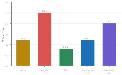
- The four secretions and their volumes: saliva 1–1.5 L, gastric juice 2–3 L, bile 0.5–1 L, pancreatic juice 1–1.5 L per day, with their pH values.
- The enzyme → substrate → product → site → optimum pH table, including enterokinase (trypsinogen → trypsin) and the brush-border enzymes (maltase, sucrase, lactase, peptidases).
- HCl (five jobs), mucus (protection), bile (emulsification — never an enzyme) and bicarbonate (neutralising chyme).
- The hormones: gastrin (G cells, pyloric antrum, acid + protein), secretin (S cells, duodenum, acid → bicarbonate), CCK (I cells, fat → bile and pancreatic enzymes).
- Absorption: monosaccharides and amino acids into blood capillaries → hepatic portal vein → liver; fats as chylomicrons into lacteals → thoracic duct → subclavian vein.
- The applied trio: peptic ulcer and H. pylori, jaundice (three types), appendicitis.
5.1 The alimentary canal and the digestive glands
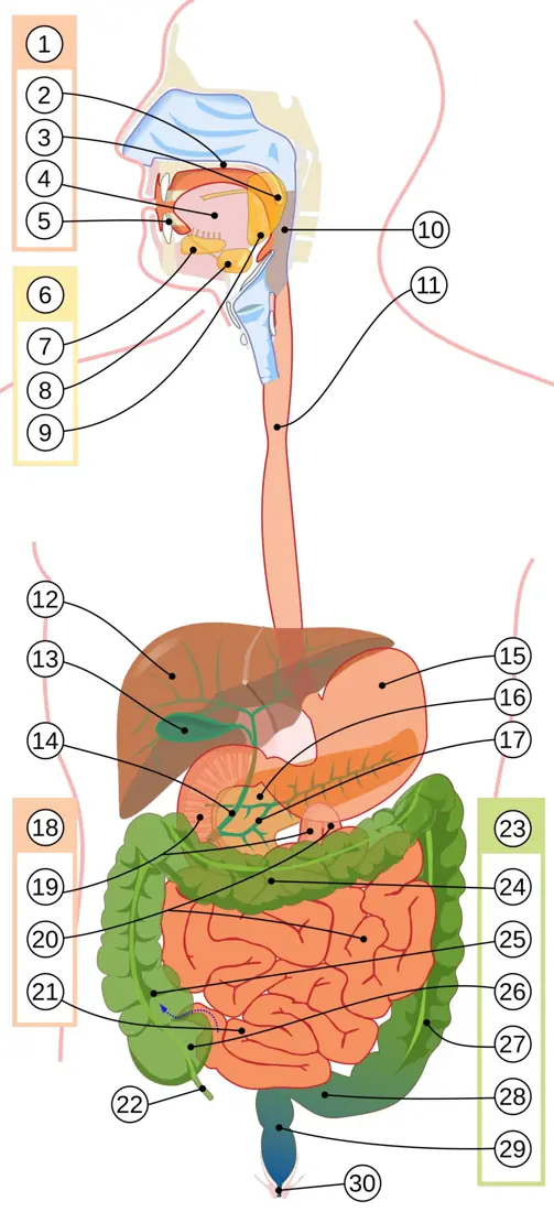
The alimentary canal is a single muscular tube about 8–9 m long, open at both ends, from mouth to anus. Its digestive glands — salivary, gastric, intestinal, liver and pancreas — pour their secretions into it. The liver, pancreas and salivary glands are exocrine here because their ducts open into the canal.
The plan of the canal
Every region of the canal is built on the same four coats, from the lumen outwards:
| Coat | Tissue | Job |
|---|---|---|
| Mucosa (mucous membrane) | epithelium + lamina propria (loose connective tissue with capillaries and lymphatics) + muscularis mucosae | secretion, absorption, a first line of defence |
| Submucosa | loose connective tissue, blood and lymph vessels, Meissner's plexus | carries away absorbed food; supplies the mucosa |
| Muscularis externa | inner circular and outer longitudinal smooth muscle, with Auerbach's (myenteric) plexus between them | peristalsis and segmentation |
| Serosa / adventitia | mesothelium + connective tissue | reduces friction; suspends the gut |
The two nerve plexuses together form the enteric nervous system, the "little brain" of the gut; it can run peristalsis on its own, but the parasympathetic (vagus) speeds it up and the sympathetic slows it down.
The mouth (buccal cavity)
- Lined by stratified squamous non-keratinised epithelium, which resists the abrasion of food.
- Teeth: an adult has 32 permanent teeth; the dental formula is 2·1·2·3 / 2·1·2·3 = 16 + 16 (incisors, canine, premolars, molars per half jaw). The deciduous (milk) set has 20 teeth, formula 2·1·0·2 / 2·1·0·2. Teeth crush and grind food (mastication), giving the surface area that chemical digestion needs.
- Tongue: skeletal muscle covered with a mucous membrane; it mixes food, helps swallowing, and carries the taste buds.
- Saliva moistens the food, dissolves sapid substances so that they can reach the taste buds, and begins starch digestion. The ball of chewed food is the bolus.
The salivary glands are three pairs, and the paper asks which is which:
| Gland | Position | Duct | Secretion |
|---|---|---|---|
| Parotid (largest) | in front of the ear, below the zygomatic arch | Stensen's duct opens beside the upper second molar | serous — watery, amylase-rich |
| Submandibular | under the lower jaw | Wharton's duct opens under the tongue | mixed (serous + mucous); most of the resting saliva |
| Sublingual (smallest) | floor of the mouth | ducts of Rivinus | mainly mucous |
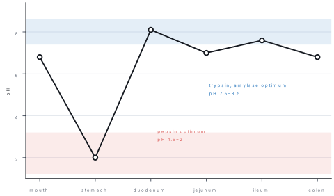
Saliva is 1–1.5 L a day, pH 6.8 (range 6.2–7.4), about 99.5 % water. It contains salivary amylase (ptyalin), maltase in small amount, the glycoprotein mucin (which becomes mucus), lysozyme (kills bacteria), immunoglobulin A, and the enzyme lingual lipase from the serous glands of the tongue. Salivary amylase converts starch → maltose (plus dextrins) and works best at pH 6.8; it is stopped by the acid of the stomach.
Lingual lipase is trivial in adults. Options such as "saliva digests protein" or "saliva contains pepsin" are wrong. Saliva's protein enzyme is absent; its fat enzyme is negligible.
The pharynx and the oesophagus
The pharynx is the shared passage for food and air; the epiglottis closes the glottis during swallowing so that the bolus enters the oesophagus and not the trachea.
The oesophagus is a muscular tube about 25 cm long passing through the diaphragm at the cardiac sphincter. It is lined by stratified squamous non-keratinised epithelium, has no digestive glands of its own and secretes no enzymes. Food moves down by peristalsis — a wave of contraction behind the bolus — helped by gravity; the whole trip takes about 5–8 seconds. The cardiac sphincter stops the acid contents of the stomach from running back; when it is incompetent the result is gastro-oesophageal reflux (heartburn).
The stomach
The stomach is a J-shaped muscular bag holding 1.5–2 L. Its regions are the cardia (around the opening), the fundus (the dome above the opening), the body and the pyloric antrum, which ends at the pyloric sphincter.
The mucosa is folded into gastric pits lined by simple columnar epithelium made of several cell types:
| Cell | Secretion | Function |
|---|---|---|
| Surface mucous cells | mucus + bicarbonate | a 1 mm gel layer that keeps HCl off the wall |
| Parietal (oxyntic) cells | HCl (pH 0.9–1.5 as secreted) and intrinsic factor | acid; intrinsic factor is needed to absorb vitamin B₁₂ |
| Chief (zymogenic) cells | pepsinogen, rennin (infants), gastric lipase | protein and milk digestion |
| G cells (pyloric antrum) | gastrin | hormone — stimulates acid secretion |
| Enterochromaffin-like cells | histamine | amplifies acid secretion |
| D cells | somatostatin | inhibits acid secretion |
Gastric juice is 2–3 L a day at pH 1.5–2.5. It contains HCl (about 0.5 %), pepsinogen, rennin in infants, gastric lipase, mucus and intrinsic factor.
HCl is not an enzyme. It does five jobs:
- Converts pepsinogen → pepsin (removes part of the molecule).
- Gives pepsin its acid optimum (pH 1.5–2).
- Kills most bacteria swallowed with food — a chemical barrier.
- Denatures dietary protein, unfolding it so that enzymes can reach the peptide bonds.
- Stimulates the release of secretin and CCK when the acid chyme reaches the duodenum, and helps absorb a little calcium and iron.
Pepsin is the only proteolytic enzyme of the stomach. Secreted as inactive pepsinogen, it is switched on by HCl and then activates more pepsinogen (autocatalysis). It breaks proteins down to peptones and polypeptides — never to amino acids. Rennin (chymosin) is present in the stomach of infants; it curdles milk by converting casein → calcium paracaseinate, so that the milk stays long enough to be digested. Adults have little rennin and curdle milk with pepsin instead.
The stomach produces chyme — a soupy, acidic mixture — and passes it into the duodenum in squirts through the pyloric sphincter. Water, alcohol, some salts and a few drugs (aspirin) are absorbed here, but almost no food.
A favourite wrong option is "most absorption occurs in the stomach". The stomach absorbs water, alcohol and some drugs only. Over 90 % of the absorption of the products of digestion happens in the small intestine, mainly the ileum.
The small intestine
The small intestine is about 6–7 m long and is the organ of digestion and absorption. It has three parts:
| Part | Length | Special features |
|---|---|---|
| Duodenum | about 25 cm, C-shaped | receives the bile duct and the pancreatic duct at the hepatopancreatic ampulla; Brunner's glands in the submucosa secrete alkaline mucus |
| Jejunum | about 2.5 m | longest villi, most digestion and absorption per cm |
| Ileum | about 3.5 m | Peyer's patches (aggregated lymphoid follicles); the main site of absorption |
Its wall multiplies the absorptive surface three times over: circular folds (plicae circulares), then villi about 1 mm long, then microvilli (the brush border) on every columnar cell. The total area becomes about 200 m², roughly a tennis court.
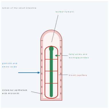
The epithelium is simple columnar with goblet cells (mucus) and, at the base of the glands, Paneth cells (antibacterial lysozyme). Between the villi lie the crypts of Lieberkühn, simple tubular glands that supply new epithelial cells every 3–5 days. Each villus holds a blind-ended lymph vessel (the lacteal) in its centre, a network of blood capillaries, and smooth muscle that makes it sway.
Intestinal juice (succus entericus) is about 2 L a day, pH 7.8–8.2. It is watery and alkaline, with mucus and the brush-border enzymes. The enzymes are not really poured into the lumen — they sit in the membrane of the microvilli, so that the last step of digestion happens as the nutrient touches the cell that will absorb it.
The large intestine
The large intestine is about 1.5 m long and about 6 cm wide. It runs caecum → ascending colon → transverse colon → descending colon → sigmoid colon → rectum → anal canal. Its epithelium is simple columnar with very numerous goblet cells; there are no villi and no digestive enzymes.
- Caecum: a blind pouch below the ileocaecal junction, with the ileocaecal valve that stops backflow. In man it is small (about 6 cm) and its worm-like extension, the vermiform appendix (8–10 cm), is a vestigial organ — in herbivores the caecum is huge and digests cellulose.
- Colon: absorbs water and mineral salts (about 1.5 L of water a day) and compacts the residue into faeces. The resident bacteria ferment leftovers and synthesise vitamin K and some B vitamins (biotin, folate, B₁₂), which are absorbed here.
- Rectum: stores faeces until defaecation; the anal canal is lined by stratified squamous epithelium and guarded by two sphincters, the internal (involuntary, smooth muscle) and the external (voluntary, skeletal muscle).
The liver and the gall bladder
The liver is the largest gland of the body — about 1.5 kg, under the diaphragm on the right. It is built of about a million lobules, each with a central vein and plates of hepatocytes separated by sinusoids lined with Kupffer cells. It has a double blood supply: the hepatic artery (oxygen) and the hepatic portal vein (nutrients from the gut).
Its functions, in the order the paper asks them:
| Function | Detail |
|---|---|
| Bile secretion | 0.5–1 L a day; bile salts emulsify fat |
| Carbohydrate metabolism | stores glycogen (glycogenesis), breaks it down (glycogenolysis), makes glucose from amino acids and fat (gluconeogenesis) |
| Protein metabolism | deamination of amino acids, urea formation (ornithine cycle), synthesis of albumin, globulin, prothrombin, fibrinogen |
| Fat metabolism | oxidation of fat, formation of lipoproteins, cholesterol synthesis |
| Storage | glycogen, vitamin A, D, E, K and B₁₂, iron (as ferritin) |
| Detoxification | converts ammonia to urea, destroys drugs and alcohol, conjugates bilirubin |
| Defence | Kupffer cells phagocytose bacteria and old red cells |
| Haemopoiesis | in the fetus; stores iron for it later |
Bile is a golden-green, alkaline (pH 7.6–8.6) fluid, 0.5–1 L a day, with no enzymes at all. Its composition:
| Component | Amount / note | Role |
|---|---|---|
| Bile salts (sodium glycocholate, taurocholate) | the important part | emulsify fat; help absorb fatty acids and vitamins A, D, E, K |
| Bile pigments — bilirubin (yellow) and biliverdin (green) | from the breakdown of haemoglobin in the spleen | excretion only; give faeces their colour |
| Cholesterol | may crystallise into gallstones | excreted |
| Lecithin, water, bicarbonate | solvent, neutraliser |
The gall bladder is a pear-shaped sac under the liver. It stores bile and concentrates it 5–10 times by absorbing water. Cholecystokinin (CCK) from the duodenum makes its smooth muscle contract and the sphincter of Oddi relax, so bile squirts into the duodenum when fatty food arrives.
Bile emulsifies fat — it breaks a large drop into thousands of tiny ones, so that the surface area for lipase rises enormously. The digestion itself is done by lipase. "Bile contains lipase", "bile digests fat into fatty acids" and "bile is acidic" are all wrong: bile is alkaline and has no enzymes.
The pancreas
The pancreas is a soft, lobulated gland lying behind the stomach, with its head in the curve of the duodenum and its tail touching the spleen. It is heterocrine — both exocrine and endocrine:
- Exocrine part (about 99 %): acini of serous cells drain into ducts that join to form the main pancreatic duct (duct of Wirsung), which opens with the bile duct at the hepatopancreatic ampulla.
- Endocrine part: the islets of Langerhans — β cells (insulin), α cells (glucagon), δ cells (somatostatin), PP cells.
Pancreatic juice is 1–1.5 L a day, pH 8.0–8.3, and is the most powerful digestive secretion. It contains:
| Enzyme | Action |
|---|---|
| Pancreatic amylase | starch → maltose |
| Trypsin (from trypsinogen) | proteins and peptones → peptides |
| Chymotrypsin (from chymotrypsinogen) | proteins → peptides |
| Carboxypeptidase (from procarboxypeptidase) | peptides → amino acids, from the C-terminal end |
| Pancreatic lipase (steapsin) | emulsified fats → fatty acids + glycerol |
| Ribonuclease and deoxyribonuclease | RNA and DNA → nucleotides |
| Bicarbonate | neutralises the acid chyme; brings the duodenal pH to about 8 |
The three proteases are secreted as inactive zymogens. Trypsinogen is switched on by enterokinase; trypsin then switches on chymotrypsinogen and procarboxypeptidase. This chain is a safety device: if the enzymes were made active inside the pancreas they would digest it — the cause of acute pancreatitis.
Enterokinase → trypsin → (chymotrypsin + carboxypeptidase). Pepsinogen is switched by HCl; trypsinogen by enterokinase. Never the other way round.
The glands and their secretions — one table
| Gland | Secretion | Volume / day | pH | Enzymes | Hormone that drives it |
|---|---|---|---|---|---|
| Salivary (parotid, submandibular, sublingual) | saliva | 1–1.5 L | 6.8 | salivary amylase, maltase, lingual lipase | parasympathetic reflex (sight, smell, taste of food) |
| Gastric (parietal, chief, G cells) | gastric juice | 2–3 L | 1.5–2.5 | pepsin, rennin, gastric lipase | gastrin |
| Liver | bile | 0.5–1 L | 7.6–8.6 | none | secretin (volume), CCK (gall bladder contraction) |
| Pancreas | pancreatic juice | 1–1.5 L | 8.0–8.3 | amylase, trypsin, chymotrypsin, carboxypeptidase, lipase, nucleases | secretin (bicarbonate), CCK (enzymes) |
| Intestinal (crypts, Brunner's) | succus entericus | about 2 L | 7.8–8.2 | maltase, sucrase, lactase, peptidases, enterokinase, intestinal lipase | local and vagal reflexes |
The enzyme table
Learn this table by rows: the paper rarely asks more than one cell of it, but it can ask any cell.
| Enzyme | Source | Substrate → product | Site | Optimum pH |
|---|---|---|---|---|
| Salivary amylase (ptyalin) | salivary glands | starch → maltose + dextrin | mouth | 6.8 |
| Gastric lipase | chief cells | butterfat → fatty acids + glycerol | stomach | 5–6 |
| Pepsin | chief cells (as pepsinogen) | proteins → peptones, polypeptides | stomach | 1.5–2 |
| Rennin (chymosin) | chief cells (infants) | casein → calcium paracaseinate | stomach | about 4 |
| Pancreatic amylase | pancreas | starch → maltose | duodenum | 7.1–8.2 |
| Trypsin | pancreas (as trypsinogen) | proteins, peptones → peptides | duodenum | 7.5–8.5 |
| Chymotrypsin | pancreas | proteins → peptides | duodenum | 7.5–8 |
| Carboxypeptidase | pancreas | peptides → amino acids | duodenum | 7.5 |
| Pancreatic lipase | pancreas | emulsified fat → fatty acids + glycerol | duodenum | 7–8 |
| Nucleases | pancreas | DNA, RNA → nucleotides | duodenum | 7.5 |
| Enterokinase | intestinal mucosa | trypsinogen → trypsin | duodenum | 7.5 |
| Maltase | intestinal brush border | maltose → 2 glucose | jejunum, ileum | 6.5–7 |
| Sucrase (invertase) | intestinal brush border | sucrose → glucose + fructose | jejunum, ileum | 6.5 |
| Lactase | intestinal brush border | lactose → glucose + galactose | jejunum, ileum | 6 |
| Peptidases (aminopeptidase, dipeptidase) | intestinal brush border | peptides → amino acids | jejunum, ileum | 7.5–8 |
| Intestinal lipase | intestinal mucosa | fats → fatty acids + glycerol | ileum | 7.5–8 |
5.2 Physiology of digestion and absorption
Digestion is the breakdown of large, insoluble food molecules into small, soluble ones that can cross the gut wall. It is mechanical (cutting, grinding, churning, emulsifying) and chemical (hydrolysis by enzymes). Absorption is the passage of the products through the mucosa into blood or lymph. Assimilation is their use by the cells.
Mechanical digestion
| Process | Where | What it does |
|---|---|---|
| Mastication (chewing) | mouth | breaks food up, mixes it with saliva, increases surface area |
| Swallowing (deglutition) | pharynx | the bolus is pushed into the oesophagus; the epiglottis guards the airway |
| Peristalsis | all of the canal | a wave of contraction behind the bolus moves it forward; it works even against gravity |
| Segmentation | small intestine | local rings of contraction chop and mix chyme with the enzymes; it does not move food along |
| Churning | stomach | the muscular wall kneads chyme and pushes it through the pylorus |
| Emulsification | duodenum | bile salts break fat into droplets of about 1 μm |
Chemical digestion of carbohydrates
| Step | Enzyme | Site | Product |
|---|---|---|---|
| starch → maltose + dextrin | salivary amylase | mouth | maltose, dextrin |
| starch → maltose | pancreatic amylase | duodenum | maltose |
| maltose → 2 glucose | maltase | brush border | glucose |
| sucrose → glucose + fructose | sucrase | brush border | glucose, fructose |
| lactose → glucose + galactose | lactase | brush border | glucose, galactose |
Cellulose is not digested at all: man has no cellulase, so it passes through as roughage (dietary fibre) — which is not useless, because it gives the colon something to work on and keeps the faeces soft.
Chemical digestion of proteins
| Step | Enzyme | Site | Product |
|---|---|---|---|
| protein → peptones, polypeptides | pepsin | stomach | peptones |
| peptones → peptides | trypsin, chymotrypsin | duodenum | peptides |
| peptides → amino acids (from the C-terminal end) | carboxypeptidase | duodenum | amino acids |
| peptides → amino acids (from the N-terminal end) | aminopeptidase | brush border | amino acids |
| dipeptides → amino acids | dipeptidase | brush border | amino acids |
Protein digestion therefore needs three pH environments in turn: acid in the stomach, alkaline in the duodenum, and neutral at the brush border. This is why a person whose pancreatic duct is blocked loses most protein digestion, and why the faeces of such a patient contain undigested muscle fibres.
Chemical digestion of fats
Fats are not digested in the mouth and hardly at all in the stomach (gastric lipase acts only on already-emulsified butterfat, at pH 5–6). The sequence is:
- Emulsification by bile salts in the duodenum — one large drop becomes many small ones, so the surface for lipase increases perhaps a thousandfold.
- Hydrolysis by pancreatic lipase: triglyceride → fatty acids + monoglycerides (and some glycerol).
- Micelle formation: bile salts gather round the fatty acids and monoglycerides and make water-soluble micelles about 5 nm across, which carry them to the brush border.
- Absorption by simple diffusion into the epithelial cell.
Chemical digestion of nucleic acids
Pancreatic ribonuclease and deoxyribonuclease split RNA and DNA into nucleotides; intestinal nucleosidases and phosphatases complete the job to pentose sugar, phosphate and nitrogenous bases.
The parts played by HCl, mucus, bile and enterokinase
| Substance | Secreted by | Part played | What it is NOT |
|---|---|---|---|
| HCl | parietal cells | activates pepsinogen, gives pepsin its acid pH, kills bacteria, denatures protein, triggers secretin/CCK | not an enzyme; it digests nothing itself |
| Mucus | mucous cells of the whole canal | a viscous glycoprotein gel that lubricates and, with bicarbonate, keeps a pH of about 7 at the cell surface; protects against acid, pepsin and mechanical injury | not a digestive juice |
| Bile | liver, stored in gall bladder | emulsifies fat; forms micelles; helps absorb vitamins A, D, E, K; neutralises acid chyme; excretes bilirubin and cholesterol | no enzymes |
| Enterokinase (enteropeptidase) | duodenal mucosa | converts trypsinogen → trypsin, the master switch of pancreatic proteolysis | not a digestive enzyme of food; it activates an enzyme |
| Bicarbonate | pancreas, Brunner's glands, bile | neutralises acid chyme, raises duodenal pH to about 8 | not an enzyme |
The stomach wall survives its own acid by a mucosal barrier: a thick mucus layer, bicarbonate trapped in it, tight junctions between the surface cells so that acid cannot leak between them, and a brisk blood flow that carries away any H⁺ that gets through. Prostaglandins maintain this barrier — which is why aspirin and other NSAIDs, which block prostaglandin synthesis, cause ulcers.
The hormones of digestion
| Hormone | Secreted by | Stimulus | Action |
|---|---|---|---|
| Gastrin | G cells of the pyloric antrum | food in the stomach — especially protein and distension; vagal stimulation | stimulates parietal cells to secrete HCl and chief cells to secrete pepsinogen; thickens the gastric mucosa |
| Secretin | S cells of the duodenum | acid chyme (pH below about 4.5) entering the duodenum | stimulates the pancreas to pour bicarbonate-rich juice; stimulates the liver to secrete bile; slows gastric emptying |
| CCK (cholecystokinin) | I cells of the duodenum and jejunum | fatty acids and amino acids in the duodenum | gall bladder contraction and relaxation of the sphincter of Oddi; pancreatic enzyme secretion; slows gastric emptying; causes a feeling of fullness |
| Gastric inhibitory peptide (GIP) | duodenal K cells | glucose, fat | inhibits gastric secretion and motility; stimulates insulin |
| Motilin, somatostatin | gut | — | motility; inhibition |
The order to remember: acid → secretin → bicarbonate; fat → CCK → bile and enzymes; food in the stomach → gastrin → acid.
Secretin answers acid and makes the pancreas secrete bicarbonate (it has almost no effect on enzymes). CCK answers fat and makes the gall bladder contract. A stem that says "CCK stimulates bicarbonate secretion" is testing exactly this swap.
Absorption
Absorption happens mostly in the small intestine — about 90 % of it, and most of that in the ileum, whose villi are numerous and whose transit is slow. The duodenum absorbs iron and calcium; the jejunum absorbs most of the monosaccharides and amino acids; the ileum absorbs vitamin B₁₂ (with intrinsic factor), bile salts and the rest; the colon absorbs water, sodium and chloride.
| Nutrient | Absorbed as | Site | Mechanism | Route |
|---|---|---|---|---|
| Carbohydrates | monosaccharides — glucose, galactose, fructose | jejunum, ileum | glucose and galactose by secondary active transport with Na⁺ (SGLT-1); fructose by facilitated diffusion (GLUT-5) | blood capillary → hepatic portal vein → liver |
| Proteins | amino acids, di- and tripeptides | jejunum, ileum | Na⁺-dependent co-transport (active) | blood capillary → hepatic portal vein → liver |
| Fats | fatty acids and monoglycerides (as micelles) | jejunum, ileum | simple diffusion into the epithelial cell, where they are re-esterified to triglyceride and packed with protein into chylomicrons | lacteal → lymph → thoracic duct → left subclavian vein |
| Water | water | small intestine (about 8 L a day), colon (1.5 L) | osmosis, following salts | blood capillary |
| Salts | Na⁺, Cl⁻, K⁺, HCO₃⁻ | mostly ileum, some colon | active transport and exchange | blood capillary |
| Iron | Fe²⁺ | duodenum | active transport, helped by vitamin C | blood → transferrin |
| Calcium | Ca²⁺ | duodenum | active transport, requires vitamin D (calcitriol) | blood |
| Vitamins A, D, E, K | with the fat | ileum | in micelles | lacteal |
| Vitamin B₁₂ | with intrinsic factor | ileum | receptor-mediated endocytosis | blood |
| Bile salts | — | ileum | active transport (enterohepatic circulation) | portal vein → liver |
The mechanism of absorption is worth one more line, because it explains the "against the gradient" questions: the epithelial cell keeps its internal Na⁺ low by pumping Na⁺ out into the blood with the Na⁺/K⁺ ATPase of its basolateral membrane. Na⁺ therefore keeps entering the cell from the lumen, and it drags glucose or an amino acid in with it. The energy is spent on the pump, not on the entry step — that is what "secondary active transport" means.
The hepatic portal route
Every capillary in a villus drains into veins that join to form the superior mesenteric vein, which unites with the splenic vein to form the hepatic portal vein. This vein enters the liver and breaks up again into sinusoids, so the liver sees the absorbed glucose and amino acids before any other organ — the first-pass effect. The liver stores or converts what is needed and releases the rest into the hepatic veins → inferior vena cava → heart.
Fats take a different road. A chylomicron is far too large to enter a blood capillary, so it enters the lacteal, is carried through lymph nodes to the thoracic duct, and joins the blood at the left subclavian vein — bypassing the liver at first.
The single most examined absorption fact: fats → lacteals (lymph), everything else water-soluble → blood capillaries → hepatic portal vein. "Fatty acids are absorbed into the capillaries of the villus" is the standard wrong option.
Applied notes: ulcer, jaundice and appendicitis
Peptic ulcer. An ulcer is an open sore where acid and pepsin have eaten through the mucosa. It occurs in the stomach (gastric ulcer) or, more often, the first part of the duodenum (duodenal ulcer). For decades the cause was thought to be stress and excess acid; in 1982 Barry Marshall and Robin Warren showed that most ulcers are caused by the bacterium Helicobacter pylori, which lives in the mucus layer, splits urea with its urease and neutralises acid around itself, and damages the barrier. The 2005 Nobel Prize followed. Other causes are NSAIDs (aspirin, ibuprofen), smoking, alcohol and severe stress. Symptoms: burning epigastric pain, worse when hungry in duodenal ulcer and after food in gastric ulcer; complications are bleeding, perforation and obstruction. Treatment: acid suppression (proton-pump inhibitors), antibiotics against H. pylori, and stopping NSAIDs. The ulcer is not caused by spicy food.
Jaundice. Yellowing of the skin, sclera and mucous membranes when plasma bilirubin rises above about 1 mg/dL (normal 0.3–1.0 mg/dL). Bilirubin is the yellow pigment made when haemoglobin is broken down in the spleen; the liver conjugates it and passes it into bile, and the gut bacteria turn it into stercobilin, which colours the faeces brown.
| Type | Cause | Plasma bilirubin | Urine | Faeces |
|---|---|---|---|---|
| Pre-hepatic (haemolytic) | too much haemoglobin broken down — malaria, incompatible transfusion, haemolytic anaemia of the newborn | unconjugated rises | no bilirubin, excess urobilinogen | normal or dark |
| Hepatic (hepatocellular) | liver cells damaged — viral hepatitis, alcohol, cirrhosis | both fractions rise | bilirubin present | pale |
| Post-hepatic (obstructive) | bile duct blocked — gallstone, cancer of the head of the pancreas | conjugated rises | dark, bilirubin present | pale, clay-coloured |
The obstructive type is the one to recognise: dark urine, pale stools, yellow skin and itch. In the newborn, physiological jaundice appears in the first week because the immature liver conjugates bilirubin slowly; it is treated with blue light (phototherapy).
Appendicitis. Inflammation of the vermiform appendix, usually because its lumen is blocked by a faecolith or lymphoid swelling. The classic story is pain that begins around the umbilicus and settles in the right iliac fossa at McBurney's point, with nausea, low fever and tenderness with guarding. The danger is perforation and peritonitis, so the treatment is appendicectomy. The appendix has no digestive function in man; it is a vestige of the large caecum of herbivores, though it does contain lymphoid tissue.
Worked examples
Problem. A fed person has 2.0 L of gastric juice at pH 2.0. How many moles and how many grams of HCl does it contain?
Solution. pH = 2.0 means [\mathrm{H^+}] = 10^{-2} mol L⁻¹. Since HCl is a strong acid, it is fully dissociated, so the amount of HCl equals the amount of H⁺:
About 0.73 g of pure HCl, which is why the stomach needs a mucus barrier.
Problem. The same 0.020 mol of HCl reaches the duodenum. What mass of sodium bicarbonate is needed to neutralise it?
Solution. NaHCO₃ + HCl → NaCl + H₂O + CO₂, so one mole of bicarbonate neutralises one mole of acid. Required = 0.020 mol. M(NaHCO₃) = 23 + 1 + 12 + 3(16) = 84 g mol⁻¹.
The pancreas secretes far more than this in a day (about 1–1.5 L of juice at pH 8.3), because acid keeps arriving with every meal.
Problem. Treat the small intestine as a cylinder of radius 1.5 cm and length 5.0 m. Folds, villi and microvilli multiply its area about 450 times. Find the plain area and the absorptive area.
Solution. Plain area = 2\pi rL = 2\pi(0.015)(5.0) = 0.47\ \text{m}^2.
Absorptive area = 450 \times 0.47 = 2.1 \times 10^2\ \text{m}^2, about 200 m² — the figure the textbooks quote, and roughly a tennis court. A cylinder alone would give less than half a square metre, which is why the folds matter.
Problem. A meal contains 250 g carbohydrate, 70 g protein and 60 g fat. Using 4 kcal g⁻¹ for carbohydrate and protein and 9 kcal g⁻¹ for fat, find its energy in kcal and kJ.
Solution. Carbohydrate 250 × 4 = 1000; protein 70 × 4 = 280; fat 60 × 9 = 540. Total = 1820 kcal. In joules, 1820 × 4.184 = 7.6 × 10³ kJ (7.6 MJ).
Three questions settle almost every stem. (1) Which enzyme? — and therefore which pH. (2) Where? — mouth, stomach, duodenum or brush border. (3) Into what? — blood capillary or lacteal. If the stem mentions bile, the answer is emulsification, never digestion; if it mentions enterokinase, the answer is trypsinogen → trypsin.
Formula and fact sheet
Lengths, volumes and numbers
| Fact | Value |
|---|---|
| Alimentary canal | 8–9 m |
| Oesophagus | 25 cm |
| Stomach capacity | 1.5–2 L |
| Small intestine | 6–7 m (duodenum 25 cm, jejunum 2.5 m, ileum 3.5 m) |
| Large intestine | 1.5 m |
| Appendix | 8–10 cm, attached to the caecum |
| Absorptive area of small intestine | about 200 m² |
| Permanent teeth | 32 (2·1·2·3 / 2·1·2·3) |
| Deciduous teeth | 20 (2·1·0·2 / 2·1·0·2) |
| Liver mass | about 1.5 kg — largest gland |
| Saliva | 1–1.5 L/day, pH 6.8 |
| Gastric juice | 2–3 L/day, pH 1.5–2.5 |
| Bile | 0.5–1 L/day, pH 7.6–8.6, no enzymes |
| Pancreatic juice | 1–1.5 L/day, pH 8.0–8.3 |
| Intestinal juice | about 2 L/day, pH 7.8–8.2 |
| Normal plasma bilirubin | 0.3–1.0 mg/dL; jaundice above about 1 mg/dL |
Names to place
| Item | Name |
|---|---|
| Ducts | parotid — Stensen's; submandibular — Wharton's; sublingual — Rivinus; pancreas — Wirsung |
| Cells | HCl and intrinsic factor — parietal (oxyntic); pepsinogen — chief; gastrin — G cells; secretin — S cells; CCK — I cells |
| Sphincters | cardiac, pyloric, ileocaecal, sphincter of Oddi, internal and external anal |
| Glands | Brunner's in the duodenal submucosa; crypts of Lieberkühn in the intestine; Peyer's patches in the ileum |
| Discoverers | H. pylori and peptic ulcer — Marshall and Warren (1982) |
The enzymes and their pH optima
| Enzyme | Optimum pH | One-line job |
|---|---|---|
| Salivary amylase | 6.8 | starch → maltose in the mouth |
| Pepsin | 1.5–2 | protein → peptones in the stomach |
| Rennin | about 4 | milk casein curdling in infants |
| Pancreatic amylase | 7.1–8.2 | starch → maltose |
| Trypsin | 7.5–8.5 | peptides, alkaline only |
| Chymotrypsin, carboxypeptidase | 7.5 | peptides → amino acids |
| Pancreatic lipase | 7–8 | fat → fatty acids + glycerol |
| Enterokinase | 7.5 | trypsinogen → trypsin |
| Maltase, sucrase, lactase | 6–7 | disaccharides → monosaccharides |
| Peptidases | 7.5–8 | peptides → amino acids |
Absorption in one line each
- Monosaccharides and amino acids → blood capillary → hepatic portal vein → liver.
- Fats → micelles → epithelial cell → chylomicrons → lacteal → thoracic duct → left subclavian vein.
- Vitamin B₁₂ needs intrinsic factor and is absorbed in the ileum.
- Iron and calcium are absorbed in the duodenum; calcium needs vitamin D.
- Water and salts are absorbed in the colon.
Hormones
| Hormone | Source | Stimulus | Effect |
|---|---|---|---|
| Gastrin | G cells, pyloric antrum | protein, distension, vagus | HCl + pepsinogen |
| Secretin | S cells, duodenum | acid chyme | pancreatic bicarbonate |
| CCK | I cells, duodenum | fat, amino acids | gall bladder contraction, pancreatic enzymes |
Traps
- Bile has no enzymes — it emulsifies.
- Pepsin only in acid; trypsin only in alkali.
- Most absorption is in the ileum, not the stomach.
- HCl is not an enzyme.
- Fat goes to the lacteal, not the blood capillary.
- Enterokinase activates trypsinogen, not pepsinogen.
- Secretin → bicarbonate; CCK → bile and enzymes.
- The colon has no villi and no enzymes.
Chapter summary
- The canal is a 8–9 m tube of four coats (mucosa, submucosa, muscularis, serosa); the enteric plexus runs peristalsis, modified by vagus (up) and sympathetic (down).
- The mouth has 32 permanent teeth, the bolus is made there, and salivary amylase (pH 6.8) starts starch digestion; the three gland pairs are parotid (Stensen's), submandibular (Wharton's) and sublingual (Rivinus).
- The stomach secretes 2–3 L of juice at pH 1.5–2.5: parietal cells give HCl and intrinsic factor, chief cells pepsinogen, rennin and lipase, G cells gastrin. HCl activates pepsinogen, kills bacteria and denatures protein; pepsin works only at pH 1.5–2 and stops at peptones.
- The small intestine (duodenum 25 cm, jejunum 2.5 m, ileum 3.5 m) receives bile and pancreatic juice, and its folds, villi and microvilli raise the absorptive area to about 200 m².
- The liver (1.5 kg, largest gland) makes 0.5–1 L of bile a day at pH 7.6–8.6 with no enzymes; bile salts emulsify fat. The gall bladder stores and concentrates bile and contracts under CCK.
- Pancreatic juice (1–1.5 L, pH 8.0–8.3) supplies amylase, trypsin, chymotrypsin, carboxypeptidase, lipase, nucleases and bicarbonate. Enterokinase converts trypsinogen to trypsin, which activates the other two zymogens.
- Hormones: gastrin (food → acid), secretin (acid → bicarbonate), CCK (fat → bile + enzymes).
- Absorption: monosaccharides and amino acids into blood capillaries → hepatic portal vein → liver; fats as chylomicrons into lacteals → thoracic duct; B₁₂ in the ileum with intrinsic factor; water and salts in the colon.
- Applied: peptic ulcer is mostly caused by H. pylori; obstructive jaundice gives dark urine and pale stools; appendicitis gives right iliac fossa pain at McBurney's point.
Practice questions
Set A — Recall (24 questions)
- The pH of gastric juice is abouta6.8b7.8–8.2c1.5–2.5d8.0–8.3
- Hydrochloric acid of the gastric juice is secreted byachief cellsbparietal cellscgoblet cellsdG cells
- The enzyme that converts trypsinogen into trypsin isaenterokinasebpepsincrennindcarboxypeptidase
- Bile is stored and concentrated in thealiverbduodenumcpancreasdgall bladder
- Bile helps the digestion of fat byahydrolysing triglyceridesbemulsifying fat into tiny dropletscconverting fat into glyceroldactivating pepsin
- The dental formula of an adult man isa2·1·0·2 / 2·1·0·2b3·1·4·2 / 3·1·4·2c2·1·2·3 / 2·1·2·3d2·2·2·3 / 2·2·2·3
- The largest gland of the human body is thealiverbpancreascthyroiddparotid
- Intrinsic factor, needed for the absorption of vitamin B₁₂, comes fromachief cellsbPaneth cellscBrunner's glandsdparietal cells
- Digested fat is absorbed from the villus into theablood capillariesbhepatic portal veinclactealsdcolonic crypts
- The duodenal hormone released by acid chyme isagastrinbsecretincinsulindcholecystokinin
- Gastrin is secreted by theaS cells of the duodenumbI cells of the jejunumcacinar cells of the pancreasdG cells of the pyloric antrum
- Cholecystokinin causesacontraction of the gall bladderbsecretion of bicarbonate by the pancreascsecretion of HCl by the stomachdclosure of the ileocaecal valve
- The milk-curdling enzyme of the infant's stomach isapepsinbtrypsincrennindlactase
- Most of the absorption of digested food occurs in theastomachbileumcduodenumdcolon
- The optimum pH for pepsin is abouta2b5c7d8
- Bile pigments are formed from the breakdown ofacholesterolbbile saltsclecithindhaemoglobin
- The number of teeth in the deciduous set of man isa16b20c28d32
- The vitamin synthesised by the bacteria of the human colon isavitamin Abvitamin Ccvitamin Kdvitamin D
- Peyer's patches are characteristic of theaileumbjejunumcduodenumdcolon
- Brunner's glands lie in theamucosa of the stomachbcrypts of the coloncpancreasdsubmucosa of the duodenum
- The volume of gastric juice secreted in 24 hours is abouta0.5–1 Lb2–3 Lc1–1.5 Ld5–6 L
- The duct of Wharton is the duct of theaparotid glandbsublingual glandcsubmandibular glanddpancreas
- Which of the following is NOT a function of hydrochloric acid?adigestion of protein into amino acidsbactivation of pepsinogenckilling of swallowed bacteriaddenaturation of dietary protein
- The vermiform appendix is attached to theacolonbrectumcileumdcaecum
Answers and explanations — Set A
1. (c) — a fed stomach is at pH 1.5–2.5. 6.8 is saliva, 7.8–8.2 intestinal juice and 8.0–8.3 pancreatic juice.
2. (b) — parietal (oxyntic) cells secrete HCl and intrinsic factor. Chief cells make pepsinogen, G cells gastrin.
3. (a) — enterokinase (enteropeptidase) of the duodenal mucosa is the only activator of trypsinogen; trypsin then activates the other zymogens.
4. (d) — the gall bladder stores bile and concentrates it 5–10 times; the liver makes it.
5. (b) — bile salts lower surface tension and break fat into droplets, raising the area for lipase. Bile has no lipase, so (a) is wrong.
6. (c) — 2 incisors, 1 canine, 2 premolars, 3 molars per half jaw, 16 + 16 = 32. Option (a) is the deciduous formula.
7. (a) — the liver, about 1.5 kg; the pancreas is a mixed gland but smaller.
8. (d) — parietal cells. Without intrinsic factor, B₁₂ is not absorbed in the ileum and pernicious anaemia follows.
9. (c) — fatty acids are rebuilt into triglycerides and packed as chylomicrons, which are too large for a capillary and enter the lacteal.
10. (b) — secretin, from S cells, when duodenal pH falls below about 4.5; it drives bicarbonate secretion.
11. (d) — G cells of the pyloric antrum. S cells make secretin, I cells cholecystokinin.
12. (a) — CCK contracts the gall bladder and relaxes the sphincter of Oddi; bicarbonate is secretin's job.
13. (c) — rennin (chymosin) converts casein to calcium paracaseinate. Adults have little of it.
14. (b) — the ileum, the longest part of the small intestine, with the greatest number of villi.
15. (a) — pepsin is a strongly acid protease, optimum pH 1.5–2; it is inactive at neutrality.
16. (d) — haemoglobin is broken down to haem and globin; haem gives biliverdin and bilirubin.
17. (b) — 20 milk teeth (2·1·0·2 per half jaw); the permanent set has 32.
18. (c) — colonic bacteria make vitamin K (and some B vitamins), which is absorbed in the colon.
19. (a) — aggregated lymphoid follicles in the ileum. They are part of the gut's immune defence.
20. (d) — Brunner's glands sit in the submucosa of the duodenum and pour alkaline mucus into it.
21. (b) — 2–3 L a day at pH 1.5–2.5; 1–1.5 L is saliva or pancreatic juice.
22. (c) — Wharton's duct drains the submandibular gland; Stensen's is the parotid, Rivinus the sublingual.
23. (a) — HCl does not split peptide bonds; pepsin does that, and only as far as peptones.
24. (d) — the appendix opens from the caecum, below the ileocaecal junction.
Set B — Understanding (13 questions)
- Hydrochloric acid must be present before pepsin can digest protein because HClasupplies the energy for hydrolysisbconverts pepsinogen to pepsin and keeps the pH at pepsin's optimumcemulsifies the proteindactivates enterokinase
- A patient with a blocked pancreatic duct passes fatty stools. The best explanation is thatapancreatic lipase does not reach the intestinebthe stomach stops making pepsincthe liver stops making biledthe villi stop absorbing glucose
- The stomach wall is not digested by its own juice becauseapepsin is secreted in its active formbthe wall contains no proteincsaliva neutralises the acidda mucus-bicarbonate layer and tight junctions protect the cells
- Bile contains no enzymes, yet fat digestion fails when the bile duct is blocked. This is becauseabile contains a hidden lipasebbile digests fat directlycbile salts emulsify fat and form micelles that lipase needsdbile neutralises pepsin
- Glucose uptake by the intestinal cell is called secondary active transport becauseathe Na⁺ gradient, maintained by the Na⁺/K⁺ pump, drives itbATP is split at the brush border for every glucosecglucose simply diffuses down its gradientdthe lacteal pulls glucose in
- Secretin secretion stops soon after acid chyme is neutralised becauseathe pancreas becomes exhaustedbgastrin switches it offcthe stimulus, a duodenal pH below about 4.5, has gonedbile destroys secretin
- The hepatic portal vein matters because itacarries oxygen to the liverbcarries absorbed nutrients from the gut to the liver firstcdrains bile from the liverdcarries fat to the thoracic duct
- In obstructive jaundice the faeces are pale becauseabile salts are not absorbedbthe liver stops making bile pigmentscfat is not digesteddno bilirubin reaches the gut, so no stercobilin forms
- After a total gastrectomy a patient needs vitamin B₁₂ injections becauseathe ileum has been removedbpepsin is needed to release B₁₂cintrinsic factor is no longer secreteddcolonic bacteria are lost
- In lactose intolerance, bloating and diarrhoea occur becauseacolonic bacteria ferment the lactose and hold water osmoticallyblactose is absorbed too quicklyclactase digests lactose inside the colondlactose destroys the villi
- The duodenum needs pancreatic bicarbonate in order toaactivate pepsinbemulsify fatckill bacteriadraise the pH so that pancreatic enzymes can work
- Pepsin is inactive at pH 7 becauseawater destroys itbits active site loses shape and the molecule is denaturedcit needs enterokinasedit acts only on fat
- The villus is the unit of absorption because it hasaa thin epithelium, a capillary net and a lacteal close to the surfaceball the digestive enzymes of the gutcPeyer's patches inside itdstratified squamous epithelium
Answers and explanations — Set B
1. (b) — HCl removes part of the pepsinogen molecule to make pepsin and holds the pH at 1.5–2, where pepsin works. It supplies no energy.
2. (a) — lipase, amylase and the proteases are all in pancreatic juice; without them fat passes out undigested (steatorrhoea).
3. (d) — the mucosal barrier: thick mucus with trapped bicarbonate, tight junctions, and good blood flow. Prostaglandins maintain it, which is why NSAIDs cause ulcers.
4. (c) — emulsification and micelle formation are physical, not chemical, but without them lipase has almost no surface to act on.
5. (a) — the cell spends ATP on the basolateral Na⁺/K⁺ pump, not on the entry step; the Na⁺ gradient does the work.
6. (c) — S cells respond to H⁺; once bicarbonate has raised the pH the stimulus disappears. This is negative feedback.
7. (b) — first-pass delivery: the liver stores, converts or releases what the gut absorbed before the rest of the body sees it.
8. (d) — stercobilin, from bilirubin acted on by gut bacteria, is the brown pigment. No bile in the gut, no colour.
9. (c) — parietal cells are lost with the stomach, so B₁₂ cannot be absorbed in the ileum even if it is eaten.
10. (a) — unabsorbed lactose is a substrate for colonic bacteria; the gas and the osmotic load cause the symptoms.
11. (d) — pancreatic enzymes have optima near pH 8; the acid chyme would otherwise inactivate them.
12. (b) — pepsin is adapted to acid; at neutral pH its shape and charge change and it stops working.
13. (a) — short diffusion distance plus two exit routes, one for water-soluble and one for fat-soluble products.
Set C — Application (9 questions)
- A person has 2.0 L of gastric juice at pH 2.0. The amount of HCl present isa0.002 molb0.20 molc0.020 mold2.0 mol
- Taking the same juice as in the previous question, the mass of HCl is abouta0.37 gb0.73 gc1.46 gd7.3 g
- A meal contains 250 g carbohydrate, 70 g protein and 60 g fat. Taking 4, 4 and 9 kcal g⁻¹, its energy isa1820 kcalb1120 kcalc2120 kcald860 kcal
- A cylinder of radius 1.5 cm and length 5.0 m has folds that multiply its area 450 times. The absorptive area is abouta0.47 m²b4.7 m²c47 m²d210 m²
- A man has burning epigastric pain, and a urea breath test is positive. The most likely cause isatoo much spicy foodbHelicobacter pylorica low-acid stomachdvitamin B₁₂ deficiency
- A 20-year-old has pain that began near the umbilicus and is now in the right iliac fossa, with fever and tenderness at McBurney's point. The diagnosis isagastric ulcerbgallstonescviral hepatitisdacute appendicitis
- A patient has yellow sclera, dark urine and clay-coloured stools. The cause isaexcessive haemolysisbviral hepatitiscobstruction of the bile ductdiron deficiency
- A child with cystic fibrosis has bulky fatty stools. The step most affected isapancreatic enzymes failing to reach the duodenumbfailure of bile salt synthesiscloss of gastrindloss of intrinsic factor
- A diet contains 100 g of protein. Taking protein as 16 % nitrogen, the nitrogen intake isa6.25 gb16 gc100 gd1.6 g
Answers and explanations — Set C
1. (c) — pH 2 means [H⁺] = 10⁻² mol L⁻¹, and HCl is fully dissociated, so n = 0.01 × 2.0 = 0.020 mol.
2. (b) — m = nM = 0.020 × 36.5 = 0.73 g of HCl. Option (c) doubles the amount by mistake.
3. (a) — 250 × 4 = 1000, 70 × 4 = 280, 60 × 9 = 540; total 1820 kcal (about 7.6 MJ).
4. (d) — plain area 2πrL = 2π(0.015)(5) = 0.47 m²; × 450 = 211 m², the textbook figure of about 200 m².
5. (b) — the urea breath test detects H. pylori urease; the bacterium, not spice, causes most peptic ulcers.
6. (d) — migration of pain from the umbilicus to the right iliac fossa with localised tenderness is the classic picture; perforation is the danger.
7. (c) — dark urine with pale stools means conjugated bilirubin is in the blood but cannot reach the gut: obstructive jaundice.
8. (a) — thick secretions block the pancreatic ducts, so lipase and the other enzymes never arrive; the fat is not hydrolysed.
9. (b) — 16 % of 100 g = 16 g N. Option (a) is the reverse calculation (100/16), the commonest slip.