Chapter 8 · Human Biology and Physiology · 2 questions in the paper · 36 min read
Excretory System
What the syllabus asks for 3
- Excretory organs and the structure of the nephron
- Formation of urine and its regulation
- Renal disorders
The excretory system carries 2 of the 40 Zoology questions. The paper builds them from three blocks: the organs and the nephron (what each part is and does), the formation of urine (filtration, reabsorption, secretion, the counter-current multiplier and the three hormones that set urine volume), and the disorders (nephritis, stones, failure, dialysis). Almost every question turns on a direction, a number or a permeability: which limb lets water through, which hormone makes urine small, which value is the renal threshold. The three traps that decide marks are the descending limb is permeable to water and the ascending limb is not, ADH increases water reabsorption so its deficiency gives a large dilute urine, and the posterior pituitary stores ADH but the hypothalamus makes it.
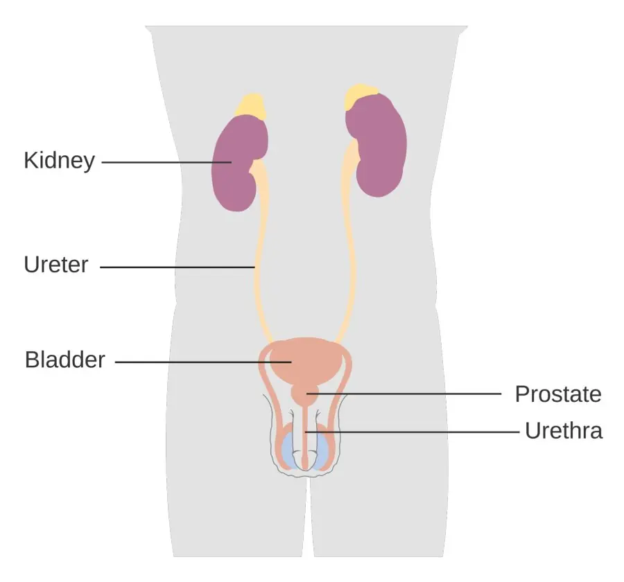
- Organs: a pair of kidneys, two ureters, the urinary bladder and the urethra, plus the accessory routes — skin (sweat), lungs (CO₂ and water vapour) and liver (urea from deamination, bile pigments).
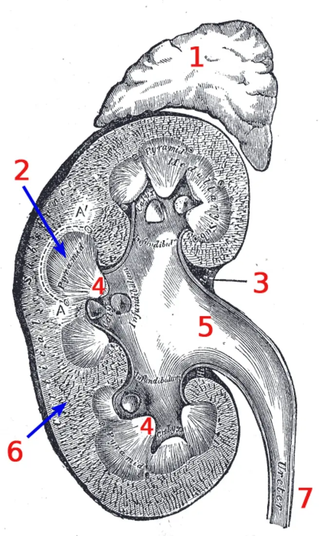
- Kidney in section: capsule → cortex (glomeruli, convoluted tubules) → medulla (pyramids, loops, collecting ducts) → renal pelvis → ureter; about 1 million nephrons per kidney.
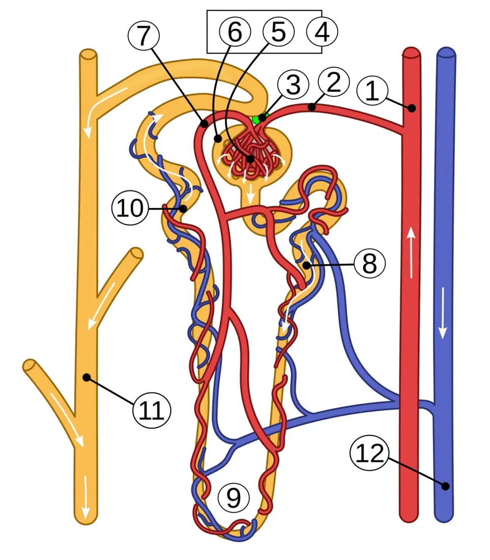
- Nephron: Bowman's capsule with podocytes, PCT with a brush border, the loop of Henle, DCT with the macula densa, the collecting duct, and the peritubular capillaries/vasa recta.
- Ultrafiltration: pores and negative charge hold back cells and plasma proteins; net filtration pressure ≈ 10 mmHg; GFR ≈ 125 mL min⁻¹ = 180 L day⁻¹.
- Reabsorption: 100 % of glucose and amino acids by active transport in the PCT; water by osmosis; the counter-current multiplier builds a gradient of 300 → 1200 mOsm L⁻¹.
- Secretion: H⁺, K⁺, NH₃ and drugs; the tubule's route for acid–base control.
- Hormones: ADH (water, collecting duct), aldosterone (Na⁺ in, K⁺ out), ANP (Na⁺ and water out); each changes both the volume and the concentration of urine.
- Disorders: nephritis, calculi, acute and chronic failure, haemodialysis versus peritoneal dialysis, transplantation.
8.1 Excretory organs and the structure of the nephron
Excretion, egestion and secretion
Excretion is the removal of the waste products of metabolism from the body — substances that were made inside cells and can no longer be used. It is not the same as egestion, which is the removal of unabsorbed food from the gut, nor as secretion, which is the release of a useful substance such as a hormone or an enzyme.
The nitrogenous waste depends on the animal's habitat. Ammonotelic animals (bony fish, aquatic invertebrates, tadpoles) excrete ammonia, which is very toxic but dissolves easily in plentiful water. Ureotelic animals (man, mammals, adult frogs, cartilaginous fish) spend energy converting ammonia to urea, which is far less toxic and can be carried in a small volume of water. Uricotelic animals (birds, reptiles, insects) convert it to uric acid, which is almost insoluble and is passed as a paste — the best saving of water, at the greatest energy cost.
The organs
| Organ | Structure | What it removes |
|---|---|---|
| Kidney (a pair) | bean-shaped, retroperitoneal, T12–L3 | urea, uric acid, creatinine, excess salts, water, H⁺; also makes renin and erythropoietin |
| Ureter (a pair) | muscular tube, 25–30 cm, peristaltic | carries urine from the renal pelvis to the bladder |
| Urinary bladder | muscular sac with transitional epithelium and the detrusor muscle | stores urine (300–400 mL, up to 600–800 mL) |
| Urethra | female ≈ 4 cm, male ≈ 20 cm | carries urine out; in the male it also carries semen |
Accessory excretory organs
- Skin — sweat glands remove water, sodium chloride, urea and lactic acid; sebaceous glands remove sebum. Sweat is not a major route for nitrogen in man, but it matters in fever and heavy work.
- Lungs — remove carbon dioxide and water vapour at every breath.
- Liver — deaminates surplus amino acids and hands the ammonia to the urea cycle (ornithine cycle), so urea is made here; it also breaks down haemoglobin and excretes bile pigments (bilirubin, biliverdin) into bile, and detoxifies drugs and alcohol.
- Large intestine — carries bile pigments and cholesterol out in faeces.
A question that asks "which organ removes nitrogenous waste" may offer the liver as well as the kidney. The liver makes urea; the kidney excretes it. Both are correct answers to different stems, so read which verb is used. The spleen is not an excretory organ — it destroys old red cells, but the pigment it produces is passed to the liver.
Position, coverings and blood supply
Each kidney is about 10–12 cm long, 5–7 cm wide and 3 cm thick, and weighs 130–150 g. It lies behind the peritoneum on the posterior abdominal wall. The right kidney is slightly lower than the left because the liver pushes it down. The medial border has a notch, the hilum, through which the renal artery enters and the renal vein and ureter leave. The hilum opens into a cavity, the renal sinus, filled with fat and the pelvis.
Three coverings protect it: the inner fibrous capsule, a middle perirenal fat cushion and the outer renal fascia.
The blood supply is a trap in itself:
Renal artery → segmental → interlobar → arcuate → interlobular → afferent arteriole → glomerulus → efferent arteriole → peritubular capillaries or vasa recta → interlobular vein → arcuate → interlobar → renal vein.
The kidney receives about 1.2 L of blood per minute, roughly a quarter of the cardiac output. The efferent arteriole is narrower than the afferent, so blood damps up in the glomerulus and the pressure there stays high — that is what drives filtration. The second capillary bed (peritubular and vasa recta) is at low pressure, which is what allows reabsorption.
The kidney in section
| Region | Contents | Job |
|---|---|---|
| Cortex | renal corpuscles (glomerulus + Bowman's capsule), PCT and DCT, peritubular capillaries | filtration and most reabsorption |
| Medulla | 8–18 renal pyramids, loops of Henle, collecting ducts, vasa recta | builds the osmotic gradient; concentrates urine |
| Renal columns (of Bertin) | cortical tissue between the pyramids | carry interlobar vessels |
| Papilla | the apex of each pyramid | drips urine into a minor calyx |
| Renal pelvis | funnel formed by major calyces | collects urine and passes it to the ureter |
The nephron
A nephron is the structural and functional unit of the kidney; each kidney holds about one million. There are two kinds. Cortical nephrons (about 85 %) sit high in the cortex with short loops that barely enter the medulla. Juxtamedullary nephrons (about 15 %) lie near the medulla and have long loops running deep into it; only they can build the steep gradient needed for concentrated urine.
| Segment | Epithelium | Main job |
|---|---|---|
| Bowman's capsule | outer parietal layer of squamous cells; inner visceral layer of podocytes | receives the filtrate |
| Proximal convoluted tubule (PCT) | cuboidal with a brush border of microvilli and many mitochondria | 65–70 % of reabsorption: all glucose and amino acids, most Na⁺, water and HCO₃⁻ |
| Descending limb | thin squamous | water leaves by osmosis; solutes stay |
| Ascending limb | thin then thick cuboidal | impermeable to water; Na⁺ and Cl⁻ pumped out (the "diluting segment") |
| Distal convoluted tubule (DCT) | cuboidal, no brush border | Na⁺ reabsorption under aldosterone, Ca²⁺ under PTH, K⁺ and H⁺ secretion |
| Collecting duct | principal cells and intercalated cells | water under ADH; final acid–base adjustment; urea recycling |
| Peritubular capillaries / vasa recta | endothelium | take back water and solutes; the vasa recta is a counter-current exchanger |
The juxtaglomerular apparatus (JGA) sits where the DCT touches its own afferent arteriole. It has three parts: the macula densa (tall, crowded cells of the DCT that sense Na⁺ and Cl⁻ in the filtrate), the juxtaglomerular cells (smooth-muscle-like cells of the afferent arteriole that store renin) and the extraglomerular mesangial cells between them. This is the sensor that starts the renin–angiotensin system when blood pressure or sodium falls.
8.2 Formation of urine and its regulation
Urine is made in three processes, in this order: ultrafiltration in the renal corpuscle, selective reabsorption along the tubule, and tubular secretion from the blood into the tubule. A fourth process, concentration, happens in the collecting duct under ADH.
Step 1 — ultrafiltration
Blood pressure forces water and small solutes out of the glomerular capillaries into Bowman's capsule. The barrier has three layers:
| Layer | Structure | What it holds back |
|---|---|---|
| Fenestrated endothelium | pores 70–100 nm | blood cells |
| Basement membrane | glycoproteins and collagen, negatively charged | large proteins; the charge repels albumin |
| Podocyte slit diaphragms | foot processes with slits about 4–7 nm | medium proteins |
So the filtrate is almost protein-free: water, glucose, amino acids, urea, uric acid, creatinine, ions and small peptides pass; cells and plasma proteins do not. Albumin (69 kDa) is held back by both its size and its negative charge — a question that offers only "size" is incomplete when "size and charge" is available.
The pressures at the glomerulus are:
| Pressure | Value |
|---|---|
| Glomerular capillary hydrostatic pressure | 55 mmHg (high because the efferent arteriole is narrow) |
| Plasma oncotic pressure (proteins pull water back) | 30 mmHg |
| Capsular hydrostatic pressure | 15 mmHg |
| Net filtration pressure | 10 mmHg |
GFR (glomerular filtration rate) is about 125 mL min⁻¹, i.e. 180 L day⁻¹ — the whole plasma volume filtered about 60 times a day. The filtration fraction is GFR ÷ renal plasma flow = 125 ÷ 625 = 20 %.
Problem. A student has a GFR of 125 mL min⁻¹ and passes 1.5 L of urine a day. What volume is reabsorbed, and what percentage is that?
Solution. Filtrate per day = 125 \times 60 \times 24 = 180\,000 mL = 180 L. Reabsorbed = 180 - 1.5 = 178.5 L. Percentage = 178.5/180 \times 100 = 99.2\ \%. So about 99 % of the filtrate is taken back; only 1 % leaves as urine.
The filtrate contains glucose, but normal urine does not, because the PCT reabsorbs it. A stem that says "glucose is not filtered" is wrong; glucose is filtered and then completely reabsorbed. The same is true of amino acids and most bicarbonate.
Problem. A patient collects 1.5 L of urine in 24 hours. Urine creatinine is 100 mg/dL and plasma creatinine is 1.0 mg/dL. What is the creatinine clearance, and is it normal?
Solution. Amount excreted per day = 100\ \text{mg/dL} \times 15\ \text{dL} = 1500 mg. Clearance = excretion ÷ plasma concentration = 1500 \div 1.0 = 1500 dL/day = 150 L/day. Divide by 1440 min: 150\,000/1440 = 104 mL/min. A normal GFR is about 125 mL/min, so 104 mL/min is mildly reduced but near normal. Creatinine is used because it is filtered and hardly reabsorbed, so its clearance approximates the GFR.
Step 2 — selective reabsorption
Reabsorption is selective: the tubule takes back what the body needs and leaves the waste. About 99 % of the water and 99.5 % of the sodium are recovered, along with all the glucose and amino acids.
| Substance | Main site | Mechanism | Reabsorbed |
|---|---|---|---|
| Glucose | PCT | secondary active transport with Na⁺ (SGLT), then facilitated diffusion out | 100 % below the threshold |
| Amino acids | PCT | active transport with Na⁺ | 100 % |
| Na⁺ | PCT (65 %), ascending limb (25 %), DCT/CD (aldosterone) | Na⁺/K⁺ ATPase on the basolateral side drives it | 99.5 % |
| Water | PCT (65–70 %), descending limb, collecting duct (ADH) | osmosis | 99 % |
| HCO₃⁻ | PCT | indirectly, as H⁺ is secreted | about 90 % |
| Urea | PCT and collecting duct | diffusion (some facilitated) | 40–50 % |
| Creatinine | — | not reabsorbed; some secreted | 0 % |
Glucose and the renal threshold. The carrier proteins can move only so much glucose per minute; their limit is the transport maximum (Tm), about 375 mg min⁻¹. As long as the filtered load stays below it, all glucose is recovered. The plasma level at which the load reaches the Tm is the renal threshold, about 180 mg/dL. Above that, the carriers saturate and glucose appears in the urine — glycosuria.
Problem. Plasma glucose is 300 mg/dL and GFR is 125 mL min⁻¹. What mass of glucose is filtered each minute, and does any appear in the urine if the Tm is 375 mg min⁻¹?
Solution. 300 mg/dL = 3 mg/mL. Load = 3 \times 125 = 375 mg min⁻¹. This is exactly the Tm, so the carriers are just saturated and glucose begins to spill. At 400 mg/dL the load would be 4 \times 125 = 500 mg min⁻¹, and about 500 - 375 = 125 mg min⁻¹ would be lost.
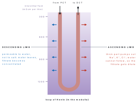
Water and the counter-current multiplier. Water is not pumped; it follows solute by osmosis. The loop of Henle makes that possible by creating a gradient of osmolarity in the medulla, from 300 mOsm L⁻¹ at the corticomedullary junction to 1200 mOsm L⁻¹ (up to 1400) at the papilla. The mechanism has four steps:
- The filtrate entering the descending limb is isotonic (300).
- The descending limb is permeable to water but not to solute. As it runs down into the salty medulla, water leaves by osmosis and the filtrate becomes steadily more concentrated, reaching about 1200 at the tip.
- The ascending limb is impermeable to water but actively pumps Na⁺ and Cl⁻ into the interstitium. Solute leaves, water cannot, so the filtrate becomes dilute — about 100 at the top.
- The NaCl left behind in the medulla keeps the interstitium hypertonic, and the vasa recta carry away the water without washing the salt out. Because the two limbs carry fluid in opposite directions and have opposite permeabilities, the gradient is multiplied along the length of the loop.
| Level | Interstitial osmolarity | Filtrate in the loop |
|---|---|---|
| Cortex / PCT | 300 | 300 (isotonic) |
| Outer medulla | 400–600 | descending 400–600 |
| Inner medulla | 900 | descending 900 |
| Papilla (tip) | 1200 | 1200 at the bend |
| Top of ascending limb | 300 | 100 (dilute) |
The length of the loop sets the steepness of the gradient. Desert mammals such as the kangaroo rat have very long loops and make urine of 5000 mOsm L⁻¹; birds have short loops and concentrate weakly; fish have no loop at all. The same logic explains why a long loop means a dry habitat.
The DCT and collecting duct. The DCT receives hypotonic fluid (about 100 mOsm L⁻¹). Here Na⁺ is reabsorbed under aldosterone and Ca²⁺ under parathyroid hormone. The collecting duct is where the final decision is made: if ADH is present, its cells insert aquaporin-2 channels and water leaves into the hypertonic medulla, so the urine becomes concentrated; if ADH is absent, the duct stays almost impermeable and a large volume of dilute urine is passed.
The descending limb is permeable to water and not to solute; the ascending limb is permeable to neither water nor (freely) solute, but pumps salt. Any option that says "the ascending limb reabsorbs water" or "the descending limb pumps sodium" is wrong. If both limbs behaved alike, the gradient would vanish and no mammal could concentrate urine.
Step 3 — tubular secretion
Secretion moves substances from the blood of the peritubular capillaries into the tubule, the opposite direction to reabsorption. It is the tubule's way of getting rid of things that filtration alone cannot handle.
| Substance | Where secreted | Why it matters |
|---|---|---|
| H⁺ | PCT and intercalated cells of the collecting duct | regulates blood pH; can acidify urine to pH 4.5 |
| K⁺ | principal cells of the DCT and collecting duct | the main route for potassium loss; increased by aldosterone |
| NH₃ | PCT | combines with H⁺ to form NH₄⁺, trapping acid in the urine |
| Creatinine | PCT | a waste that is also filtered |
| Drugs | PCT | penicillin, aspirin, morphine, diuretics — many are protein-bound and never filtered |
| Organic acids and bases | PCT | e.g. para-aminohippuric acid (PAH), used to measure renal blood flow |
Secretion matters for two reasons. First, some wastes (K⁺, H⁺) are almost entirely removed this way. Second, drugs bound to plasma proteins are too large to filter, so secretion is their only exit.
Hormonal regulation
Three hormones set the final volume and concentration of urine.
ADH (vasopressin). Made in the supraoptic and paraventricular nuclei of the hypothalamus, carried down axons and stored in the posterior pituitary, from which it is released. Osmoreceptors in the hypothalamus detect a rise in plasma osmolarity (as little as 2 %), and ADH is released. It binds V₂ receptors on the collecting duct and inserts aquaporin-2 water channels, so water is reabsorbed and the urine becomes small and concentrated. A fall in osmolarity switches ADH off and a large dilute urine is produced.
Diabetes insipidus. A deficiency of ADH (or a failure of the kidney to respond to it) gives polyuria — up to 15–20 L a day of pale urine with a specific gravity of 1.001–1.005 — and intense polydipsia. The urine is dilute because the water is not reabsorbed, not because glucose is pulling water into the tubule.
Aldosterone. A steroid from the zona glomerulosa of the adrenal cortex. It increases Na⁺ reabsorption and K⁺ secretion in the DCT and collecting duct; water follows the sodium. The result is a small volume of urine with little sodium and more potassium, and a rise in blood pressure.
The renin–angiotensin–aldosterone system (RAAS). When blood pressure, blood volume or Na⁺ falls (or K⁺ rises), the juxtaglomerular cells release renin. Renin converts angiotensinogen (from the liver) to angiotensin I; angiotensin-converting enzyme (ACE), mostly in the lungs, converts it to angiotensin II. Angiotensin II is a strong vasoconstrictor and also stimulates aldosterone release, ADH release and thirst. The net effect is to retain sodium and water and raise blood pressure.
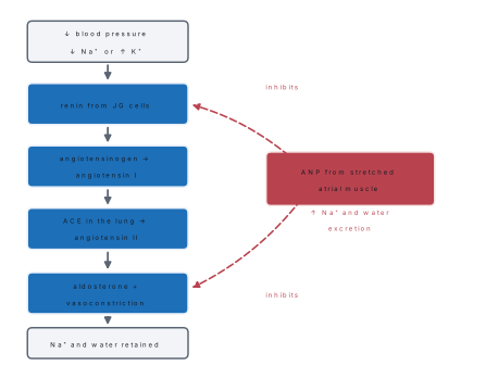
ANP (atrial natriuretic peptide). Stretch of the atria — as when blood volume rises — releases this 28-amino-acid peptide from atrial muscle cells. ANP inhibits Na⁺ reabsorption in the collecting duct, inhibits renin and aldosterone, dilates the afferent arteriole and constricts the efferent one, so GFR rises. The result is a large volume of sodium-rich, dilute urine and a fall in blood pressure. It is the physiological opponent of the RAAS.
| Hormone | Source | Main action | Urine volume | Urine concentration |
|---|---|---|---|---|
| ADH | hypothalamus → posterior pituitary | ↑ water permeability of DCT/CD (aquaporin-2) | ↓ | ↑ (up to 1200 mOsm L⁻¹) |
| Aldosterone | adrenal cortex, zona glomerulosa | ↑ Na⁺ reabsorption, ↑ K⁺ secretion | ↓ | ↑ (water follows Na⁺) |
| ANP | atrial muscle | ↓ Na⁺ reabsorption, ↓ renin, ↑ GFR | ↑ | ↓ (dilute) |
| PTH | parathyroid | ↑ Ca²⁺ reabsorption, ↓ phosphate reabsorption | little change | — |
ADH saves water, aldosterone saves salt (and wastes potassium), ANP throws both away. For each, ask two questions: does urine volume go up or down? and does the urine get more or less concentrated?
Composition of urine
| Property | Normal value |
|---|---|
| Volume | 1–2 L per day (1.5 L average) |
| Colour | pale yellow (urochrome) |
| pH | 4.6–8.0, average about 6 |
| Specific gravity | 1.003–1.035 (average 1.020) |
| Osmolarity | 300–1200 mOsm L⁻¹ |
| Water | about 95 % |
| Urea | 25–35 g per day — the most abundant solute |
| Uric acid | 0.5–1 g per day |
| Creatinine | 1–1.5 g per day |
| Others | Na⁺, K⁺, Cl⁻, sulphate, phosphate, ammonia, urobilinogen, hormones, vitamins |
Abnormal constituents are the quickest route to a diagnosis, and the paper likes them as one-line matches.
| Constituent | What it signals |
|---|---|
| Glucose | diabetes mellitus; also renal glycosuria (low threshold) and pregnancy |
| Protein (albumin) | glomerulonephritis, nephrotic syndrome, pre-eclampsia; transient after heavy exercise or fever |
| Ketones | starvation, uncontrolled type 1 diabetes (ketoacidosis), persistent vomiting |
| Blood / red cells | glomerulonephritis, stones, trauma, tumour, urinary infection |
| Casts (hyaline, granular, red-cell) | renal tubular damage; red-cell casts point to glomerulonephritis |
| Pus cells and nitrite | urinary tract infection (cystitis, pyelonephritis) |
| Bilirubin | obstructive jaundice, hepatitis |
| Crystals | oxalate or urate stones, gout |
| hCG | pregnancy (basis of the pregnancy test) |
Glycosuria means glucose has exceeded the renal threshold. It happens in diabetes mellitus because the plasma level is high, but also in renal glycosuria, where the carriers themselves are faulty and the threshold is low even though blood glucose is normal. The stem usually gives the blood glucose to separate the two.
8.3 Renal disorders
Nephritis
Nephritis is inflammation of the kidney. The commonest form is glomerulonephritis, an immune-complex (type III) reaction: antibodies made against a β-haemolytic streptococcus (after a throat or skin infection) cross-react with the glomerular basement membrane, or antigen–antibody complexes lodge there. It usually appears 1–3 weeks after the infection.
Features: haematuria (smoky or cola-coloured urine), proteinuria, oedema (especially around the eyes in the morning), hypertension and oliguria (less urine). Most children recover; some progress to chronic renal failure. Nephrotic syndrome is a related picture with heavy protein loss, low blood albumin and generalised oedema.
Kidney stones (renal calculi)
Stones form when salts in concentrated urine crystallise. About 75–80 % are calcium oxalate; others are uric acid, cystine and struvite (the last is associated with infection). Risk factors are a low fluid intake, a hot climate, hypercalcaemia, a diet high in oxalate (spinach, rhubarb, nuts, tea), gout and repeated urinary infection.
A stone causes renal colic — severe pain from the loin to the groin as it moves down the ureter — with haematuria and sometimes obstruction and infection. Small stones pass with fluids and analgesia; larger ones are broken by extracorporeal shock-wave lithotripsy (ESWL) or removed through a ureteroscope.
Renal failure
Renal failure is a fall in GFR severe enough to retain waste.
| Acute renal failure | Chronic renal failure | |
|---|---|---|
| Onset | hours to days | months to years |
| Common causes | shock, severe dehydration, blood loss, nephrotoxic drugs, obstruction | diabetes mellitus and hypertension (the two commonest), glomerulonephritis, polycystic kidney |
| Reversible? | often, if the cause is corrected | no; progressive |
| Features | oliguria or anuria, rising urea and creatinine, high K⁺ | uraemia, anaemia (less erythropoietin), acidosis, oedema, bone disease |
When the kidneys fail, urea and creatinine rise (uraemia), K⁺ rises (hyperkalaemia, dangerous for the heart), H⁺ accumulates (acidosis) and erythropoietin falls (anaemia). Treatment is by dialysis or transplantation.
Dialysis
Dialysis filters the blood through a semipermeable membrane against a dialysate whose concentration is set so that wastes diffuse out while useful substances stay.
| Haemodialysis | Peritoneal dialysis | |
|---|---|---|
| Membrane | cellulose or synthetic membrane in a dialyser | the peritoneum |
| Blood flow | pumped from an arteriovenous fistula | the patient's own capillaries |
| Schedule | 3–4 hours, 2–3 times a week, in hospital | 4–6 hour dwells, about 4 exchanges a day, at home |
| Efficiency | high | lower |
| Main risk | clotting (heparin used), hypotension, infection of the access | peritonitis |
In haemodialysis the blood and the dialysate flow in opposite directions (counter-current), which keeps a concentration gradient along the whole membrane so that diffusion does not stop halfway.
Kidney transplantation
A healthy kidney from a living related donor or a cadaver is placed in the iliac fossa and joined to the iliac vessels and the bladder. The donor and recipient are matched for ABO and HLA antigens. The recipient takes immunosuppressant drugs (ciclosporin, tacrolimus, steroids) for life to prevent rejection. A working transplant gives a better quality of life and longer survival than dialysis, but the supply of organs is far short of the need.
125 mL min⁻¹ (GFR) → 180 L day⁻¹ (filtrate) → 1.5 L day⁻¹ (urine): 99 % reabsorbed. 180 mg/dL is the renal threshold for glucose, 1200 mOsm L⁻¹ the maximum urine concentration. These three facts answer a large share of the numerical questions.
Formula and fact sheet
Organs and structure
| Fact | Value |
|---|---|
| Kidneys | a pair, T12–L3, right slightly lower; 10–12 × 5–7 × 3 cm; 130–150 g each |
| Coverings | fibrous capsule, perirenal fat, renal fascia |
| Regions | cortex, medulla (8–18 pyramids), renal columns, pelvis, calyces |
| Nephron count | about 1 million per kidney |
| Cortical / juxtamedullary nephrons | 85 % / 15 % (long loops) |
| Ureter | 25–30 cm, peristaltic |
| Bladder | transitional epithelium, detrusor muscle, 300–400 mL |
| Urethra | female 4 cm, male 20 cm |
| Blood flow | 1.2 L min⁻¹, about 25 % of cardiac output |
| Efferent vs afferent arteriole | efferent narrower → high glomerular pressure |
Filtration
| Fact | Value |
|---|---|
| Filtration barrier | fenestrated endothelium (70–100 nm), basement membrane (negative), slit diaphragms (4–7 nm) |
| Filtered | water, glucose, amino acids, urea, uric acid, creatinine, ions |
| Not filtered | cells, plasma proteins (albumin 69 kDa) |
| Pressures | 55 − (30 + 15) = 10 mmHg net |
| GFR | 125 mL min⁻¹ = 180 L day⁻¹ |
| Filtration fraction | 125/625 = 20 % |
| Reabsorbed | about 99 % of water, 99.5 % of Na⁺ |
Reabsorption and secretion
| Fact | Value |
|---|---|
| PCT | 65–70 % of reabsorption; brush border; glucose and amino acids 100 % |
| Glucose transport | secondary active (Na⁺ cotransport, SGLT) |
| Renal threshold for glucose | 180 mg/dL |
| Transport maximum (Tm) | about 375 mg min⁻¹ |
| Descending limb | permeable to water, not solute |
| Ascending limb | impermeable to water; pumps Na⁺/Cl⁻ |
| Gradient | 300 → 1200 mOsm L⁻¹ (max 1400) |
| Filtrate leaving the loop | about 100 mOsm L⁻¹ (dilute) |
| Secreted | H⁺, K⁺, NH₃, creatinine, drugs (penicillin, aspirin), PAH |
Hormones
| Hormone | Source | Effect on urine |
|---|---|---|
| ADH (vasopressin) | hypothalamus → posterior pituitary | ↑ water reabsorption → small, concentrated |
| Aldosterone | adrenal cortex, zona glomerulosa | ↑ Na⁺ reabsorption, ↑ K⁺ secretion → small volume |
| ANP | atrial muscle | ↑ Na⁺ and water excretion → large, dilute |
| PTH | parathyroid | ↑ Ca²⁺ reabsorption, ↑ phosphate excretion |
| Renin | JG cells | starts RAAS; converts angiotensinogen → angiotensin I |
| ACE | lung endothelium | angiotensin I → angiotensin II |
Urine
| Fact | Value |
|---|---|
| Volume | 1–2 L day⁻¹ |
| pH | 4.6–8.0 (average 6) |
| Specific gravity | 1.003–1.035 |
| Water | 95 % |
| Most abundant solute | urea (25–35 g day⁻¹) |
| Diabetes insipidus | 15–20 L day⁻¹, SG 1.001–1.005, dilute |
| Diabetes mellitus | polyuria with glucose in urine, high SG |
Disorders
| Disorder | Key fact |
|---|---|
| Glomerulonephritis | immune complex after streptococcal infection; haematuria, proteinuria, oedema |
| Stones | calcium oxalate commonest; renal colic; lithotripsy |
| Acute failure | reversible; shock, dehydration, toxins |
| Chronic failure | diabetes and hypertension commonest; uraemia, anaemia, acidosis |
| Haemodialysis | dialyser, counter-current, 3–4 h, 2–3 times a week |
| Peritoneal dialysis | peritoneum as membrane, done at home, risk of peritonitis |
| Transplant | HLA matching, immunosuppression for life |
Chapter summary
- The excretory organs are the kidneys, ureters, bladder and urethra; the skin, lungs and liver are accessory routes, and the liver makes urea while the kidney excretes it.
- Each kidney has about one million nephrons, in a cortex (corpuscles, convoluted tubules) and a medulla (pyramids, loops, collecting ducts) that drains through the pelvis into the ureter.
- Ultrafiltration at the glomerulus passes water and small solutes but holds back cells and protein by pore size and negative charge; net pressure ≈ 10 mmHg, GFR ≈ 125 mL min⁻¹ (180 L day⁻¹).
- Reabsorption recovers about 99 % of the filtrate: glucose and amino acids by active transport in the PCT (threshold 180 mg/dL, Tm 375 mg min⁻¹), water by osmosis.
- The counter-current multiplier works because the descending limb is permeable to water and the ascending limb is not but pumps salt; the medulla is graded 300 → 1200 mOsm L⁻¹, and the vasa recta preserve the gradient.
- Secretion adds H⁺, K⁺, NH₃, creatinine and drugs to the tubule and is the main route for acid removal.
- ADH (made in the hypothalamus, stored in the posterior pituitary) concentrates urine; aldosterone retains Na⁺ and loses K⁺; ANP does the opposite. Diabetes insipidus = ADH problem = large dilute urine; diabetes mellitus = insulin problem = large glucose-containing urine.
- Abnormal constituents: glucose (diabetes), protein (nephritis), ketones (starvation, ketoacidosis), blood (nephritis, stones), casts (tubular damage).
- Disorders: nephritis, calcium oxalate stones, acute and chronic failure; haemodialysis uses a dialyser, peritoneal dialysis uses the peritoneum, and transplantation needs HLA matching and lifelong immunosuppression.
Practice questions
Set A — Recall (24 questions)
- The structural and functional unit of the kidney is theaneuronbrenal corpusclecnephrondmalpighian tubule
- Each human kidney contains abouta10 000 nephronsb1 million nephronsc100 million nephronsd1000 nephrons
- The glomerulus is enclosed byathe loop of Henlebthe collecting ductcthe renal pelvisdBowman's capsule
- Which part of the nephron is freely permeable to water but not to solutes?adescending limb of the loop of Henlebascending limb of the loop of Henlecdistal convoluted tubuledcollecting duct
- The greatest part of the reabsorption of the filtrate takes place in theaBowman's capsulebloop of Henlecproximal convoluted tubuledcollecting duct
- Glucose is reabsorbed from the filtrate byasimple diffusionbactive transport in the PCTcosmosis in the loopdfiltration
- Glucose first appears in the urine when its plasma concentration exceeds abouta100 mg/dLb80 mg/dLc300 mg/dLd180 mg/dL
- Antidiuretic hormone is released from theaposterior pituitarybanterior pituitarycadrenal cortexdthyroid
- ADH makes the collecting duct more permeable toaglucosebsodiumcwaterdproteins
- A person who passes 15–20 litres of dilute urine a day most probably lacksainsulinbADHcaldosteronedthyroxine
- Aldosterone is secreted by theaadrenal medullabposterior pituitarycjuxtaglomerular cellsdadrenal cortex
- Renin is secreted by theajuxtaglomerular cellsbmacula densacliverdadrenal cortex
- Angiotensin I is converted to angiotensin II mainly in theakidneybliverclungsdheart
- Atrial natriuretic peptide is secreted byathe adrenal medullabstretched atrial muscle cellscthe juxtaglomerular cellsdthe posterior pituitary
- The normal glomerular filtration rate in an adult is abouta125 mL/minb1250 mL/minc25 mL/mind125 mL/hour
- The normal daily volume of urine in an adult is abouta15 Lb0.1 Lc5 Ld1–2 L
- The most abundant nitrogenous waste in urine isauric acidbcreatininecureadammonia
- The specific gravity of normal urine lies betweena1.000 and 1.001b1.003 and 1.035c1.040 and 1.060d0.900 and 0.950
- Which of the following is NOT an excretory organ?askinblungscliverdspleen
- The urinary bladder is lined byatransitional epitheliumbsimple squamous epitheliumcstratified keratinised epitheliumdciliated columnar epithelium
- Which substance is almost completely reabsorbed from the filtrate?aureabcreatininecglucoseduric acid
- The vasa recta arealymph vessels of the cortexbbranches of the renal vein in the cortexcthe afferent arteriolesdcapillary loops running beside the loop of Henle
- The commonest type of kidney stone is made ofauric acidbcalcium oxalateccystinedstruvite
- In peritoneal dialysis the membrane used is theaperitoneumbglomerular membraneccellulose tubing of a dialyserdbladder wall
Answers and explanations — Set A
1. (c) — the nephron is the functional unit; the renal corpuscle (b) is only its first part, the glomerulus plus Bowman's capsule.
2. (b) — about one million per kidney, giving roughly two million for the pair.
3. (d) — Bowman's capsule is the cup-shaped beginning of the tubule; the glomerulus is the capillary tuft inside it.
4. (a) — the descending limb has aquaporins but no solute carriers, so only water leaves. The ascending limb (b) is the opposite.
5. (c) — the PCT recovers 65–70 % of the filtrate because of its brush border and dense mitochondria.
6. (b) — glucose is moved by secondary active transport with Na⁺ (SGLT) in the PCT; it is never reabsorbed by diffusion alone.
7. (d) — the renal threshold is about 180 mg/dL; above it the carriers are saturated and glucose spills into the urine.
8. (a) — ADH is made in the hypothalamus but released from the posterior pituitary, which stores it.
9. (c) — ADH inserts aquaporin-2 channels, so water is reabsorbed and the urine becomes concentrated.
10. (b) — 15–20 L of dilute urine is the classic picture of diabetes insipidus, an ADH deficiency.
11. (d) — aldosterone comes from the zona glomerulosa of the adrenal cortex; the medulla (a) makes adrenaline.
12. (a) — the juxtaglomerular cells of the afferent arteriole store and release renin.
13. (c) — angiotensin-converting enzyme sits on the endothelium of the lung capillaries.
14. (b) — ANP is released by atrial muscle cells when they are stretched by a rise in blood volume.
15. (a) — 125 mL/min, which is 180 L a day. Option (b) is ten times too high and (d) mixes minutes with hours.
16. (d) — 1–2 L a day, about 1 % of the filtrate; 15 L (a) suggests diabetes insipidus.
17. (c) — urea, 25–35 g a day, is the most abundant solute; uric acid and creatinine are much smaller in amount.
18. (b) — 1.003–1.035; a fixed low value suggests diabetes insipidus, a fixed high one diabetes mellitus.
19. (d) — the spleen destroys old red cells but does not excrete waste; skin, lungs and liver all do.
20. (a) — transitional epithelium stretches as the bladder fills and is impermeable to urine.
21. (c) — glucose is completely reabsorbed below the threshold; creatinine (b) is not reabsorbed at all.
22. (d) — the vasa recta are the long capillary loops of the juxtamedullary nephrons that run beside the loop of Henle.
23. (b) — calcium oxalate accounts for about 75–80 % of stones.
24. (a) — the peritoneum is the dialysing membrane in peritoneal dialysis; the dialyser membrane (c) is used in haemodialysis.
Set B — Understanding (14 questions)
- Plasma proteins are not filtered at the glomerulus mainly because ofatheir small sizebtheir rapid reabsorptionctheir size and negative chargedtheir absence from plasma
- The efferent arteriole is narrower than the afferent arteriole, whichakeeps glomerular pressure highblowers glomerular pressurecstops filtrationdincreases glucose reabsorption
- If the ascending limb of the loop of Henle became permeable to water, the medullary gradient wouldadoublebstay the samecriseddisappear
- ADH reduces urine volume because itaincreases the GFRbincreases water reabsorption in the collecting ductcincreases sodium excretiondconstricts the ureter
- Diabetes insipidus and diabetes mellitus both cause polyuria, but in diabetes insipidus the urine isarich in glucosebrich in proteinclarge in volume and dilutedsmall in volume and concentrated
- Aldosterone raises blood pressure mainly byaincreasing Na⁺ and water reabsorptionbincreasing glucose reabsorptioncdilating the afferent arterioledinhibiting renin
- ANP lowers blood pressure byaincreasing renin releasebpromoting sodium and water excretioncconstricting the afferent arterioledincreasing ADH release
- Tubular secretion of H⁺ is important because itafilters proteinbreabsorbs glucosecconcentrates urinedhelps regulate blood pH
- The counter-current mechanism works because the two limbs of the loopahave opposite permeabilities and opposite flowsbcarry fluid in the same directioncboth absorb waterdboth secrete glucose
- Glucose appears in the urine in uncontrolled diabetes mellitus becauseathe PCT stops workingbthe GFR risescthe filtered load exceeds the transport maximumdADH falls
- A patient with glomerulonephritis usually showsaglucose and ketones in urinebprotein and red cells in urinecbile and urobilinogendno abnormality at all
- Compared with haemodialysis, peritoneal dialysisais fasterbneeds a dialysercneeds heparinduses the peritoneum and can be done at home
- The renal cortex containsaglomeruli and convoluted tubulesbonly collecting ductscthe renal pelvisdthe renal pyramids
- For urine to be concentrated in the collecting duct, the medullary interstitium must beahypotonicbisotonicchypertonicdfree of sodium
Answers and explanations — Set B
1. (c) — the basement membrane is negatively charged and the slits are narrow, so albumin is repelled as well as blocked. Size alone (a) is the tempting half-answer.
2. (a) — a narrow outlet holds blood back, so glomerular pressure stays around 55 mmHg and filtration continues.
3. (d) — water would follow the salt out of the ascending limb, the salt would be diluted, and no gradient could be maintained.
4. (b) — ADH inserts water channels in the collecting duct; it does not change GFR or sodium handling.
5. (c) — no ADH means no water reabsorption, so the urine is dilute; glucose is the marker of diabetes mellitus (a).
6. (a) — sodium is retained and water follows, expanding blood volume; aldosterone does not affect glucose.
7. (b) — ANP is a natriuretic hormone: it increases sodium and water loss and inhibits renin and aldosterone.
8. (d) — secreting H⁺ removes acid from the blood; the urine can reach pH 4.5.
9. (a) — water leaves the descending limb and salt leaves the ascending limb, while the flows run in opposite directions.
10. (c) — above 180 mg/dL the filtered load passes the Tm of about 375 mg/min, so the excess is excreted.
11. (b) — glomerular damage lets protein and red cells through; glucose (a) points to diabetes.
12. (d) — the peritoneum is the membrane, so no dialyser or heparin is needed and the patient can treat himself at home.
13. (a) — the cortex holds the renal corpuscles and the convoluted tubules; the pyramids (d) are medullary.
14. (c) — water can leave the duct only if the surrounding medulla is saltier than the filtrate.
Set C — Application (10 questions)
- A person has a GFR of 125 mL/min. The volume of filtrate formed in 24 hours is abouta18 Lb180 Lc1.8 Ld1800 L
- Of the 180 L of filtrate formed daily, about 1.5 L is voided. The percentage reabsorbed is abouta50 %b75 %c90 %d99 %
- Plasma glucose is 300 mg/dL and GFR is 125 mL/min. The filtered load of glucose per minute isa375 mgb37.5 mgc300 mgd2400 mg
- Plasma glucose is 400 mg/dL, GFR 125 mL/min and the Tm for glucose 375 mg/min. Glucose excreted per minute is abouta0 mgb500 mgc125 mgd375 mg
- Urine creatinine is 100 mg/dL, urine flow 1.5 L/day and plasma creatinine 1.0 mg/dL. Creatinine clearance is abouta15 mL/minb104 mL/minc150 mL/mind1.5 mL/min
- A patient passes 18 L of urine a day with an osmolarity of 60 mOsm/L and no glucose. The likely diagnosis isadiabetes insipidusbdiabetes mellituscglomerulonephritisda kidney stone
- In a water-deprivation test the urine stays dilute even after ADH is injected. This shows thatathe pituitary makes too much ADHbthe patient has diabetes mellituscthe kidneys are normaldthe kidney does not respond to ADH
- A man on a very low salt diet has high renin and high aldosterone. His urine is likely to bearich in sodium and largebrich in glucoseclow in sodium and smallddilute and large
- In metabolic acidosis the urine becomes more acidic because the tubules secrete moreaK⁺bH⁺cglucosedprotein
- In haemodialysis the blood and the dialysate flow in opposite directions so thataa concentration gradient is kept along the whole membranebthe blood is warmedcglucose can be addeddclotting is prevented
Answers and explanations — Set C
1. (b) — 125 \times 60 \times 24 = 180\,000 mL = 180 L. Option (a) forgets the 60.
2. (d) — (180 - 1.5)/180 = 0.992, about 99 %.
3. (a) — 300 mg/dL = 3 mg/mL; 3 \times 125 = 375 mg/min. This equals the Tm, so glucose just begins to spill.
4. (c) — load = 4 \times 125 = 500 mg/min; excreted = 500 - 375 = 125 mg/min. Option (b) is the load, not the loss.
5. (b) — clearance = (100 \times 1500)/1.0 = 150\,000 mL/day = 104 mL/min, close to the normal GFR.
6. (a) — a large volume of very dilute urine without glucose is diabetes insipidus; in diabetes mellitus the urine contains glucose and is concentrated.
7. (d) — nephrogenic diabetes insipidus: the hormone is present but the collecting duct does not respond, so the urine stays dilute.
8. (c) — low salt raises aldosterone, which reclaims sodium; water follows, so the volume is small.
9. (b) — the tubule secretes H⁺ to remove acid; potassium secretion (a) is increased in alkalosis, not acidosis.
10. (a) — counter-current flow keeps the concentration difference across the membrane all the way along, so diffusion continues.