Chapter 9 · Human Biology and Physiology · 2 questions in the paper · 31 min read
Nervous System and Sense Organs
What the syllabus asks for 3
- Central, peripheral and autonomic nervous systems
- The nerve impulse and the synapse
- The eye and the ear
The nervous system and sense organs are rated at 2 questions in this book's estimate of the 40-question Zoology paper. The examinable pattern is mostly “which structure does this job?”: medulla for vital centres, cerebellum for coordination, hypothalamus for homeostasis, nodes of Ranvier for saltatory conduction, rods and cones for vision, and the organ of Corti for hearing. The second pattern is directional logic — sensory fibres enter by the dorsal root, motor fibres leave by the ventral root, and chemical synapses transmit in one direction only.
- CNS = brain + spinal cord. PNS = 12 pairs of cranial nerves + 31 pairs of spinal nerves.
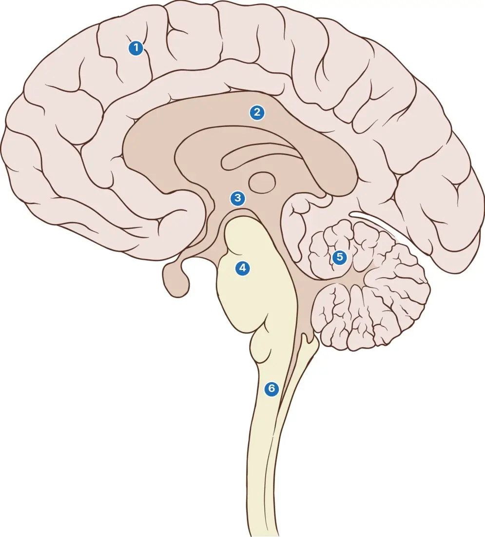
- Cerebrum: conscious thought, memory and voluntary movement; cerebellum: balance and coordination; hypothalamus: temperature, hunger, thirst and pituitary control; medulla: breathing, heart and blood-pressure centres.
- Sympathetic outflow is thoracolumbar, usually adrenergic and “fight or flight”; parasympathetic outflow is craniosacral, cholinergic and “rest and digest”.
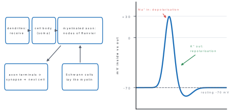
- Resting potential is about −70 mV; threshold about −55 mV; depolarisation is Na⁺ entry, repolarisation is K⁺ exit; the Na⁺/K⁺ pump moves 3 Na⁺ out for 2 K⁺ in.
- Myelin and nodes of Ranvier give saltatory conduction — faster and cheaper in ATP. At a chemical synapse, Ca²⁺ entry releases transmitter vesicles.
- Eye: rods for dim light, cones for colour and detail, fovea for sharpest vision, blind spot where the optic nerve leaves.
- Ear: malleus → incus → stapes → oval window → cochlea; semicircular canals detect rotation, utricle and saccule detect gravity and linear acceleration.
9.1 Central, peripheral and autonomic nervous systems
The nervous system is the body's rapid communication network. Its signals travel as electrical impulses along neurons and act within milliseconds. Unlike hormones, whose effects may persist for minutes or hours, a nervous signal is brief, precisely addressed and quickly switched off.
The neuron: the signalling cell
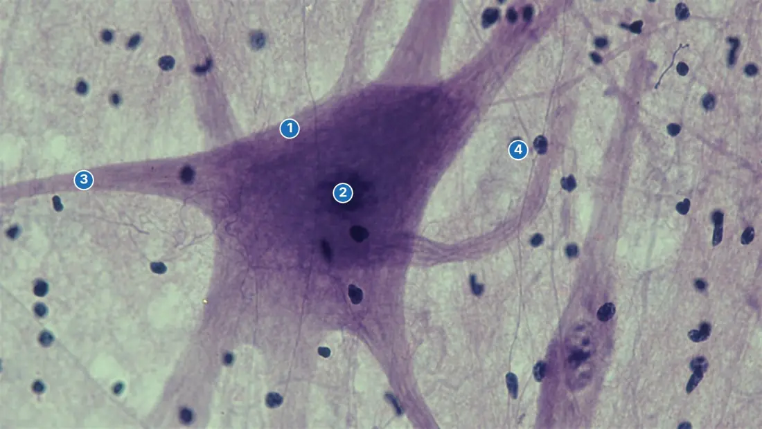
A neuron is the structural and functional unit of the nervous system. Its cell body (cyton/perikaryon) contains the nucleus and Nissl substance (rough endoplasmic reticulum) for protein synthesis. Short branched dendrites receive signals and conduct them toward the cell body. A single long axon conducts the action potential away from the cell body to axon terminals or synaptic knobs. Most mature neurons have lost the capacity for ordinary cell division, so their metabolic cell body must maintain processes that may be more than a metre long.
Neurons are classified functionally as:
- Sensory (afferent) neurons — receptors to CNS.
- Motor (efferent) neurons — CNS to muscles or glands.
- Interneurons/relay neurons — connect neurons within the CNS and are responsible for integration.
Supporting neuroglial cells protect and maintain neurons. In the PNS, Schwann cells form myelin; in the CNS, oligodendrocytes do so. Astrocytes help maintain the blood–brain barrier and extracellular ion balance, while microglia act as immune/phagocytic cells.
Organisation at a glance
| Division | Main parts | Basic role |
|---|---|---|
| Central nervous system (CNS) | brain and spinal cord | receives, integrates and stores information; issues commands |
| Peripheral nervous system (PNS) | cranial and spinal nerves | carries impulses between CNS, receptors and effectors |
| Somatic nervous system | sensory input and motor output to skeletal muscle | voluntary movement and conscious sensation |
| Autonomic nervous system (ANS) | sympathetic and parasympathetic divisions | involuntary control of viscera, glands, cardiac and smooth muscle |
| Enteric nervous system | nerve plexuses in gut wall | largely local control of digestion, modulated by ANS |
Brain: protection and major centres
The human brain weighs about 1.3–1.4 kg and lies in the cranium. It is surrounded by three meninges: tough outer dura mater, middle arachnoid mater, and delicate inner pia mater. Cerebrospinal fluid (CSF), formed mainly by the choroid plexuses in the ventricles, cushions the CNS, reduces its effective weight and helps maintain a stable chemical environment.
| Brain region | High-yield functions |
|---|---|
| Cerebrum/cerebral cortex | intelligence, learning, memory, language, conscious sensation and voluntary movement |
| Frontal lobe | motor cortex, planning, personality; Broca's area for speech production |
| Parietal lobe | touch, pressure, pain, temperature and body position |
| Temporal lobe | hearing; Wernicke's area for language comprehension |
| Occipital lobe | visual processing |
| Corpus callosum | large commissural tract joining the two cerebral hemispheres |
| Thalamus | major relay and sorting station for sensory pathways to cortex |
| Hypothalamus | body temperature, hunger, thirst, sleep–wake rhythm, autonomic control and regulation of pituitary secretion |
| Midbrain | visual/auditory reflexes, eye movements and relay pathways |
| Pons | relay between cerebrum and cerebellum; helps regulate breathing |
| Cerebellum | posture, balance, muscle tone and fine coordination of voluntary movement |
| Medulla oblongata | cardiac, respiratory, vasomotor, swallowing, coughing, sneezing and vomiting centres |
The cerebrum does not itself “feel” every sensation in a simple point-to-point way; perception is constructed by networks. Nevertheless, for school and CEE purposes the lobe associations above are the tested first approximation. Damage to the medulla is immediately life-threatening because breathing and cardiovascular reflexes fail; cerebellar damage characteristically produces an unsteady gait, poor coordination and intention tremor rather than paralysis or loss of consciousness.
Spinal cord and reflexes
The spinal cord extends from the medulla through the vertebral canal to about the level of the first or second lumbar vertebra in adults. In cross-section, grey matter is central and H-shaped, containing neuron cell bodies, dendrites and synapses; white matter lies outside and consists mainly of myelinated ascending and descending tracts. This is the reverse of the cerebrum, where grey matter forms the outer cortex.
Each spinal nerve is mixed. Its dorsal root carries sensory fibres into the cord and bears the dorsal-root ganglion containing sensory cell bodies. Its ventral root carries motor fibres out of the cord. A useful memory is SAME DAVE: Sensory Afferent, Motor Efferent; Dorsal Afferent, Ventral Efferent.
A reflex arc is the shortest functional pathway of an involuntary reflex:
receptor → sensory neuron → CNS integration → motor neuron → effector
In withdrawal from a hot object, nociceptors excite sensory neurons; spinal interneurons activate flexor motor neurons and inhibit antagonistic extensors; the hand moves before ascending signals produce conscious pain. The knee-jerk is a simpler monosynaptic stretch reflex: muscle-spindle sensory neurons synapse directly on quadriceps motor neurons. Reflexes are rapid, stereotyped and protective, but higher centres can modify many of them.
Cranial and spinal nerves
Humans have 12 pairs of cranial nerves and 31 pairs of spinal nerves (8 cervical, 12 thoracic, 5 lumbar, 5 sacral and 1 coccygeal). Especially testable cranial nerves are:
| Nerve | Main school-level association |
|---|---|
| I Olfactory | smell |
| II Optic | vision |
| III Oculomotor | most eye movements; parasympathetic pupil constriction and accommodation |
| V Trigeminal | facial sensation and muscles of mastication; largest cranial nerve |
| VII Facial | facial expression, taste from anterior tongue, salivary/lacrimal glands |
| VIII Vestibulocochlear | hearing and balance |
| X Vagus | extensive parasympathetic supply to thoracic and abdominal viscera |
| XII Hypoglossal | tongue movements |
Autonomic nervous system
Autonomic pathways usually contain two neurons in series: a preganglionic neuron from CNS to an autonomic ganglion and a postganglionic neuron from ganglion to effector. Both divisions often innervate the same organ and act in a balanced, frequently antagonistic manner.
| Feature | Sympathetic | Parasympathetic |
|---|---|---|
| CNS origin | thoracic and upper lumbar cord (thoracolumbar) | brainstem cranial nerves III, VII, IX, X and sacral cord (craniosacral) |
| Preganglionic fibres | usually short | usually long |
| Postganglionic fibres | usually long | usually short; ganglia near or in target organ |
| Main postganglionic transmitter | noradrenaline (norepinephrine), with notable cholinergic exceptions such as most sweat glands | acetylcholine |
| General state | emergency, exercise, “fight or flight” | recovery, digestion, “rest and digest” |
| Heart | rate and force increase | rate decreases, mainly through vagus |
| Pupil | dilates | constricts |
| Bronchial smooth muscle | relaxes; airways widen | contracts; airways narrow |
| Gut motility/secretion | decreases | increases |
| Urinary bladder | detrusor relaxes; storage favoured | detrusor contracts; emptying favoured |
The adrenal medulla is functionally a modified sympathetic ganglion: its chromaffin cells release adrenaline and noradrenaline into blood, prolonging and generalising sympathetic effects. Most systemic blood vessels receive important sympathetic vasoconstrictor tone but little or no parasympathetic innervation, so “every organ receives equal and opposite supply” is too simple.
The vagus supplies much of the thorax and abdomen, but the eye, glands of the head and pelvic viscera depend on other cranial or sacral parasympathetic pathways. Likewise, acetylcholine is not exclusively parasympathetic: it is the transmitter at autonomic ganglia, at the neuromuscular junction and in many CNS synapses.
Cerebrum thinks, thalamus relays, hypothalamus keeps the house (heat, hunger, thirst, pituitary), cerebellum balances, medulla runs the vital reflexes (heart, breathing, swallowing). Five rooms, one line each.
9.2 The nerve impulse and the synapse
Resting potential and action potential
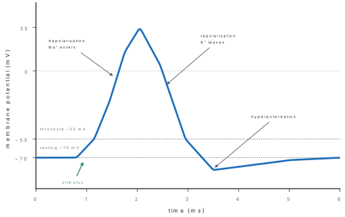
An unstimulated axon maintains a resting membrane potential of about −70 mV inside relative to outside. The sodium–potassium ATPase pumps 3 Na⁺ out and 2 K⁺ in, while leakage of K⁺ through potassium channels makes the inside negative. Large intracellular anions that cannot cross the membrane also contribute.
When a stimulus depolarises the membrane to threshold, roughly −55 mV, voltage-gated Na⁺ channels open regeneratively. Na⁺ rushes in and the inside becomes positive, peaking around +30 to +40 mV. This is depolarisation. Na⁺ channels then inactivate and voltage-gated K⁺ channels open; K⁺ leaves, restoring negativity in repolarisation. A brief excess K⁺ outflow may cause hyperpolarisation. The pump maintains long-term ionic gradients; it does not create each individual spike beat by beat.
All-or-none behaviour, refractoriness and coding
Below threshold, only a local graded potential occurs. At or above threshold, a full action potential of characteristic amplitude is produced: the all-or-none law. Increasing stimulus intensity therefore does not make a single impulse larger; it increases its frequency and recruits additional sensory fibres.
During the absolute refractory period, inactivated Na⁺ channels cannot reopen, so no second action potential can be triggered. This limits maximum firing frequency and helps ensure forward propagation. During the following relative refractory period, a stronger-than-usual stimulus may succeed.
Conduction and saltatory transmission
Local currents depolarise adjacent membrane, propagating the action potential without decrement. Diameter and myelination determine speed. Large myelinated mammalian fibres may conduct at about 100 m s⁻¹; thin unmyelinated pain fibres may conduct below 2 m s⁻¹. In a myelinated axon, voltage-gated channels are concentrated at the nodes of Ranvier; the signal is regenerated from node to node in saltatory conduction. Myelin also reduces ion leakage across internodes, saving pump ATP.
A motor nerve is stimulated at the wrist and then at the elbow. The muscle responds after 3.2 ms and 9.2 ms respectively. The stimulation sites are 42 cm apart. The neuromuscular delay is common to both trials, so subtract:
This is characteristic of a myelinated motor fibre. The subtraction is the examinable trick: do not divide by either raw latency alone.
Chemical synaptic transmission
At a chemical synapse, the presynaptic terminal and postsynaptic membrane are separated by a synaptic cleft of roughly 20 nm. Transmission follows a strict sequence:
- An action potential depolarises the presynaptic knob.
- Voltage-gated Ca²⁺ channels open and Ca²⁺ enters.
- Synaptic vesicles fuse and release neurotransmitter by exocytosis.
- Transmitter diffuses across the cleft and binds postsynaptic receptors.
- Ion channels open or intracellular signalling changes, producing an excitatory or inhibitory postsynaptic potential.
- Transmitter is removed by enzymatic breakdown, reuptake or diffusion.
An EPSP depolarises the postsynaptic cell toward threshold; an IPSP hyperpolarises it or stabilises it away from threshold. EPSPs may add by temporal summation (rapid signals from one terminal) or spatial summation (signals from several terminals). Major transmitters include acetylcholine, glutamate, GABA, glycine, dopamine, noradrenaline and serotonin. The same molecule can excite one target and inhibit another because the receptor, not the transmitter alone, determines the response.
Because vesicles are presynaptic and receptors postsynaptic, a chemical synapse is normally one-way. Electrical synapses, joined by gap junctions, are faster and may be bidirectional, but they are less flexible.
Neuromuscular junction and applied synapse facts
At the skeletal neuromuscular junction, acetylcholine opens nicotinic receptors in the motor end plate, generating an end-plate potential and then a muscle action potential. Acetylcholinesterase rapidly hydrolyses ACh to terminate contraction.
| Agent/condition | Synaptic lesion | Functional result |
|---|---|---|
| Botulinum toxin | blocks vesicle release of ACh | flaccid paralysis |
| Curare-like blockers | block nicotinic ACh receptors | muscle cannot be excited; flaccid weakness |
| Organophosphate insecticides | inhibit acetylcholinesterase | ACh persists; excessive glandular/muscle stimulation, then depolarisation block |
| Myasthenia gravis | autoimmune loss/blockade of ACh receptors | fatigable skeletal-muscle weakness |
When the threshold (~ −55 mV) is reached, voltage-gated Na⁺ channels open and Na⁺ rushes in — that upstroke is depolarisation (towards +30 mV). Then K⁺ channels open and K⁺ leaves, restoring the resting potential (~ −70 mV) — that downstroke is repolarisation. MCQs that make K⁺ entry cause the impulse have the traffic backwards. Two permanent companions: the impulse is all-or-none, and saltatory conduction at the nodes of Ranvier is why myelinated fibres are faster.
9.3 The eye and the ear
A receptor is a specialised cell or ending that transduces one form of energy into receptor potentials. The eye contains photoreceptors for light; the ear contains mechanoreceptors for sound pressure, head rotation, gravity and linear acceleration.
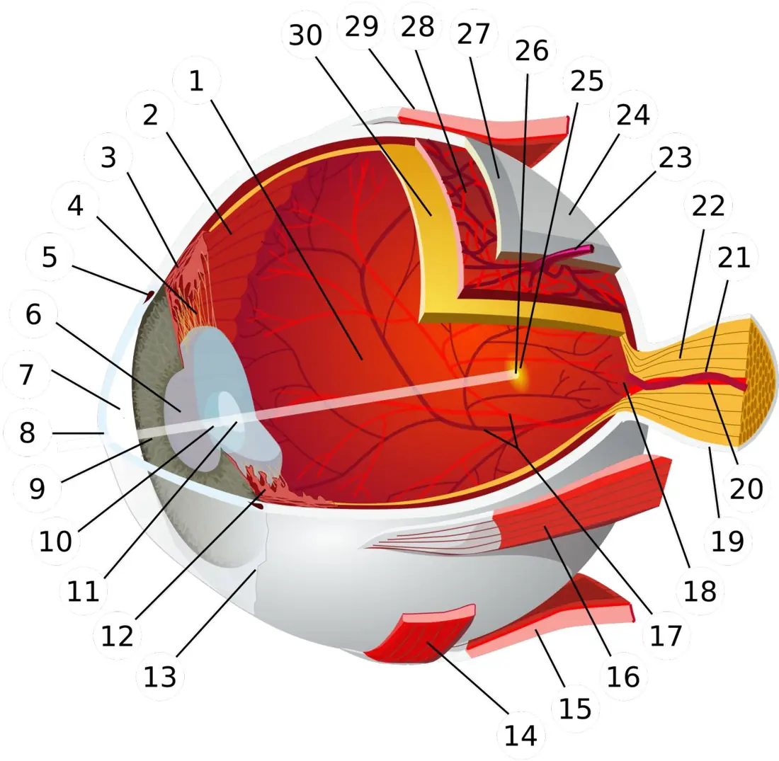
Structure of the eye
The eyeball has three coats:
- Sclera — tough outer protective layer; its transparent anterior part is the cornea, which supplies most of the eye's refractive power.
- Choroid/uvea — vascular, pigmented middle layer that absorbs stray light; anteriorly it forms the ciliary body and pigmented iris.
- Retina — inner neural layer containing photoreceptors and processing neurons.
The pupil is not a structure but the opening in the iris. Circular sphincter muscles under parasympathetic control constrict it in bright light; radial dilator muscles under sympathetic control widen it in dim light or arousal. Aqueous humour, secreted by ciliary processes into the anterior segment, nourishes the avascular cornea and lens and drains mainly through the canal of Schlemm. Blocked drainage can raise intraocular pressure in glaucoma. The jelly-like vitreous humour fills the posterior cavity and helps maintain shape.
Focusing and accommodation
The cornea provides the greatest fixed bending of light because of the large refractive-index change at the air–cornea surface. The lens provides fine adjustment. For a distant object, ciliary muscles relax, suspensory zonules are taut, and the lens is relatively thin. For a near object, the ciliary muscle contracts, the zonular fibres slacken, and the elastic lens becomes more convex, increasing its power. This is accommodation. The retinal image is real, inverted and smaller; the brain interprets it as upright.
| Defect | Optical problem | Usual correction |
|---|---|---|
| Myopia (short sight) | distant rays focus in front of retina; eyeball too long or optics too strong | diverging/concave lens (negative power) |
| Hypermetropia (long sight) | near rays would focus behind retina; eyeball too short or optics too weak | converging/convex lens (positive power) |
| Presbyopia | ageing lens/capsule loses accommodative flexibility | reading or bifocal/progressive lens |
| Astigmatism | unequal corneal or lens curvature in different meridians | cylindrical lens |
| Cataract | lens becomes opaque | surgical lens replacement |
Retina: rods, cones and visual pathway
Light passes through retinal neurons before reaching photoreceptors near the pigmented epithelium. Photoreceptor signals pass to bipolar cells and then ganglion cells; ganglion-cell axons form the optic nerve. At the optic disc, where these axons and retinal vessels exit, no photoreceptors exist, creating the blind spot.
| Feature | Rods | Cones |
|---|---|---|
| Approximate number | about 120 million | about 6 million |
| Pigment | rhodopsin | three classes of photopsin/iodopsin |
| Best in | dim light | bright light |
| Colour | no colour discrimination | red, green and blue channels for colour vision |
| Detail | poorer acuity; extensive convergence increases sensitivity | high acuity, especially in fovea |
| Distribution | abundant in peripheral retina | concentrated centrally; fovea almost exclusively cones |
Rhodopsin contains retinal derived from vitamin A; severe vitamin-A deficiency can therefore impair dim-light vision (night blindness) and damage the ocular surface. In the fovea, each cone has a relatively private pathway to the brain, preserving fine spatial detail. Peripheral rods converge heavily, sacrificing detail for sensitivity.
At the optic chiasm, fibres from the nasal half of each retina cross. Each cerebral hemisphere therefore processes the opposite visual field. A pituitary tumour pressing the central chiasm characteristically damages crossing nasal fibres and causes loss of both temporal visual fields (bitemporal hemianopia).
Structure and working of the ear
The ear has three divisions:
- Outer ear: pinna and external auditory canal collect and direct sound to the tympanic membrane.
- Middle ear: air-filled tympanic cavity containing malleus, incus and stapes. The Eustachian (auditory) tube connects it to the nasopharynx and equalises pressure across the eardrum.
- Inner ear: bony and membranous labyrinth containing the cochlea for hearing and vestibular apparatus for balance.
The ossicles solve an impedance-matching problem: sound in air would otherwise reflect from the fluid-filled inner ear. Force collected over the relatively large tympanic membrane is delivered by a lever system to the much smaller oval window, increasing pressure roughly twentyfold. The stapes moves the oval window; pressure waves travel through perilymph, displace the basilar membrane, and are relieved at the round window.
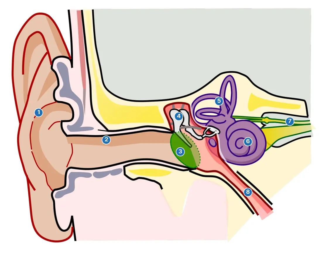
Hair cells of the organ of Corti, resting on the basilar membrane, bend against the tectorial membrane. This bending opens mechanically gated channels and excites fibres of the cochlear division of cranial nerve VIII. The stiff narrow base of the cochlea responds best to high frequencies; the flexible wide apex responds best to low frequencies. Loudness is coded mainly by amplitude of vibration, firing rate and recruitment of neurons.
Balance
The three semicircular canals lie approximately in three perpendicular planes. At the base of each, hair cells in the crista ampullaris project into a gelatinous cupula. Head rotation makes endolymph lag behind, bends the cupula and signals angular acceleration — dynamic equilibrium.
The utricle and saccule contain maculae whose hair cells are embedded in a gelatinous sheet weighted by calcium-carbonate otoliths. Gravity or linear acceleration shifts the sheet, signalling head position and straight-line movement — static/linear equilibrium.
Middle-ear disease that dampens the drum or ossicles causes conductive hearing loss. Damage to cochlear hair cells or the auditory nerve causes sensorineural hearing loss. Excessive noise characteristically destroys hair cells; unlike the ossicles, mature human cochlear hair cells regenerate poorly.
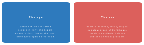
Three swaps the paper lives on: rods = dim, cones = colour (not the reverse); myopia = concave lens, hypermetropia = convex (image before vs behind the retina); cochlea hears, canals balance (both inner ear, different rooms).
Problem. A myelinated fibre conducts at 100 m/s and an unmyelinated one at 1 m/s. How long does one impulse take to travel a 1 m pathway in each? What is the total change in membrane voltage from rest (−70 mV) to the peak (+30 mV)? Solution. Myelinated: t = 1 ÷ 100 = 0.01 s (10 ms). Unmyelinated: t = 1 ÷ 1 = 1 s — a hundred times slower, which is why myelin matters. Voltage swing: +30 − (−70) = 100 mV.
Problem. A sensory impulse travels 0.60\text{ m} up an A\alpha afferent fibre at 60\text{ m s}^{-1}, crosses one central chemical synapse (synaptic delay 0.5\text{ ms}), and returns 0.60\text{ m} along a motor axon at 60\text{ m s}^{-1}. What is the total reflex-arc conduction time?
Solution. Afferent time = 0.60 / 60 = 0.010\text{ s} = 10\text{ ms}; efferent time = 10\text{ ms}; plus 0.5\text{ ms} synaptic delay gives \mathbf{20.5\text{ ms}}.
Formula and fact sheet
- CNS: brain and spinal cord. PNS: 12 cranial + 31 spinal nerve pairs.
- Meninges outside → inside: dura → arachnoid → pia (“DAP”).
- Vital centres: medulla. Coordination: cerebellum. Homeostasis and pituitary link: hypothalamus. Sensory relay: thalamus.
- Spinal roots: dorsal = sensory/afferent; ventral = motor/efferent.
- Sympathetic = thoracolumbar; parasympathetic = craniosacral.
- Resting potential about −70 mV, threshold about −55 mV, peak about +35 mV.
- Depolarisation: Na⁺ inflow. Repolarisation: K⁺ outflow. Pump: 3 Na⁺ out, 2 K⁺ in, using ATP.
- Conduction velocity: v = \dfrac{\text{distance}}{\text{time difference}}; subtract common delays in clinical two-point stimulation.
- Synaptic trigger: Ca²⁺ entry. Neuromuscular transmitter: acetylcholine. Terminating enzyme: acetylcholinesterase.
- Rods: rhodopsin, dim light, peripheral, low acuity. Cones: colour, bright light, fovea, high acuity.
- Near accommodation: ciliary contraction → zonules slacken → lens more convex.
- Myopia: concave/negative lens. Hypermetropia: convex/positive lens.
- Ear sequence: pinna → auditory canal → tympanum → malleus → incus → stapes → oval window → cochlea → organ of Corti → nerve VIII → temporal cortex.
- Semicircular canals: rotation. Utricle/saccule: gravity and linear motion.
Rapid-revision tables and high-yield facts
| Quantity | Value |
|---|---|
| Resting potential / peak | ≈ −70 mV / ≈ +30 mV (swing 100 mV) |
| Pump ratio | 3 Na⁺ out : 2 K⁺ in |
| Conduction speed | myelinated up to ~120 m/s; unmyelinated ~1 m/s |
| Synaptic cleft / delay | ~20 nm / ~0.5 ms |
| Cranial / spinal nerve pairs | 12 / 31 |
| Rods / cones | ~120 million / ~7 million |
| Eye defects | myopia concave, hypermetropia convex, astigmatism cylindrical |
| Ear ossicles | malleus, incus, stapes |
| Hearing / balance | cochlea (organ of Corti) / semicircular canals + vestibule |
Chapter summary
- The CNS integrates information; the PNS connects it with receptors and effectors through cranial and spinal nerves.
- Cerebral cortex supports conscious perception and voluntary action, while the hypothalamus, cerebellum and medulla have sharply testable special roles.
- Spinal reflexes use dorsal sensory entry and ventral motor exit and can act before conscious perception.
- Sympathetic and parasympathetic divisions differ in origin, fibre length, transmitter and typical visceral effects.
- An action potential is an all-or-none, self-propagating reversal of membrane potential driven first by Na⁺ entry and then by K⁺ exit.
- Myelination permits rapid saltatory conduction between nodes of Ranvier.
- Chemical synapses use Ca²⁺-triggered transmitter release and are one-way, summable and pharmacologically vulnerable.
- In the eye, the cornea supplies most fixed refraction, the lens accommodates, rods favour sensitivity and foveal cones favour acuity and colour.
- In the ear, ossicles amplify pressure into cochlear fluids; hair cells in the organ of Corti transduce sound, while vestibular hair cells signal rotation, gravity and linear acceleration.
Practice questions
Set A — Recall (25 questions)
- The structural and functional unit of the nervous system is theaneuroglial cellbaxoncnephrondneuron
- The central nervous system consists of theabrain and cranial nervesbbrain and spinal cordcspinal cord and gangliadreceptors and effectors
- The number of pairs of human spinal nerves isa12b21c30d31
- The outermost meninx surrounding the brain is theapia materbdura matercarachnoid materdchoroid
- Cerebrospinal fluid is produced mainly by theameningesbchoroid plexusccorpus callosumdarachnoid villi
- The principal respiratory and cardiovascular centres are located in theamedulla oblongatabcerebellumccerebrumdthalamus
- Balance, posture and fine coordination are controlled chiefly by theacerebellumbmedullacponsdfrontal lobe
- Hunger, thirst and body temperature are regulated mainly by theathalamusbmidbrainchypothalamusdcerebellum
- The primary visual cortex is located in theafrontal lobebtemporal lobecoccipital lobedparietal lobe
- The corpus callosum connects theatwo cerebellar hemispheresbbrain and spinal cordcthalamus and cortexdtwo cerebral hemispheres
- Sympathetic preganglionic neurons arise from theabrainstem onlybsacral cord onlycthoracolumbar corddcervical cord only
- Most parasympathetic postganglionic neurons releaseanoradrenalinebdopaminecserotonindacetylcholine
- The normal resting potential of a neuron is abouta+35 mVb−70 mVc−55 mVd0 mV
- Rapid depolarisation of an axon is caused mainly byaK⁺ influxbCl⁻ influxcCa²⁺ effluxdNa⁺ influx
- Myelin around a peripheral nerve fibre is produced byaastrocytesbSchwann cellscmicrogliadependymal cells
- Release of synaptic vesicles is triggered directly by entry ofaNa⁺bK⁺cCa²⁺dCl⁻
- Photoreceptors most important for vision in dim light are thearodsbconescbipolar cellsdganglion cells
- The sharpest visual image is formed at theafoveabblind spotcpupildora serrata
- The shape of the eye lens during accommodation is changed by theairisbretinacoptic nervedciliary muscle
- The optical image in a normal eye is formed on theacorneablenscretinadiris
- The correct sequence of ear ossicles from eardrum inward isaincus → malleus → stapesbmalleus → incus → stapescstapes → incus → malleusdmalleus → stapes → incus
- Sound receptor hair cells are located in theaorgan of Cortibutriclecsacculedcrista ampullaris
- The semicircular canals are concerned mainly withahearing low notesbhearing high notescstatic balancedrotational balance
- The Eustachian tube equalises pressure between theamiddle ear and pharynxbouter ear and cochleactwo cochleaedeye and nose
- The cranial nerve carrying smell impulses is theaoptic nervebfacial nervecolfactory nervedvagus nerve
Answers and explanations — Set A
1. (d) — A neuron integrates and conducts the nervous signal; axon is only one part of it.
2. (b) — The CNS is the brain and spinal cord; nerves and most ganglia belong to the PNS.
3. (d) — There are 31 spinal pairs: 8 cervical, 12 thoracic, 5 lumbar, 5 sacral and 1 coccygeal.
4. (b) — From outside inward the meninges are dura, arachnoid and pia mater.
5. (b) — Choroid plexuses in the ventricles secrete most cerebrospinal fluid.
6. (a) — The medulla contains cardiac, vasomotor and respiratory centres.
7. (a) — The cerebellum coordinates movement and maintains posture and balance.
8. (c) — The hypothalamus is the principal homeostatic and autonomic integrating centre.
9. (c) — Visual cortex lies mainly in the occipital lobe.
10. (d) — Corpus callosum fibres interconnect corresponding regions of the cerebral hemispheres.
11. (c) — Sympathetic outflow leaves the thoracic and upper lumbar spinal cord.
12. (d) — Acetylcholine is the usual parasympathetic postganglionic transmitter.
13. (b) — The inside of a resting neuron is about 70 mV negative to the outside.
14. (d) — Opening voltage-gated sodium channels produces rapid Na⁺ entry and depolarisation.
15. (b) — Schwann cells myelinate peripheral axons; oligodendrocytes do so in the CNS.
16. (c) — Ca²⁺ influx into the presynaptic terminal causes vesicle fusion and transmitter release.
17. (a) — Rods are highly sensitive and function best in dim light, without colour discrimination.
18. (a) — The cone-packed fovea gives maximum visual acuity.
19. (d) — Ciliary muscle changes zonular tension and therefore lens convexity.
20. (c) — The cornea and lens focus a real, inverted image on the retina.
21. (b) — Vibration passes malleus → incus → stapes to the oval window.
22. (a) — Hair cells of the organ of Corti transduce cochlear fluid movement into impulses.
23. (d) — Endolymph movement in semicircular canals signals angular acceleration.
24. (a) — The auditory tube opens between the middle ear and nasopharynx.
25. (c) — Cranial nerve I, the olfactory nerve, carries smell.
Set B — Understanding (15 questions)
- Saltatory conduction is fast and economical chiefly because the action potential is regeneratedacontinuously under the myelinbonly in the cell bodycby Schwann-cell nucleidonly at nodes of Ranvier
- A stronger stimulus is distinguished from a weaker one mainly by thealarger size of each impulseblonger refractory periodchigher impulse frequencydslower conduction velocity
- A chemical synapse normally transmits in one direction becauseaaxons have no membrane potentialbthe cleft contains myelincimpulses cannot enter dendritesdvesicles and receptors are on opposite sides
- Damage to the dorsal root of a spinal nerve would most directly cause loss ofasensation from its dermatomebmotor supply to all limbscsympathetic outflow onlydcerebrospinal fluid flow
- In a sudden fright, sympathetic activity would tend to produceabronchodilation and reduced gut motilitybpupil constriction and slow heartcincreased digestion and urinationdbronchoconstriction and salivation
- After a meal, increased parasympathetic activity most directlyadilates the pupilbincreases gut secretion and motilitycraises metabolic ratedrelaxes the bladder detrusor
- Demyelination slows or blocks impulses because it increasesatransmitter release at nodesbaxon diameter greatlycsodium channel density everywheredleakage and loss of saltatory conduction
- A hand is withdrawn before pain from a hot pan is consciously felt because theareceptor stops firing at oncebmotor fibres bypass the CNSccortex inhibits all paindspinal reflex is completed before cortical perception
- For near vision, the correct change isaciliary relaxation and flatter lensbzonules tighten and lens thinscciliary contraction and more convex lensdpupil dilation and flatter cornea
- The blind spot has no vision because it containsaonly conesbno rods or conescno lens tissuedexcessive pigment
- A faint star may be seen better when viewed slightly off-centre because the peripheral retina hasamore sensitive rods than the foveaba thicker lenscno bipolar cellsdonly colour cones
- The ossicles are essential mainly because theyachange sound into light signalsbincrease pressure delivered to inner-ear fluidcproduce endolymphddetect head rotation
- High-frequency sounds produce maximum basilar-membrane movement near theabase of the cochleabapex of the cochleacround window onlydEustachian tube
- Otoliths make the utricle and saccule sensitive toasound pitchbblood pressurecgravity and linear accelerationdcolour intensity
- Inhibition of acetylcholinesterase at the neuromuscular junction causes acetylcholine toadisappear instantlybpersist and overstimulate the end platecbecome noradrenalinedstop being released
Answers and explanations — Set B
1. (d) — Ion exchange and regeneration are concentrated at exposed nodes; internodes are insulated.
2. (c) — Action potentials are all-or-none, so intensity is frequency- and recruitment-coded.
3. (d) — Transmitter stores are presynaptic and the relevant receptors are postsynaptic.
4. (a) — Dorsal roots carry afferent sensory fibres into the spinal cord.
5. (a) — Sympathetic activation prepares for action: airways widen and digestive activity is reduced.
6. (b) — Parasympathetic activity promotes digestion through secretion and peristalsis.
7. (d) — Loss of insulation leaks local current and prevents efficient node-to-node regeneration.
8. (d) — The spinal motor response is launched before ascending pathways complete conscious processing.
9. (c) — Ciliary contraction slackens zonules, allowing the elastic lens to round up.
10. (b) — The optic disc contains exiting axons and vessels but no photoreceptors.
11. (a) — Peripheral retina is rod-rich and highly convergent; the fovea lacks rods.
12. (b) — Area reduction and leverage overcome the air-to-fluid impedance mismatch.
13. (a) — The stiff basal basilar membrane is tuned to high frequencies.
14. (c) — Dense calcium-carbonate crystals shift under gravity or linear acceleration.
15. (b) — Failure to hydrolyse ACh prolongs end-plate stimulation and may produce spasm or depolarisation block.
Set C — Application (10 questions)
- A pain impulse travels 1.2 m at 60 m s⁻¹. The axonal conduction time is abouta20 msb50 msc72 msd2 ms
- If a neuron has an absolute refractory period of 2 ms, its theoretical maximum firing rate is abouta50 s⁻¹b100 s⁻¹c500 s⁻¹d5000 s⁻¹
- A myopic student is prescribed a lens of power −2.0 D. Its focal length and type area+50 cm, convexb+20 cm, convexc−20 cm, concaved−50 cm, concave
- A patient has an unsteady gait and overshoots when reaching for objects, but muscle power is normal. The likeliest lesion is in theacerebellumbmedullacoptic nervedolfactory bulb
- Botulinum toxin prevents acetylcholine release at motor terminals. The expected paralysis is mainlyaspastic, with continuous contractionbflaccid, from failure of muscle excitationcabsent because ACh is inhibitorydconfined to smooth muscle
- The tympanic membrane has area 55 mm², the oval window 3.2 mm², and the ossicular lever ratio is 1.3. Pressure amplification is abouta4 timesb8 timesc13 timesd22 times
- When a student shifts gaze from a distant mountain to this page, the lens change is produced byaradial iris muscles pulling the lens flatbincreased vitreous pressure thinning the lenscciliary contraction slackening the zonulesdcorneal muscles increasing corneal curvature
- Fluid behind the eardrum during middle-ear infection most directly causes hearing loss byadestroying cochlear hair cellsbcutting the auditory nervecdamping movement of drum and ossiclesdblocking the optic pathway
- A beta-blocker reduces sympathetic beta-receptor stimulation of the heart. Its expected cardiac effect isafaster and stronger beatingbslower and less forceful beatingccomplete absence of vagus actiondincreased acetylcholine release
- A pituitary tumour compresses the centre of the optic chiasm. The characteristic visual defect is loss of theanasal fields of both eyesbtemporal fields of both eyescentire field of one eye onlydcolour vision with normal fields
Answers and explanations — Set C
1. (a) — Time = distance/speed = 1.2/60 = 0.020 s = 20 ms, excluding receptor and synaptic delays.
2. (c) — Maximum rate is at most 1/(2\times10^{-3}) = 500 impulses per second.
3. (d) — f=1/P=1/(-2.0)=-0.50 m; the negative sign denotes a diverging concave lens.
4. (a) — Cerebellar damage impairs coordination and balance while basic muscle strength may remain normal.
5. (b) — Without ACh release the end plate cannot excite skeletal muscle, producing flaccid weakness.
6. (d) — Amplification ≈ (55/3.2)\times1.3\approx22.
7. (c) — Near accommodation requires ciliary contraction, slack zonules and a more convex lens.
8. (c) — Middle-ear fluid impairs mechanical conduction through the tympanic membrane and ossicles.
9. (b) — Blocking cardiac beta receptors opposes sympathetic increases in rate and contractility.
10. (b) — Crossing nasal-retinal fibres carry temporal-field information; central chiasmal damage causes bitemporal hemianopia.