Chapter 7 · Human Biology and Physiology · 3 questions in the paper · 36 min read
Circulatory System
What the syllabus asks for 4
- The heart, the heartbeat, the cardiac cycle and cardiac output
- Arterial and venous systems; blood pressure
- Blood and blood groups
- Cardiovascular disorders
The circulatory system carries 3 of the 40 Zoology questions — the largest share of the human physiology units. The paper draws on four blocks: the heart (chambers, valves, conducting system, cardiac cycle and output), the vessels and blood pressure, the blood (cells, clotting, ABO and Rh groups), and the disorders (atherosclerosis, heart attack, angina, varicose veins, anaemia). Almost every question is a name, a number or a direction of flow. The three traps that decide marks are: the left ventricle has the thickest wall, the pulmonary artery carries deoxygenated blood, and O negative is the universal donor for red cells.
- Anatomy: four chambers, the tricuspid and bicuspid (mitral) valves, the two semilunar valves, and the great vessels; the left ventricle wall is the thickest because it drives the systemic circuit.
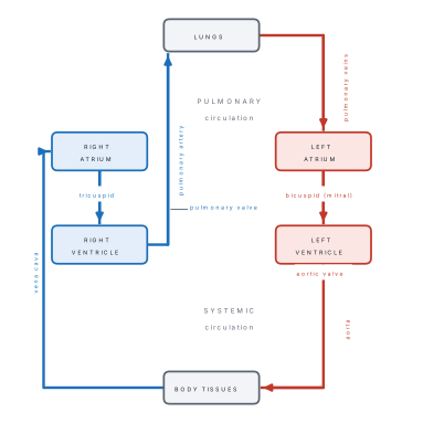
- Double circulation: right heart → lungs → left heart (pulmonary); left heart → body → right heart (systemic); the coronary arteries come from the ascending aorta.
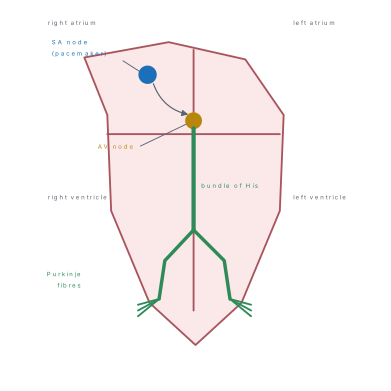
- Conducting system: SA node → AV node → bundle of His → right and left branches → Purkinje fibres; the SA node is the pacemaker because it depolarises fastest.
- Cardiac cycle (0.8 s at 72/min): atrial systole 0.1 s, ventricular systole 0.3 s, diastole 0.5 s; S1 = AV valves closing, S2 = semilunar valves closing.
- Cardiac output = stroke volume × heart rate ≈ 70 mL × 72 ≈ 5 L min⁻¹; ejection fraction ≈ 60 %.
- ECG: P = atrial depolarisation, QRS = ventricular depolarisation, T = ventricular repolarisation.
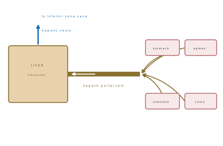
- Vessels: arteries thick-walled, no valves, high pressure; veins thin-walled, valves, low pressure; the hepatic portal vein runs gut → liver.
- Blood pressure 120/80; hypertension ≥ 140/90; baroreceptors in the carotid sinus and aortic arch.
- Blood: plasma 55 %, cells 45 %; RBC 5 million/mm³, 120 days; WBC 4000–11 000/mm³; platelets 1.5–4 lakh/mm³; ABO and Rh groups; O− donor, AB+ recipient; erythroblastosis fetalis.
7.1 The heart, the heartbeat, the cardiac cycle and cardiac output
Position and coverings
The heart is a hollow muscular organ of about 250–300 g, roughly the size of the owner's fist, lying in the mediastinum between the lungs, behind the sternum. Two-thirds of it lies to the left of the midline, and its apex points down and to the left, reaching the fifth intercostal space about 9 cm from the midline.
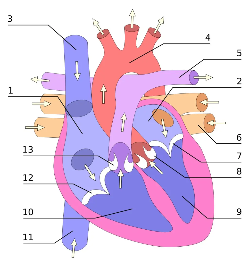
It is enclosed in the pericardium: a tough outer fibrous layer and a serous layer of two sheets — the parietal lining the sac and the visceral stuck to the heart (the epicardium). Between them is a film of pericardial fluid that lets the heart move without friction.
The wall has three layers:
| Layer | Structure | Note |
|---|---|---|
| Epicardium | visceral serous pericardium + fat | carries the coronary vessels |
| Myocardium | cardiac muscle | thickest in the left ventricle |
| Endocardium | endothelium + connective tissue | lines the chambers and valves |
The chambers and valves
| Chamber | Receives | Sends blood to | Wall |
|---|---|---|---|
| Right atrium | superior and inferior vena cava, coronary sinus | right ventricle | thin |
| Right ventricle | right atrium | pulmonary artery | moderate |
| Left atrium | four pulmonary veins | left ventricle | thin |
| Left ventricle | left atrium | aorta | thickest |
The two atria are separated by the interatrial septum, which bears the fossa ovalis — the remnant of the fetal opening. The ventricles are separated by the thick interventricular septum.
There are four valves, all of which open and close passively with the pressure difference across them:
| Valve | Position | Opens when | Prevents |
|---|---|---|---|
| Tricuspid (right AV) | between right atrium and ventricle | atrial pressure > ventricular | backflow into the right atrium |
| Bicuspid / mitral (left AV) | between left atrium and ventricle | atrial pressure > ventricular | backflow into the left atrium |
| Pulmonary semilunar | at the exit of the right ventricle | ventricular pressure > pulmonary artery | backflow into the right ventricle |
| Aortic semilunar | at the exit of the left ventricle | ventricular pressure > aorta | backflow into the left ventricle |
The AV valves are held by chordae tendineae attached to papillary muscles, which stop them turning inside out during ventricular contraction.
It pumps blood round the whole body, so it must generate about 120 mmHg; the right ventricle only pumps to the lungs, at about 25 mmHg. A question that offers "the right ventricle, because it is larger" is testing this.
The double circulation and the coronary circulation
Blood passes through the heart twice in one complete circuit:
- Pulmonary circulation: right ventricle → pulmonary trunk → pulmonary arteries → lungs → pulmonary veins → left atrium. Short, low pressure.
- Systemic circulation: left ventricle → aorta → all the tissues → superior and inferior vena cava → right atrium. Long, high pressure.
This is why the heart is described as a double pump and why the two sides never mix in a healthy adult.
The heart muscle itself is fed by the coronary circulation. The right and left coronary arteries arise from the ascending aorta just above the aortic valve, and their branches run in the epicardium. Blood returns by the cardiac veins into the coronary sinus, which opens into the right atrium. The coronary arteries fill mainly during diastole, when the ventricle is relaxed — one reason a very fast heart rate is dangerous.
The conducting system
Cardiac muscle contracts without any nerve: it is myogenic. A special patch of muscle, the sinoatrial (SA) node, in the wall of the right atrium near the opening of the superior vena cava, depolarises spontaneously about 70–80 times a minute and sets the rhythm. The wave then travels:
SA node → atria → AV node → bundle of His → right and left bundle branches → Purkinje fibres → ventricular muscle.
| Part | Rate if it acts alone (per min) | Conduction velocity |
|---|---|---|
| SA node | 70–80 (the pacemaker) | slow |
| AV node | 40–60 | 0.05 m s⁻¹ — the slowest, giving a 0.1 s delay |
| Bundle of His and branches | 30–40 | 1–2 m s⁻¹ |
| Purkinje fibres | 20–40 | 4 m s⁻¹ — the fastest |
The SA node sets the rhythm because its membrane leaks ions so that it reaches threshold first; it therefore drives the other, slower pacemakers before they can fire on their own. They are latent pacemakers, ready to take over if the SA node fails.
The AV nodal delay matters: it lets the atria finish emptying before the ventricles contract, so the ventricles are properly filled.
The cardiac cycle
One complete beat — contraction (systole) and relaxation (diastole) of the four chambers — is the cardiac cycle. At a rate of 72–75 beats a minute it lasts 0.8 s:
| Phase | Duration | What happens |
|---|---|---|
| Atrial systole | 0.1 s | the atria contract and top up the ventricles (about 20 % of filling) |
| Ventricular systole | 0.3 s | AV valves close (S1); pressure rises; semilunar valves open; blood is ejected |
| Ventricular diastole | 0.5 s | semilunar valves close (S2); ventricles relax and fill; AV valves open |
The pressure changes drive everything:
| Event | Pressure relation | Result |
|---|---|---|
| AV valves close | ventricular > atrial | first heart sound |
| Semilunar valves open | ventricular > aorta / pulmonary artery | ejection begins |
| Semilunar valves close | aorta > ventricular | second heart sound |
| AV valves open | atrial > ventricular | rapid filling |
Typical pressures: left ventricle 120/0–10 mmHg, aorta 120/80, left atrium about 8 mmHg, right atrium about 2 mmHg.
Heart sounds. S1 ("lub") is the closure of the AV valves at the start of ventricular systole — longer and lower. S2 ("dub") is the closure of the semilunar valves at the end of systole — shorter and sharper. S3 and S4 are soft filling sounds, often inaudible in health.

Cardiac output
Stroke volume (SV) is the blood ejected by one ventricle in one beat — about 70 mL. Heart rate (HR) is the number of beats per minute — about 72. Cardiac output (CO) is the volume pumped by one ventricle in a minute:
The ejection fraction is the fraction of the end-diastolic volume that is ejected:
Cardiac output rises in exercise to 20–25 L min⁻¹, by increasing both rate and stroke volume. It depends on:
| Factor | Effect |
|---|---|
| Preload (venous return) | more filling → more stretch → stronger contraction (Frank–Starling law) |
| Afterload (arterial pressure) | higher pressure → harder to eject → lower stroke volume |
| Contractility | adrenaline and sympathetic nerves increase it |
| Heart rate | up to a point raises output; too fast shortens filling and lowers it |
The electrocardiogram
The ECG records the electrical activity of the heart from the body surface. Its three waves:
| Wave | Meaning | Duration |
|---|---|---|
| P | atrial depolarisation (just before atrial systole) | 0.08–0.10 s |
| QRS | ventricular depolarisation (atrial repolarisation is hidden inside it) | 0.08–0.12 s |
| T | ventricular repolarisation | — |
The PR interval (0.12–0.20 s) shows the AV nodal delay; a long PR means heart block. ST elevation is the classic sign of a myocardial infarction.
Problem. At rest a student has a stroke volume of 70 mL and a heart rate of 72 min⁻¹. In exercise his stroke volume rises to 120 mL and his rate to 150 min⁻¹. Find both cardiac outputs.
Solution. Rest: 70 \times 72 = 5040 mL min⁻¹ ≈ 5.0 L min⁻¹. Exercise: 120 \times 150 = 18\,000 mL min⁻¹ = 18 L min⁻¹.
So the output can rise more than threefold, and most of the rise comes from the stroke volume at first, then from the rate.
Problem. A patient's end-diastolic volume is 120 mL and end-systolic volume 50 mL. Find the stroke volume and the ejection fraction.
Solution. SV = 120 - 50 = 70 mL. EF = 70/120 = 0.58 = 58 %.
A failing heart may have an ejection fraction below 40 %, which is why it tires on exertion.
7.2 Arterial and venous systems; blood pressure
Artery, vein and capillary
| Feature | Artery | Vein | Capillary |
|---|---|---|---|
| Wall | thick, three coats, much elastic and smooth muscle | thin, little muscle | one cell thick (endothelium + basement membrane) |
| Lumen | narrow, stays round | wide, collapses when empty | extremely narrow (7–10 μm) |
| Valves | none (except at the heart) | yes, especially in the limbs | none |
| Blood flow | fast, pulsatile | slow, steady | slowest — the widest total cross-section |
| Pressure | high (120/80 in the aorta) | low (near 0 in the vena cava) | falls from arteriole to venule |
| Carries | blood away from the heart | blood towards the heart | exchange between blood and tissue |
| Example | aorta, carotid, renal artery | vena cava, jugular, saphenous | in every tissue |
Arteries near the heart are elastic (the aorta expands with each systole and recoils in diastole, smoothing the flow into a pulse); arteries further out are muscular and control the distribution of blood. Arterioles are the main resistance vessels and set blood pressure.
The main vessels
| Region | Artery | Vein |
|---|---|---|
| Head and neck | common carotid → internal and external carotid | internal and external jugular |
| Upper limb | subclavian → axillary → brachial → radial and ulnar | cephalic, basilic, brachial |
| Thorax | descending thoracic aorta | azygos, hemiazygos |
| Abdomen | coeliac trunk, superior and inferior mesenteric, renal, gonadal | hepatic, renal, mesenteric |
| Lower limb | common iliac → external iliac → femoral → popliteal | great saphenous, femoral, popliteal |
| Whole body | aorta (largest artery) | vena cava (largest vein) |
The aorta has three parts: ascending, the arch (giving the brachiocephalic, left common carotid and left subclavian), and the descending thoracic and abdominal aorta, which ends by dividing into the two common iliac arteries. The superior vena cava drains the head, neck and arms; the inferior vena cava drains everything below the diaphragm.
The hepatic portal system
A portal system is a set of veins that breaks into a second capillary bed before reaching the heart. The hepatic portal vein is formed behind the neck of the pancreas by the union of the superior mesenteric vein and the splenic vein (joined by the inferior mesenteric and gastric veins). It carries absorbed glucose, amino acids, vitamins and also drugs and toxins from the gut to the sinusoids of the liver. There the liver stores, converts or detoxifies them, and the blood leaves by the hepatic veins into the inferior vena cava.
This is why the liver is the first organ to see what we eat, and why a drug taken by mouth is partly destroyed before it reaches the rest of the body (the first-pass effect).
Blood pressure
Blood pressure is the force the blood exerts on the vessel wall. It is written as systolic/diastolic: 120/80 mmHg in a healthy young adult.
- Systolic pressure (120) is the peak during ventricular ejection.
- Diastolic pressure (80) is the lowest, just before the next beat.
- Pulse pressure = systolic − diastolic = 40 mmHg.
- Mean arterial pressure ≈ diastolic + ⅓ pulse pressure ≈ 93 mmHg.
It is measured with a sphygmomanometer and a cuff on the upper arm, at the level of the heart. As the cuff deflates, the first tapping sound (Korotkoff sound) marks the systolic pressure, and the point at which the sound disappears marks the diastolic.
| Category | Systolic | Diastolic |
|---|---|---|
| Hypotension | below 90 | below 60 |
| Normal | 90–119 | 60–79 |
| Pre-hypertension / high normal | 120–139 | 80–89 |
| Hypertension | 140 or more | 90 or more |
Problem. A patient's blood pressure is 150/95 mmHg. Find the pulse pressure and the mean arterial pressure.
Solution. Pulse pressure = 150 - 95 = 55 mmHg.
The mean pressure is nearer the diastolic value because the heart spends about twice as long in diastole as in systole. A MAP below about 60 mmHg means the brain and kidneys are badly perfused.
Blood pressure depends on cardiac output and peripheral resistance (the radius of the arterioles), and also on blood volume, blood viscosity and the elasticity of the arteries. A vasoconstriction of the arterioles raises pressure; atherosclerosis stiffens the arteries and raises the systolic pressure.
Regulation is both nervous and hormonal:
| Mechanism | Where | Effect |
|---|---|---|
| Baroreceptors | carotid sinus and aortic arch | stretch signals to the medulla; a rise in pressure slows the heart and dilates vessels |
| Chemoreceptors | carotid and aortic bodies | low O₂, high CO₂ → vasoconstriction and faster heart |
| Vasomotor centre | medulla | sets sympathetic tone to arterioles |
| Adrenaline, noradrenaline | adrenal medulla, sympathetic nerves | raise rate, force and resistance |
| Renin–angiotensin–aldosterone | kidney | angiotensin II constricts vessels; aldosterone retains Na⁺ and water |
| ADH | posterior pituitary | water retention, raises volume |
| ANP | heart atria | promotes Na⁺ and water loss, lowers pressure |
The pulse is the wave felt in an artery when the aorta expands in systole. It normally equals the heart rate, but in some arrhythmias a beat is too weak to produce a wave, so the pulse rate is lower than the rate on the ECG (a pulse deficit). In an exam, "pulse rate always equals heart rate" is the wrong option.
7.3 Blood and blood groups
Blood is a fluid connective tissue: cells suspended in a liquid matrix. An adult has about 5 L (7–8 % of body mass). If a sample is spun in a centrifuge, the plasma forms about 55 % and the cells about 45 %; the percentage of cells is the haematocrit (packed cell volume).
Plasma
Plasma is 91–92 % water with 7–8 % protein and about 0.9 % inorganic salts, plus glucose, amino acids, lipids, hormones, urea and CO₂.
| Protein | Percentage of plasma protein | Function |
|---|---|---|
| Albumin | 55–60 % (most abundant) | maintains osmotic pressure; carries fatty acids and drugs |
| Globulins | about 35 % | α and β carry lipids and vitamins; γ-globulins are antibodies |
| Fibrinogen | about 4 % | converted to fibrin in clotting |
Serum is plasma without fibrinogen — that is, the fluid left after blood has clotted.
The formed elements
| Cell | Count per mm³ | Structure | Function | Life span |
|---|---|---|---|---|
| Red blood cells (erythrocytes) | 4.5–5.5 million | biconcave disc, no nucleus in man, filled with haemoglobin | carry O₂ and CO₂ | 120 days |
| Neutrophils | 3000–6000 (60–70 % of WBC) | lobed nucleus, granular | phagocytosis of bacteria — the first responders | hours to days |
| Eosinophils | 2–4 % | bilobed, red granules | attack parasites; allergy | days |
| Basophils | 0.5–1 % | lobed, blue granules | release histamine and heparin | days |
| Lymphocytes | 20–30 % | round nucleus, little cytoplasm | B cells make antibodies; T cells kill infected cells | years |
| Monocytes | 2–8 % | kidney-shaped nucleus, largest WBC | become macrophages | months |
| Platelets (thrombocytes) | 1.5–4 lakh | cell fragments from megakaryocytes | haemostasis | 8–10 days |
Erythropoiesis — the making of red cells — happens in the red bone marrow of ribs, sternum, vertebrae and pelvis, under the hormone erythropoietin from the kidney, which rises when the kidney senses low oxygen. It needs iron (for haem), vitamin B₁₂ and folic acid (for DNA in dividing cells) and vitamin B₆. Old red cells are destroyed by macrophages in the spleen, liver and bone marrow; the iron is recycled and the haem is converted to bilirubin.
Haemostasis — stopping bleeding
- Vascular spasm — the cut vessel constricts.
- Platelet plug — platelets stick to the collagen, swell and aggregate.
- Coagulation — the clotting cascade. Tissue damage exposes thromboplastin (factor III), which with Ca²⁺ and other factors converts prothrombin (II) → thrombin. Thrombin converts soluble fibrinogen (I) into insoluble fibrin threads that trap the cells into a clot.
- Clot retraction and repair — the clot shrinks, and fibroblasts repair the vessel.
- Fibrinolysis — plasmin dissolves the clot once the vessel is healed.
There are 13 clotting factors. Vitamin K is needed by the liver to make prothrombin and factors VII, IX and X, and Ca²⁺ (factor IV) is required at several steps — which is why calcium-binding drugs such as citrate stop blood clotting in a bottle. Heparin, from basophils and mast cells, is a natural anticoagulant.
Blood groups — ABO and Rh
Karl Landsteiner described the ABO groups in 1900. The red cell carries agglutinogens (antigens) and the plasma carries agglutinins (antibodies) against the antigens it does not have:
| Group | Agglutinogen on RBC | Agglutinin in plasma | Can receive red cells from |
|---|---|---|---|
| A | A | anti-B | A, O |
| B | B | anti-A | B, O |
| AB | A and B | none | A, B, AB, O |
| O | none | anti-A and anti-B | O only |
A transfusion of the wrong group makes the donor cells clump (agglutination) and then burst (haemolysis), releasing haemoglobin that can block the kidney tubules — a transfusion reaction.
- O negative is the universal donor of red cells: its cells carry no A, B or D antigen, so no recipient's antibodies can attack them.
- AB positive is the universal recipient: its plasma has no anti-A, anti-B or anti-D.
The Rh factor (rhesus antigen, D antigen) was found by Landsteiner and Wiener in 1940. About 85 % of people are Rh positive. Anti-D is not present naturally; it appears only after an Rh-negative person is exposed to Rh- positive blood.
Erythroblastosis fetalis (haemolytic disease of the newborn). An Rh-negative mother carrying an Rh-positive fetus is sensitised at delivery, when fetal cells enter her blood, and makes anti-D IgG. In a later pregnancy with another Rh-positive fetus, that IgG crosses the placenta and destroys the fetal red cells: the baby is born anaemic and jaundiced, and may die. It is prevented by giving the mother anti-D immunoglobulin after the first delivery.
"O negative is the universal donor" is true for packed red cells. If whole plasma were transfused, O plasma contains anti-A and anti-B and would attack A or B recipient cells. The paper asks the red-cell rule; answer O negative, but know why.
7.4 Cardiovascular disorders
| Disorder | What happens | Cause / risk | Key feature |
|---|---|---|---|
| Atherosclerosis | fatty streaks → plaque of cholesterol, cells and calcium in the arterial intima; lumen narrows and the wall stiffens | high LDL, smoking, hypertension, diabetes, obesity, family history | angina, heart attack, stroke, gangrene |
| Angina pectoris | reversible ischaemia of the heart muscle | narrowed coronary arteries | crushing chest pain on exertion, relieved by rest or nitrates |
| Myocardial infarction (heart attack) | a coronary artery is blocked by a thrombus; part of the muscle dies | plaque rupture, smoking, hypertension | severe chest pain radiating to the left arm and jaw, sweating, ST elevation, troponin rises |
| Hypertension | sustained BP ≥ 140/90 | age, salt, obesity, stress, kidney disease | "silent killer": stroke, heart failure, kidney damage |
| Varicose veins | dilated, tortuous, elongated veins with incompetent valves | long standing, pregnancy, heredity | aching legs, visible blue swellings; worse in the lower limb |
| Anaemia | haemoglobin below normal | iron deficiency (commonest), B₁₂ deficiency, blood loss, haemolysis | pallor, fatigue, breathlessness, fast pulse |
| Stroke | blood supply to part of the brain is cut | thrombus, embolus, haemorrhage | sudden weakness, speech loss |
Atherosclerosis begins with injury to the endothelium; LDL cholesterol enters the wall, is oxidised, and macrophages swallow it to become "foam cells". Smooth muscle and collagen form a fibrous cap over a lipid core. A plaque may rupture, exposing the core to blood, and a clot forms on it — the event that causes most heart attacks and strokes.
Anaemia is classified by the size of the red cells:
| Type | Cause | Cell size |
|---|---|---|
| Iron-deficiency | poor diet, blood loss, pregnancy | microcytic, hypochromic |
| Pernicious | lack of intrinsic factor → no B₁₂ absorption | macrocytic |
| Haemolytic | red cells destroyed (malaria, sickle cell, Rh disease) | normal or abnormal shape |
| Aplastic | bone marrow fails (drugs, radiation) | few cells of all types |
Angina: pain on exertion, lasts minutes, relieved by rest or a nitrate, no enzyme rise. Heart attack: pain at rest, lasts longer, not relieved, with sweating and nausea, ECG changes and raised troponin. That single contrast answers most clinical stems.
Formula and fact sheet
The heart
| Fact | Value |
|---|---|
| Mass | 250–300 g |
| Chambers / valves | 4 / 4 (2 AV, 2 semilunar) |
| Thickest wall | left ventricle |
| Pacemaker | SA node, 70–80 min⁻¹ |
| AV node rate / delay | 40–60 min⁻¹ / 0.1 s |
| Fastest conduction | Purkinje fibres, 4 m s⁻¹ |
| Slowest conduction | AV node, 0.05 m s⁻¹ |
| Cardiac cycle at 72–75 min⁻¹ | 0.8 s (atrial systole 0.1, ventricular systole 0.3, diastole 0.5) |
| Stroke volume | 70 mL |
| End-diastolic / end-systolic volume | 120 / 50 mL |
| Ejection fraction | ≈ 60 % |
| Cardiac output | ≈ 5 L min⁻¹ |
| S1 / S2 | AV valves close / semilunar valves close |
ECG
| Wave | Meaning |
|---|---|
| P | atrial depolarisation |
| QRS | ventricular depolarisation |
| T | ventricular repolarisation |
| PR interval | 0.12–0.20 s (AV nodal delay) |
Vessels and pressure
| Fact | Value |
|---|---|
| Normal BP | 120/80 mmHg |
| Pulse pressure | 40 mmHg |
| Mean arterial pressure | ≈ 93 mmHg |
| Hypertension | ≥ 140/90 |
| Hypotension | < 90/60 |
| Largest artery / vein | aorta / vena cava |
| Portal vein formed by | superior mesenteric + splenic veins |
Blood
| Fact | Value |
|---|---|
| Volume | about 5 L |
| Plasma / cells | 55 % / 45 % |
| Plasma water / protein | 91–92 % / 7–8 % |
| Most abundant plasma protein | albumin (55–60 %) |
| RBC count | 4.5–5.5 million/mm³ |
| RBC life span | 120 days |
| Haemoglobin | 13–17 g/dL (about 15) |
| WBC count | 4000–11 000/mm³ |
| Neutrophil / lymphocyte / monocyte | 60–70 % / 20–30 % / 2–8 % |
| Platelets | 1.5–4 lakh/mm³ |
| Clotting factors | 13; vitamin K for II, VII, IX, X |
Blood groups
| Group | Antigen | Antibody |
|---|---|---|
| A | A | anti-B |
| B | B | anti-A |
| AB | A + B | none |
| O | none | anti-A + anti-B |
- Universal donor (red cells): O negative.
- Universal recipient: AB positive.
- Rh positive: about 85 %; anti-D causes erythroblastosis fetalis.
- ABO described by Landsteiner (1900); Rh by Landsteiner and Wiener (1940).
Traps
- The left ventricle wall is thickest.
- The pulmonary artery carries deoxygenated blood; the pulmonary vein carries oxygenated blood.
- O negative is the universal donor; AB positive the universal recipient.
- S1 = AV valves, S2 = semilunar valves.
- Most CO₂ is carried as bicarbonate (unit 6), not by red cells alone.
- Veins have valves; arteries do not.
- The SA node, not the AV node, is the pacemaker.
- Cardiac output = SV × HR, not SV + HR.
Chapter summary
- The heart (250–300 g) lies in the mediastinum inside the pericardium; its wall is epicardium, myocardium (thickest in the left ventricle) and endocardium.
- Four chambers and four valves: tricuspid and bicuspid (mitral) AV valves with chordae tendineae, and the pulmonary and aortic semilunar valves; all open and close passively.
- Double circulation: right ventricle → lungs → left atrium (pulmonary); left ventricle → body → right atrium (systemic). The coronary arteries arise from the ascending aorta.
- The SA node (70–80 min⁻¹) is the pacemaker because it depolarises fastest; the impulse passes SA → AV → bundle of His → Purkinje, with a 0.1 s delay at the AV node.
- The cardiac cycle lasts 0.8 s: atrial systole 0.1 s, ventricular systole 0.3 s, diastole 0.5 s; S1 is AV valve closure, S2 semilunar closure.
- CO = SV × HR ≈ 70 × 72 ≈ 5 L min⁻¹; ejection fraction ≈ 60 %; exercise raises output to 20–25 L min⁻¹.
- ECG: P = atrial depolarisation, QRS = ventricular depolarisation, T = ventricular repolarisation.
- Arteries are thick-walled, valveless and high-pressure; veins are thin-walled, valved and low-pressure; the hepatic portal vein carries gut blood to the liver before the heart.
- BP 120/80; hypertension ≥ 140/90; baroreceptors in the carotid sinus and aortic arch; the medulla sets vasomotor tone.
- Blood is 55 % plasma and 45 % cells; RBC 5 million/mm³ and 120 days, WBC 4000–11 000/mm³, platelets 1.5–4 lakh/mm³; albumin is the commonest plasma protein.
- Haemostasis: spasm → platelet plug → prothrombin → thrombin → fibrin → clot retraction → fibrinolysis.
- ABO: A has anti-B, B has anti-A, AB has none, O has both; O− donor, AB+ recipient; Rh incompatibility causes erythroblastosis fetalis.
- Disorders: atherosclerosis (plaque), angina (reversible), MI (thrombus, troponin), varicose veins (valve failure), anaemia (low haemoglobin).
Practice questions
Set A — Recall (29 questions)
- The pacemaker of the human heart is theaAV nodebbundle of HiscSA nodedPurkinje fibre
- The left atrioventricular valve is also called theatricuspid valvebbicuspid valvecpulmonary valvedaortic valve
- The number of chambers in the human heart isatwobthreecfivedfour
- The heart is enclosed in a sac called theapericardiumbpleuracperitoneumdmesentery
- At 75 beats per minute, one cardiac cycle lastsa0.3 sb0.8 sc1.0 sd0.5 s
- The stroke volume of a resting adult is abouta70 mLb120 mLc5 Ld50 mL
- The cardiac output of a resting adult is abouta70 mLb500 mLc5 L per minuted50 L per minute
- The first heart sound is produced by closure of theasemilunar valvesbopening of the AV valvescblood entering the aortadatrioventricular valves
- The P wave of an ECG representsaatrial depolarisationbventricular depolarisationcventricular repolarisationdatrial repolarisation
- The QRS complex of an ECG representsaatrial depolarisationbatrial repolarisationcventricular repolarisationdventricular depolarisation
- The T wave of an ECG representsaatrial systolebventricular depolarisationcventricular repolarisationdclosure of the valves
- The impulse is delayed on its way from the atria to the ventricles at theaSA nodebAV nodecbundle of HisdPurkinje fibres
- The normal blood pressure of a young adult isa80/120 mmHgb140/90 mmHgc100/70 mmHgd120/80 mmHg
- Blood pressure is measured with aastethoscopebspirometercsphygmomanometerdelectrocardiograph
- The universal donor for red cells isaO negativebAB positivecA positivedB negative
- The universal recipient of blood isaO negativebAB positivecA negativedB positive
- Rh incompatibility between mother and fetus causesajaundice in the motherberythroblastosis fetalischaemophiliadleukaemia
- The most numerous formed element of blood is theawhite blood cellbplateletclymphocytedred blood cell
- The average life span of a human red blood cell isa120 daysb60 daysc30 daysd180 days
- The normal white blood cell count per mm³ isa4–5 millionb1.5–4 lakhc4000–11 000d500–1000
- The normal platelet count per mm³ isa4–5 millionb4000–11 000c1.5–4 lakhd5–10 lakh
- The most abundant protein of plasma isaalbuminbglobulincfibrinogendprothrombin
- Vitamin K is required for the synthesis ofahaemoglobinbalbumincplateletsdprothrombin
- The hepatic portal vein is formed by the union of theahepatic vein and inferior vena cavabsuperior mesenteric and splenic veinscrenal and iliac veinsdjugular and subclavian veins
- Which vessel carries deoxygenated blood?aaortabpulmonary arterycpulmonary veindcarotid artery
- The coronary arteries arise from theapulmonary trunkbarch of the aortacascending aortadbrachiocephalic artery
- A heart attack is caused by blockage of aapulmonary arterybcarotid arterycrenal arterydcoronary artery
- Varicose veins are caused mainly byaincompetent valves in the veinsbatherosclerosiscanaemiadhypertension
- Which of the following is a granulocyte?alymphocytebmonocytecneutrophildred blood cell
Answers and explanations — Set A
1. (c) — the SA node, in the wall of the right atrium, depolarises fastest and so sets the rhythm.
2. (b) — the bicuspid or mitral valve, on the left side; the tricuspid is on the right.
3. (d) — two atria and two ventricles, making the heart a double pump.
4. (a) — the pericardium, with fibrous and serous layers and a film of fluid between them.
5. (b) — 0.8 s, made up of atrial systole 0.1 s, ventricular systole 0.3 s and diastole 0.5 s.
6. (a) — about 70 mL per beat; 120 mL is the end-diastolic volume and 50 mL the end-systolic volume.
7. (c) — 70 mL × 72 ≈ 5 L per minute.
8. (d) — S1 is the closure of the AV valves at the start of ventricular systole; S2 is the closure of the semilunar valves.
9. (a) — atrial depolarisation, which just precedes atrial systole.
10. (d) — ventricular depolarisation. Atrial repolarisation is hidden inside the same complex.
11. (c) — ventricular repolarisation; the ventricles recover before the next beat.
12. (b) — the AV node conducts at only 0.05 m s⁻¹, giving a delay of about 0.1 s.
13. (d) — 120/80 mmHg. 140/90 is the threshold for hypertension.
14. (c) — the sphygmomanometer, with a cuff and a mercury or digital gauge.
15. (a) — O negative red cells carry no A, B or D antigen, so no recipient has antibodies against them.
16. (b) — AB positive plasma has no anti-A, anti-B or anti-D.
17. (b) — anti-D from a sensitised Rh-negative mother destroys fetal red cells.
18. (d) — about 5 million red cells per mm³, against 4000–11 000 white cells.
19. (a) — 120 days; about 2 million are destroyed every second and replaced by the marrow.
20. (c) — 4000–11 000 per mm³. 4–5 million is the red cell count.
21. (c) — 1.5–4 lakh per mm³, from megakaryocytes.
22. (a) — albumin, 55–60 % of plasma protein; it holds the osmotic pressure.
23. (d) — prothrombin and factors VII, IX and X are made in the liver and need vitamin K.
24. (b) — the superior mesenteric and splenic veins join behind the pancreas.
25. (b) — the pulmonary artery carries blood from the right ventricle to the lungs.
26. (c) — the right and left coronary arteries leave the ascending aorta just above the aortic valve.
27. (d) — a coronary artery blocked by a thrombus starves part of the muscle, which dies.
28. (a) — the valves fail, blood pools and the vein stretches and becomes tortuous.
29. (c) — neutrophils, eosinophils and basophils have granules; lymphocytes and monocytes do not.
Set B — Understanding (17 questions)
- The wall of the left ventricle is the thickest because itaholds the largest volume of bloodblies closest to the lungscpumps blood round the whole bodydreceives oxygenated blood
- The SA node sets the rhythm of the heart because itahas the fastest rate of spontaneous depolarisationbis the largest part of the conducting systemchas the richest nerve supplydlies in the left atrium
- The AV valves close at the start of ventricular systole becauseathe atria contract at that momentbthe semilunar valves opencblood pushes them shut from belowdventricular pressure rises above atrial pressure
- The delay at the AV node is useful because italets the atria fill with bloodblets the atria empty completely before the ventricles contractcslows the heart ratedkeeps the semilunar valves closed
- The pulmonary artery carries deoxygenated blood because itacarries blood from the right ventricle to the lungsbis really a vein in structurechas a very thin walldcarries blood towards the heart
- Blood pressure is higher in arteries than in veins because arteriesacontain more bloodbare closer to the pump and have thick elastic wallsccontain valvesdare wider than veins
- Veins in the legs have valves in order toaraise the pressure of the bloodbslow the flow of bloodcprevent backflow when pressure is lowdfilter the blood
- O negative blood can be given to anyone because its red cellsacarry both A and B antigensbcarry no antibodiescare the commonest typedcarry no A, B or D antigen
- AB positive blood can receive any group because its plasmaahas no antigensbhas all the antibodiescis Rh negativedhas no anti-A, anti-B or anti-D
- Erythroblastosis fetalis usually appears in a second pregnancy becauseathe second baby is Rh negativebthe mother was sensitised in the first pregnancycthe father is Rh negativedthe first baby was AB positive
- Cardiac muscle does not fatigue because itahas a long refractory period and rests in diastolebhas no nerve supplycdoes not use oxygendcontains no mitochondria
- The hepatic portal vein is important because itacarries oxygen to the liverbdrains blood from the heartctakes absorbed nutrients to the liver firstdcarries hormones to the brain
- The ECG shows no separate wave for atrial repolarisation because itais hidden inside the QRS complexbnever happenscis too small to recorddcomes after the T wave
- The second heart sound is shorter and sharper than the first because theaAV valves are largerbatria contract at that momentcsemilunar valves snap shut against high pressuredblood flow stops completely
- The pulse is felt as a wave because theablood flows in a continuous streambaorta expands with each systolecveins contract rhythmicallydheart moves inside the chest
- Blood pressure is measured on the arm held at heart level so thatathe cuff fits easilybthe artery is at its widestcno valves interferedthe reading is not changed by gravity
- Severe anaemia makes the pulse fast because the heartacompensates for poor oxygen carriage by raising its outputbbecomes weakercis compressed by the lungsdloses its nerve supply
Answers and explanations — Set B
1. (c) — the systemic circuit has a much higher resistance, so the left ventricle needs a much thicker muscle.
2. (a) — its membrane reaches threshold first, so it fires before the slower latent pacemakers can.
3. (d) — the valves are passive: when ventricular pressure exceeds atrial pressure they are pushed shut.
4. (b) — the 0.1 s delay allows complete filling, so stroke volume is not wasted.
5. (a) — it is an artery by structure but carries deoxygenated blood from the right ventricle to the lungs.
6. (b) — pressure falls steadily along the circuit because of resistance; the elastic aorta also smooths the flow.
7. (c) — venous pressure is too low to push blood up the leg against gravity, so valves and the muscle pump do the work.
8. (d) — with no A, B or D antigen on the red cells, no recipient's plasma has an antibody that can clump them.
9. (d) — the plasma of AB positive has no antibodies at all, so it does not attack transfused cells.
10. (b) — the first delivery sensitises the mother, who then makes anti-D IgG that crosses the placenta next time.
11. (a) — the long refractory period prevents tetanus, and the heart rests for about 0.5 s in every 0.8 s cycle.
12. (c) — first-pass delivery lets the liver store, convert or detoxify what the gut has absorbed.
13. (a) — atrial repolarisation coincides with ventricular depolarisation and is buried in the QRS.
14. (c) — the semilunar valves close suddenly against the high pressure of the aorta and pulmonary artery.
15. (b) — each systole pushes blood into the aorta, which stretches and then recoils, sending a wave along the arteries.
16. (d) — hydrostatic pressure would add to or subtract from the reading if the arm were above or below heart level.
17. (a) — with less haemoglobin the heart raises output to keep oxygen delivery up, so the pulse is fast.
Set C — Application (10 questions)
- A man has a stroke volume of 80 mL and a heart rate of 75 per minute. His cardiac output isa5.0 L min⁻¹b6.0 L min⁻¹c0.6 L min⁻¹d60 L min⁻¹
- A patient's end-diastolic volume is 120 mL and end-systolic volume 50 mL. His ejection fraction isa70 %b50 %c42 %d58 %
- A patient's blood pressure is 140/90 mmHg. His pulse pressure isa50 mmHgb230 mmHgc40 mmHgd90 mmHg
- For the same patient, the mean arterial pressure is abouta115 mmHgb90 mmHgc107 mmHgd140 mmHg
- A 55-year-old has crushing chest pain at rest, sweating, ST elevation and a raised troponin. The diagnosis isaangina pectorisbpericarditiscasthmadmyocardial infarction
- A shopkeeper who stands all day has dilated, tortuous veins in both legs that ache in the evening. The condition isavaricose veinsbmyocardial infarctioncanaemiadhypertension
- A newborn is deeply jaundiced within 24 hours; the mother is O negative and the baby A positive. The cause isairon deficiencybphysiological jaundice onlycABO incompatibility with haemolysisdcongenital heart disease
- A man loses 1 litre of blood in an accident. The immediate compensation isaa rise in red cell production within hoursbtachycardia and vasoconstrictionca fall in ADH secretiondbradycardia
- A man has readings of 160/100 mmHg on three separate visits. He hasahypotensionbnormal pressureca pulse deficitdhypertension
- A woman has fatigue and breathlessness; her haemoglobin is 8 g/dL and the red cells are microcytic and hypochromic. The likely cause isairon deficiencybvitamin B₁₂ deficiencychaemolysisdaplastic anaemia
Answers and explanations — Set C
1. (b) — CO = SV × HR = 80 × 75 = 6000 mL = 6.0 L min⁻¹.
2. (d) — SV = 120 − 50 = 70 mL; EF = 70/120 = 0.58 = 58 %.
3. (a) — pulse pressure = systolic − diastolic = 140 − 90 = 50 mmHg.
4. (c) — MAP = diastolic + ⅓ pulse pressure = 90 + 50/3 ≈ 107 mmHg.
5. (d) — pain at rest with ST elevation and raised troponin means muscle is dying: infarction, not angina.
6. (a) — long standing raises venous pressure; the valves fail and the veins dilate.
7. (c) — maternal anti-A (and anti-D if Rh negative) crossed the placenta and haemolysed the baby's cells.
8. (b) — the baroreceptor reflex raises heart rate and constricts vessels; new red cells take days, not hours.
9. (d) — 160/100 on repeated readings is hypertension (≥ 140/90).
10. (a) — microcytic, hypochromic cells with low haemoglobin are the signature of iron deficiency; B₁₂ deficiency gives macrocytic cells.