Chapter 6 · Human Biology and Physiology · 2 questions in the paper · 32 min read
Respiratory System
What the syllabus asks for 3
- Respiratory organs and the mechanism of breathing
- Exchange and transport of gases; regulation of respiration
- Respiratory disorders
The respiratory system carries 2 of the 40 Zoology questions. The paper asks for the organs and their lining, the mechanics of breathing (which muscle does what, and which way the pressure moves), the lung volumes, the partial pressures and the two transport routes for O₂ and CO₂, and the control of breathing. The applied questions are almost always one of five diseases. The traps are the same every year: most CO₂ travels as bicarbonate, not as carbaminohaemoglobin, and the respiratory centre responds to CO₂, not to O₂.
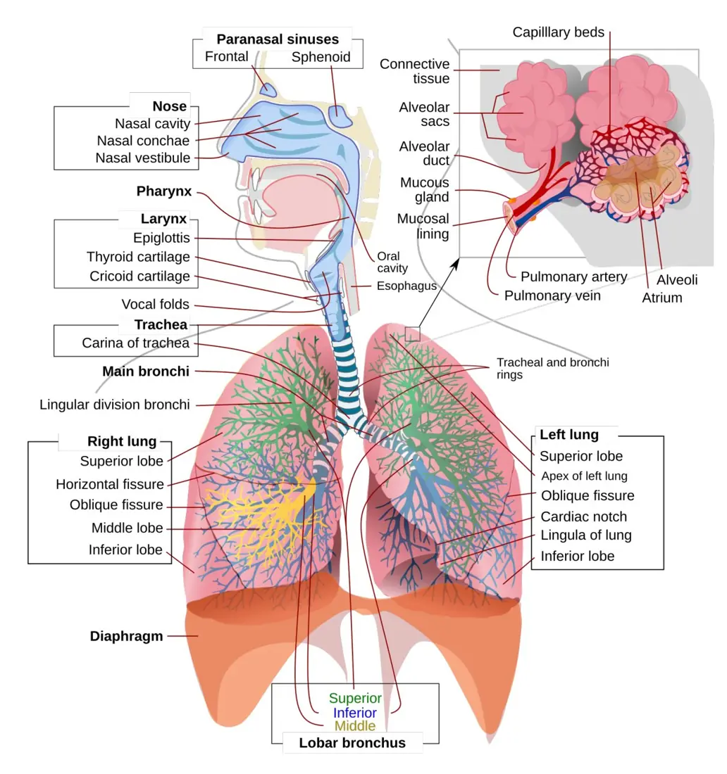
- The route nostrils → nasal cavity → pharynx → larynx → trachea → bronchi → bronchioles → alveoli, and the epithelium of each part (pseudostratified ciliated columnar in trachea and bronchi; simple squamous in the alveolus).
- Trachea: 10–12 cm, C-shaped hyaline cartilage rings open behind; the right bronchus is wider, shorter and more vertical, so foreign bodies lodge there.
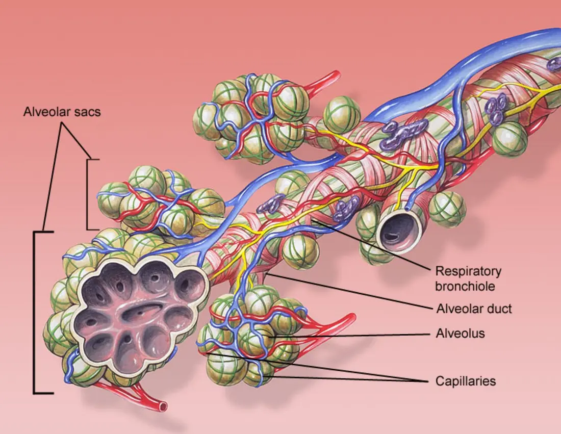
- Alveoli: 300–500 million, total surface about 70–80 m², wall 0.5 μm thick; type II cells make surfactant.
- Breathing: inspiration is active (diaphragm + external intercostals), expiration passive; intrapleural pressure is always negative.
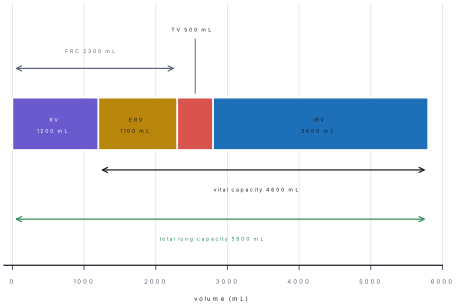
- Volumes: TV 500, IRV 3000, ERV 1100, RV 1200 mL; VC = TV + IRV + ERV; TLC = VC + RV; FRC = ERV + RV.
- Partial pressures: alveolar pO₂ 104, arterial 95–100, venous 40 mmHg; pCO₂ 40 arterial, 45 venous.
- O₂ travels 97 % on haemoglobin; CO₂ travels 70 % as bicarbonate, 23 % carbamino, 7 % dissolved.
- Control: the medulla sets the rhythm and answers a rise in CO₂/H⁺; the carotid and aortic bodies answer a fall in O₂ below about 60 mmHg.
6.1 Respiratory organs and the mechanism of breathing
Respiration is the whole process by which an animal takes in O₂, uses it to release energy from food and gives out CO₂. It has three levels:
| Level | Where | What happens |
|---|---|---|
| External respiration (breathing) | lungs | exchange of O₂ and CO₂ between alveolar air and blood |
| Internal respiration | tissues | exchange between blood and tissue cells |
| Cellular respiration | mitochondria | C₆H₁₂O₆ + 6O₂ → 6CO₂ + 6H₂O + energy (about 38 ATP) |
The organs, from nostril to alveolus
The airway is divided into a conducting part (nostrils to terminal bronchioles), which carries, warms, moistens and filters the air, and a respiratory part (respiratory bronchioles, alveolar ducts and alveoli), where exchange happens. The conducting part holds about 150 mL of air — the dead space — which takes no part in exchange.
| Organ | Structure | Epithelium | Job |
|---|---|---|---|
| Nostrils and nasal cavity | vestibule with hairs; three conchae; richly supplied with blood | stratified squamous at the vestibule, ciliated columnar with goblet cells above | filters, warms to about 37 °C, moistens, and smells |
| Pharynx (naso-, oro-, laryngo-) | muscular tube behind the mouth | stratified squamous (ciliated columnar in the nasopharynx) | common passage for air and food |
| Larynx (voice box) | thyroid, cricoid and arytenoid cartilages; vocal cords; glottis guarded by the epiglottis | stratified squamous over the cords, ciliated columnar elsewhere | produces sound; keeps food out of the airway |
| Trachea (windpipe) | 10–12 cm long, 2.5 cm wide; 16–20 C-shaped hyaline cartilage rings open behind, closed by the trachealis muscle | pseudostratified ciliated columnar with goblet cells | conducts air; mucus traps dust, cilia sweep it up |
| Primary bronchi | one to each lung; the right is wider, shorter and more vertical | same as the trachea | conducts air |
| Bronchioles | no cartilage; smooth muscle in the wall | ciliated cuboidal, then simple cuboidal | control airflow (bronchoconstriction, bronchodilation) |
| Alveoli | 300–500 million air sacs; wall 0.5 μm thick | simple squamous (type I cells), type II cells, macrophages | gas exchange; surfactant |
The mucociliary escalator is the defence of the conducting zone: goblet cells and submucosal glands make mucus, and the cilia beat upwards about 1,000 times a minute to carry trapped dust and bacteria to the pharynx, where they are swallowed. Cigarette smoke paralyses the cilia, which is why smokers cough.
The larynx and the trachea
The larynx is a box of cartilages at the top of the trachea. The thyroid cartilage forms the Adam's apple; the cricoid is a complete ring below it; the arytenoid cartilages move the vocal cords, and air passing between them produces sound. The glottis is the opening; the epiglottis folds over it during swallowing.
The trachea runs down the neck in front of the oesophagus and divides at the carina (level of the fourth to fifth thoracic vertebra) into the two primary bronchi. Its cartilage rings are C-shaped, incomplete at the back, so that a bolus of food in the oesophagus can bulge into it.
The right primary bronchus is wider, shorter and more vertical than the left, so an inhaled peanut or a coin usually lodges in the right lung. This is a standard application question, and the distractor is always "the left bronchus because it is longer".
The lungs and the pleura
Each lung lies in its own pleural cavity. The visceral pleura sticks to the lung, the parietal pleura lines the chest wall, and a thin film of pleural fluid between them holds the two surfaces together by surface tension. This is what keeps the lung inflated.
| Feature | Right lung | Left lung |
|---|---|---|
| Lobes | three (upper, middle, lower) | two (upper, lower) |
| Fissures | oblique and horizontal | oblique only |
| Notch | none | cardiac notch (room for the heart) |
| Size | shorter and wider (the liver pushes it up) | longer and narrower |
The alveolus
An alveolus is a thin-walled sac about 0.2–0.3 mm across, surrounded by a network of capillaries. Its wall has three cell types:
- Type I (squamous) cells — about 95 % of the surface; only 0.1–0.5 μm thick; they form the respiratory membrane with the capillary endothelium.
- Type II (septal) cells — cuboidal; they secrete surfactant, a mixture rich in dipalmitoyl lecithin, which lowers surface tension and stops the alveoli collapsing as they empty. A premature baby born before about 28–30 weeks may lack surfactant and develop respiratory distress syndrome.
- Alveolar macrophages — engulf dust and bacteria.
The respiratory membrane (alveolar epithelium + fused basement membrane + capillary endothelium) is only 0.5 μm thick — about 1/100 of the thickness of this page — so diffusion is very fast.
The mechanism of breathing
Inspiration (inhalation) is active. The diaphragm contracts and flattens (descending about 1.5 cm), and the external intercostal muscles contract and lift the ribs up and out. The thoracic cavity gets bigger in all three dimensions, so the pressure inside the lungs (intrapulmonary pressure) falls about 1 mmHg below atmospheric, and air flows in.
Expiration (exhalation) at rest is passive. The muscles relax, the elastic lung tissue and the surface tension of the alveoli recoil, the thoracic volume falls, intrapulmonary pressure rises about 1 mmHg above atmospheric, and air flows out. Forced expiration uses the internal intercostals and the abdominal muscles.
| Pressure | Value | Meaning |
|---|---|---|
| Atmospheric | 760 mmHg | the reference |
| Intrapulmonary (alveolar) | −1 mmHg in inspiration, +1 mmHg in expiration | the small difference that moves air |
| Intrapleural | −4 mmHg at rest, −6 to −8 mmHg in inspiration | always subatmospheric; keeps the lung stretched against the chest wall |
The intrapleural pressure is always negative because the growing chest pulls the elastic lung outward, and the fluid film resists separation. If air enters the pleural cavity — a pneumothorax from a stab wound or a burst bleb — the negative pressure is lost, the lung's own elasticity makes it collapse, and breathing on that side fails.
Compliance is the ease with which the lungs stretch: a healthy lung inflates easily because surfactant lowers surface tension and the elastic fibres are arranged to recoil gently. Elasticity is the tendency to spring back, and it is what drives quiet expiration. In emphysema elasticity is lost and compliance rises, so the lung inflates easily but empties badly; in fibrosis compliance falls and the lung is stiff, so breathing costs much more work.
| Muscle | Nerve | Action |
|---|---|---|
| Diaphragm | phrenic nerve (C3, C4, C5) | flattens, increases vertical diameter — about 75 % of quiet inspiration |
| External intercostals | intercostal nerves | raise the ribs, increase the transverse diameter |
| Sternocleidomastoid, scalenes | cervical nerves | lift the sternum and first ribs in forced inspiration |
| Internal intercostals, abdominal muscles | intercostal nerves | pull the ribs down and squeeze the abdomen in forced expiration |
Lung volumes and capacities
| Name | Definition | Typical value (young adult man) |
|---|---|---|
| Tidal volume (TV) | air moved in or out in one quiet breath | 500 mL |
| Inspiratory reserve volume (IRV) | extra air taken in after a normal inspiration | 3000 mL |
| Expiratory reserve volume (ERV) | extra air forced out after a normal expiration | 1100 mL |
| Residual volume (RV) | air left in the lungs that cannot be breathed out | 1200 mL |
| Inspiratory capacity (IC) | TV + IRV | 3500 mL |
| Functional residual capacity (FRC) | ERV + RV | 2300 mL |
| Vital capacity (VC) | TV + IRV + ERV — the largest breath possible | 4600 mL |
| Total lung capacity (TLC) | VC + RV | 5800 mL |
Two rules answer almost every volume question: vital capacity never includes the residual volume, and the residual volume cannot be measured by a simple spirometer — it needs helium dilution or body plethysmography.
Worked examples
Problem. A resting student has a tidal volume of 500 mL and breathes 12 times a minute. The dead space is 150 mL. Find the minute ventilation and the alveolar ventilation.
Solution. Minute ventilation = 500 \times 12 = 6000 mL = 6.0 L min⁻¹.
Alveolar ventilation = (500 - 150) \times 12 = 4200 mL = 4.2 L min⁻¹.
So 1.8 L of every 6 L breathed only fills the conducting airways. During exercise the depth of breathing matters more than the rate, because the dead space is fixed.
Problem. A person has 15 g of haemoglobin per 100 mL of blood, and 1 g of haemoglobin binds 1.34 mL of O₂ when saturated. How much O₂ does 5 L of arterial blood carry?
Solution. Per 100 mL: 15 \times 1.34 = 20.1 mL. Per litre: 201 mL. In 5 L: 201 \times 5 \approx 1005 mL ≈ 1.0 L of O₂.
This is why anaemia causes breathlessness even when the lungs are normal: the volume of blood is fine, but there is too little haemoglobin to fill it.
Problem. A spirometer gives TV = 500 mL, IRV = 3000 mL and ERV = 1100 mL. The residual volume is measured separately as 1200 mL. Find VC and TLC.
Solution. VC = 500 + 3000 + 1100 = 4600 mL. TLC = 4600 + 1200 = 5800 mL. A student who writes 4600 mL for TLC has forgotten the residual volume — the commonest error in this topic.
6.2 Exchange and transport of gases; regulation of respiration
Partial pressures
Gases move from a region of higher partial pressure to one of lower partial pressure. That single sentence explains all gas exchange.
| Site | pO₂ (mmHg) | pCO₂ (mmHg) |
|---|---|---|
| Atmospheric air (dry) | 159 | 0.3 |
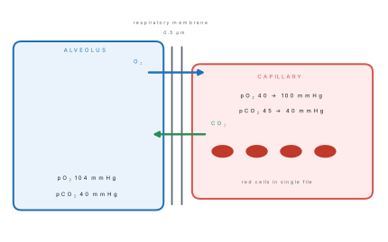
| Alveolar air | 104 | 40 |
| Arterial blood | 95–100 | 40 |
| Venous blood | 40 | 45 |
| Tissue cells | below 40 | above 45 |
Alveolar pO₂ is lower than atmospheric because the air is saturated with water vapour (which adds 47 mmHg) and because O₂ is constantly being absorbed.
Exchange across the respiratory membrane
- In the lungs: alveolar pO₂ (104) > venous pO₂ (40), so O₂ diffuses into the blood; venous pCO₂ (45) > alveolar pCO₂ (40), so CO₂ diffuses out.
- In the tissues: arterial pO₂ (95) > tissue pO₂ (<40), so O₂ leaves the blood; tissue pCO₂ (>45) > arterial pCO₂ (40), so CO₂ enters it.
Diffusion is by simple diffusion — no energy, no carrier. It is fast because the membrane is 0.5 μm thick, the area is 70–80 m², and the gradients are steep. CO₂ diffuses about 20 times faster than O₂ because it is far more soluble in the membrane, which is why hypoxia comes before hypercapnia in most lung disease.
Transport of oxygen
About 97 % of the O₂ in blood is carried by haemoglobin and 3 % is dissolved in the plasma. Each haemoglobin molecule has four haem groups, each with one Fe²⁺, and binds four O₂ molecules:
Oxyhaemoglobin is bright red; deoxyhaemoglobin is dark red-purple, which is why venous blood looks darker. Carboxyhaemoglobin — haemoglobin with carbon monoxide — cannot carry O₂ at all, and CO binds about 200–250 times more strongly than O₂, so even a little CO is dangerous.
The oxygen dissociation curve and the Bohr effect
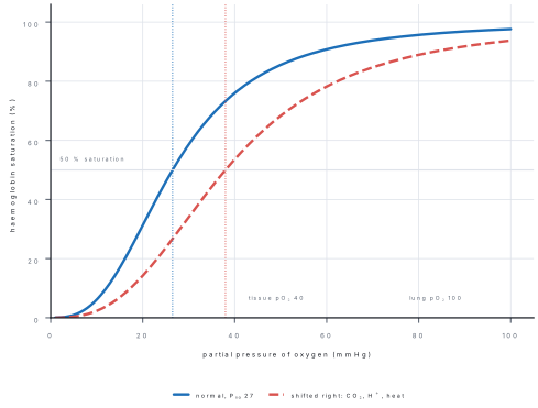
Plot the percentage saturation of haemoglobin against pO₂ and you get a sigmoid (S-shaped) curve. Its shape matters:
- At the lungs (pO₂ about 100 mmHg) haemoglobin is about 98 % saturated — it picks up O₂ easily.
- At the tissues (pO₂ about 40 mmHg) it is about 75 % saturated — it lets O₂ go.
- The P₅₀ is the pO₂ at which haemoglobin is 50 % saturated: about 26–27 mmHg. A low P₅₀ means high affinity.
The curve shifts to the right (affinity falls, O₂ is released more easily) when any of these rise:
| Factor | Why |
|---|---|
| pCO₂ | more CO₂ → more H⁺ (carbonic acid) |
| H⁺ (lower pH) | the Bohr effect proper |
| Temperature | exercising muscle is warmer |
| 2,3-BPG | made in red cells; rises at altitude and in anaemia |
A shift to the left (higher affinity) happens with a fall in CO₂, H⁺, temperature and 2,3-BPG, and in fetal haemoglobin, which must take O₂ from the mother's blood across the placenta.
The Bohr effect says that in active tissue, where CO₂ and H⁺ are high, haemoglobin releases O₂ more readily. Students often write that it "helps haemoglobin pick up oxygen" — that is the opposite. The same shift helps loading in the lung, where CO₂ is low, but the effect is named for the tissue side.
Transport of carbon dioxide
| Form | Percentage | Where / how |
|---|---|---|
| Bicarbonate (HCO₃⁻) | 70 % | CO₂ + H₂O → H₂CO₃ → H⁺ + HCO₃⁻, catalysed by carbonic anhydrase inside the red cell; HCO₃⁻ leaves into the plasma and Cl⁻ enters the cell (chloride shift) |
| Carbaminohaemoglobin | 23 % | CO₂ binds the globin (not the haem) — HbCO₂ |
| Dissolved in plasma | 7 % | simple solution; this is the part that sets the pCO₂ |
The Haldane effect is the mirror of the Bohr effect: deoxygenated haemoglobin carries more CO₂. In the tissues, where Hb has just given up O₂, it takes up CO₂ and H⁺ easily; in the lungs, oxygenation pushes CO₂ out. The two effects together make gas transport far more efficient than either alone.
The commonest wrong answer in the whole chapter
"Most CO₂ is carried as carbaminohaemoglobin" is wrong. 70 % travels as bicarbonate, 23 % as carbamino compounds and 7 % dissolved. A related trap: the enzyme that makes bicarbonate is carbonic anhydrase, and it is inside the red cell, not in the plasma.
Regulation of respiration
Breathing is rhythmic and automatic, but it can be overridden for a while by the will — which is why we can hold our breath, and why speech is possible.
| Centre | Position | Function |
|---|---|---|
| Dorsal respiratory group | medulla oblongata | sets the basic rhythm — inspiration for about 2 s, then relaxation |
| Ventral respiratory group | medulla | active only in forced breathing; drives expiration |
| Pneumotaxic centre | upper pons | switches inspiration off, so it controls the rate and depth |
| Apneustic centre | lower pons | prolongs inspiration |
| Central chemoreceptors | ventral medulla | respond to H⁺ in the cerebrospinal fluid, which rises when blood CO₂ rises |
| Peripheral chemoreceptors | carotid bodies and aortic bodies | respond to a fall in O₂ (strongly below about 60 mmHg) and to a rise in H⁺ |
| Stretch receptors | lungs | Hering–Breuer reflex — a big inflation stops inspiration |
| Cerebral cortex | cerebrum | voluntary control, speech, breath-holding |
The main drive to breathe is CO₂, not O₂. A rise in arterial pCO₂ of only a few mmHg increases ventilation greatly. CO₂ crosses the blood–brain barrier easily and forms H⁺ in the cerebrospinal fluid, and the central chemoreceptors respond to that H⁺. Oxygen lack is only a back-up stimulus, and it becomes important only when arterial pO₂ falls below about 60 mmHg — which is why a patient breathing oxygen may lose the drive to breathe if the disease has made him depend on hypoxic drive.
(1) The respiratory centre is in the medulla, not the pons, cerebellum or cortex; the pons only modifies it. (2) The centre responds chiefly to CO₂/H⁺. A stem that says "the respiratory centre is stimulated mainly by a fall in oxygen" is the standard wrong option.
Exercise and high altitude
In exercise, ventilation may rise from 6 to 100 L min⁻¹. The depth of each breath increases first, because the dead space is fixed and deep breathing is more efficient than fast shallow breathing. More CO₂ and H⁺ from the muscles stimulate the respiratory centre, and the Bohr shift delivers more O₂ where it is needed.
At high altitude the partial pressure of oxygen in the air falls, so arterial pO₂ falls and the carotid bodies increase ventilation. The person also makes more 2,3-BPG, which shifts the dissociation curve right, and over weeks produces more red cells (erythropoietin from the kidney). This is why climbers need days to acclimatise, and why acute mountain sickness — headache, breathlessness, nausea — strikes those who go up too fast.
6.3 Respiratory disorders
| Disorder | What happens | Cause | Key clinical point |
|---|---|---|---|
| Asthma | reversible narrowing of the bronchioles — smooth-muscle spasm, mucosal oedema, thick mucus | allergy (house dust, pollen), cold air, exercise, stress; histamine and leukotrienes; eosinophils | wheezing, difficulty in expiration, attacks; treated with bronchodilators and steroids |
| Bronchitis (chronic) | inflammation of bronchi with too much mucus; goblet cells enlarge | smoking, dust, pollution | cough with sputum for at least 3 months in 2 successive years |
| Emphysema | alveolar walls destroyed, air spaces enlarge, elastic recoil lost | smoking, α₁-antitrypsin deficiency | barrel chest, pursed-lip breathing, ↑ residual volume, ↓ surface area, "pink puffer" |
| Pneumonia | alveoli filled with fluid and pus — consolidation | Streptococcus pneumoniae most often; viruses | fever, cough, rusty sputum, crackles; a diffusion problem |
| Tuberculosis | caseous (cheese-like) necrosis and cavities, usually at the lung apex | Mycobacterium tuberculosis, droplet infection | chronic cough, evening fever, weight loss; Ghon complex; BCG vaccine; DOTS treatment for 6 months |
| Lung cancer | malignant growth of the bronchial epithelium | smoking (tar), asbestos, radon | persistent cough, blood in sputum, weight loss |
Emphysema and chronic bronchitis together are called COPD (chronic obstructive pulmonary disease). In both, air is trapped, so residual volume rises and vital capacity falls; the chest becomes barrel-shaped and expiration is prolonged through pursed lips, which keeps the small airways from collapsing.
| Feature | Obstructive (asthma, bronchitis, emphysema) | Restrictive (pneumonia, TB, fibrosis) |
|---|---|---|
| Main problem | air cannot get out | lung cannot fill in |
| FEV₁ | reduced | reduced |
| Residual volume | increased | normal or reduced |
| Vital capacity | normal or slightly reduced | reduced |
| Expiration | prolonged, wheeze | rapid, shallow breathing |
Effects of smoking.
- Nicotine — addictive; raises heart rate and blood pressure; constricts vessels.
- Tar — a mixture of carcinogens; causes lung cancer and paralyses the cilia.
- Carbon monoxide — binds haemoglobin about 200–250 times more strongly than O₂, forming carboxyhaemoglobin, so less O₂ is carried.
- Irritants — make the mucosa swell and the glands secrete more mucus, while the paralysed cilia cannot clear it: the smoker's cough, then chronic bronchitis, then emphysema.
Answering a respiratory disease question
Ask three things. Obstructive or restrictive? Asthma, bronchitis and emphysema obstruct the airway (air trapping, ↑ RV); pneumonia, TB and fibrosis restrict the lung (↓ VC, ↓ compliance). Reversible or not? Asthma is reversible; emphysema is not. Infective or not? Pneumonia and TB are infective; asthma, emphysema and bronchitis (chronic) are not.
Formula and fact sheet
Structure and numbers
| Fact | Value |
|---|---|
| Trachea | 10–12 cm long, 2.5 cm wide, 16–20 C-shaped hyaline rings |
| Carina | level of T4–T5 |
| Right lung / left lung | 3 lobes / 2 lobes; left has a cardiac notch |
| Alveoli | 300–500 million |
| Alveolar surface area | 70–80 m² |
| Respiratory membrane | 0.5 μm |
| Surfactant | type II alveolar cells; dipalmitoyl lecithin; deficiency → RDS |
| Dead space | about 150 mL |
| Respiratory rate (adult, rest) | 12–20 per minute |
| Diaphragm nerve | phrenic, C3–C5 |
Volumes (young adult man)
| Volume | mL |
|---|---|
| Tidal volume (TV) | 500 |
| Inspiratory reserve (IRV) | 3000 |
| Expiratory reserve (ERV) | 1100 |
| Residual volume (RV) | 1200 |
| Inspiratory capacity (TV + IRV) | 3500 |
| Functional residual capacity (ERV + RV) | 2300 |
| Vital capacity (TV + IRV + ERV) | 4600 |
| Total lung capacity (VC + RV) | 5800 |
Pressures (mmHg)
| Site | pO₂ | pCO₂ |
|---|---|---|
| Atmosphere | 159 | 0.3 |
| Alveolus | 104 | 40 |
| Arterial blood | 95–100 | 40 |
| Venous blood | 40 | 45 |
Intrapulmonary pressure: −1 mmHg in inspiration, +1 mmHg in expiration. Intrapleural pressure: −4 mmHg at rest, −6 to −8 mmHg in inspiration.
Gas transport
| Gas | Form | % |
|---|---|---|
| O₂ | oxyhaemoglobin | 97 |
| O₂ | dissolved in plasma | 3 |
| CO₂ | bicarbonate | 70 |
| CO₂ | carbaminohaemoglobin | 23 |
| CO₂ | dissolved | 7 |
- 1 g haemoglobin carries 1.34 mL O₂; 15 g/dL blood carries about 20 mL/dL.
- P₅₀ ≈ 27 mmHg; Bohr shift right with ↑CO₂, ↑H⁺, ↑temperature, ↑2,3-BPG.
- Haldane effect: deoxygenated Hb carries more CO₂.
- CO₂ diffuses about 20× faster than O₂; CO binds Hb 200–250× more strongly.
Control
| Centre | Site | Responds to |
|---|---|---|
| Dorsal respiratory group | medulla | sets rhythm |
| Pneumotaxic centre | pons | rate and depth |
| Central chemoreceptors | medulla | H⁺ from CO₂ |
| Peripheral chemoreceptors | carotid and aortic bodies | ↓O₂ (below 60 mmHg), ↑H⁺ |
Diseases
| Disease | One line |
|---|---|
| Asthma | reversible bronchospasm, wheeze, expiratory difficulty |
| Chronic bronchitis | cough + sputum ≥ 3 months for 2 years, smoking |
| Emphysema | destroyed alveolar walls, ↑ RV, barrel chest |
| Pneumonia | S. pneumoniae, consolidation, rusty sputum |
| Tuberculosis | M. tuberculosis, apical cavities, BCG, DOTS 6 months |
Traps
- Most CO₂ is carried as bicarbonate (70 %), not carbamino (23 %).
- The respiratory centre answers CO₂/H⁺, not O₂.
- Quiet expiration is passive; only forced expiration uses muscles.
- Intrapleural pressure is always negative.
- Vital capacity excludes the residual volume.
- The right bronchus takes foreign bodies.
- Type II cells make surfactant; type I cells do the exchange.
Chapter summary
- Air passes nostrils → nasal cavity → pharynx → larynx → trachea → bronchi → bronchioles → alveoli; the conducting part is lined by pseudostratified ciliated columnar epithelium, the alveolus by simple squamous.
- The trachea has 16–20 C-shaped hyaline rings and divides at the carina; the right bronchus is wider, shorter and more vertical, so it takes inhaled objects.
- The right lung has three lobes, the left two; the pleura holds the lung against the chest wall with a negative intrapleural pressure.
- Alveoli number 300–500 million, give 70–80 m² of surface and a 0.5 μm barrier; type II cells make surfactant, without which premature babies get RDS.
- Inspiration is active (diaphragm, phrenic C3–C5, plus external intercostals); quiet expiration is passive.
- Volumes: TV 500, IRV 3000, ERV 1100, RV 1200 mL; VC = TV + IRV + ERV = 4600; TLC = VC + RV = 5800; FRC = ERV + RV.
- Gas exchange is by simple diffusion down partial-pressure gradients: alveolar pO₂ 104, venous 40; venous pCO₂ 45, alveolar 40; CO₂ diffuses about 20× faster than O₂.
- O₂: 97 % on haemoglobin, 3 % dissolved; the sigmoid curve has P₅₀ ≈ 27 mmHg and shifts right with CO₂, H⁺, heat and 2,3-BPG (Bohr effect).
- CO₂: 70 % bicarbonate (carbonic anhydrase, chloride shift), 23 % carbamino, 7 % dissolved; Haldane effect.
- The medulla sets the rhythm and responds mainly to CO₂/H⁺; the carotid and aortic bodies respond to a fall in O₂.
- Asthma is reversible obstruction, chronic bronchitis is cough with sputum, emphysema destroys alveolar walls (↑ RV), pneumonia consolidates alveoli and TB causes apical caseous cavities; smoking paralyses cilia, adds CO and tar.
Practice questions
Set A — Recall (24 questions)
- The right lung of man hasaone lobebthree lobesctwo lobesdfour lobes
- The trachea is lined byasimple squamous epitheliumbstratified squamous epitheliumcpseudostratified ciliated columnar epitheliumdsimple cuboidal epithelium
- Pulmonary surfactant is secreted byatype II alveolar cellsbtype I alveolar cellscgoblet cellsdalveolar macrophages
- The C-shaped rings of the trachea are made ofaelastic cartilagebfibrocartilagecbonedhyaline cartilage
- The tidal volume of a resting adult is abouta1100 mLb3000 mLc500 mLd1200 mL
- The residual volume of the lungs is abouta500 mLb1100 mLc4800 mLd1200 mL
- Vital capacity is equal toatidal volume + residual volumebtidal + inspiratory reserve + expiratory reservecinspiratory + expiratory reserve + residualdtotal lung capacity − vital capacity
- The diaphragm is supplied by theaphrenic nervebvagus nervecintercostal nervesdsympathetic chain
- During quiet inspiration the diaphragmacontracts and flattensbrelaxes and domes upwardsccontracts and domes upwardsddoes not move at all
- The intrapleural pressure isaalways positivebalways slightly below atmosphericcequal to atmosphericdzero
- Most of the oxygen in blood is carriedadissolved in the plasmabas bicarbonatecas carbaminohaemoglobindas oxyhaemoglobin
- Most of the carbon dioxide in blood is carried asacarbaminohaemoglobinbdissolved gascbicarbonatedcarbonic acid
- The respiratory centre is situated in theacerebrumbcerebellumcpons onlydmedulla oblongata
- The chief stimulus to the respiratory centre is a rise inaCO₂ and H⁺bO₂cnitrogendwater vapour
- Carbonic anhydrase, which speeds up the formation of bicarbonate, is found in theaplasmabalveolar aircred blood celldplatelet
- The partial pressure of oxygen in alveolar air is abouta40 mmHgb104 mmHgc95 mmHgd159 mmHg
- The number of alveoli in the two lungs is abouta3000b300 millionc30 milliond3 million
- The thickness of the respiratory membrane is abouta5 μmb50 μmc0.5 μmd0.05 μm
- Which of the following is NOT part of the conducting zone?atracheabbronchuscbronchioledalveolus
- One gram of haemoglobin can combine with abouta1.34 mL of O₂b20 mL of O₂c0.3 mL of O₂d4 mL of O₂
- The P₅₀ of adult haemoglobin is abouta40 mmHgb100 mmHgc27 mmHgd10 mmHg
- Quiet expiration is produced mainly byaelastic recoil of the lungsbcontraction of abdominal musclesccontraction of internal intercostalsdcontraction of the diaphragm
- Destruction of the alveolar walls with loss of elastic recoil is the feature ofaasthmabemphysemacpneumoniadtuberculosis
- BCG vaccine gives protection againstapneumoniabasthmacinfluenzadtuberculosis
Answers and explanations — Set A
1. (b) — the right lung has upper, middle and lower lobes; the left has two, with a cardiac notch.
2. (c) — pseudostratified ciliated columnar with goblet cells; the cilia sweep the mucus upwards.
3. (a) — type II (septal) cells. Type I cells are the flat cells that form the exchange surface.
4. (d) — hyaline cartilage, incomplete at the back so that the oesophagus can bulge into the trachea.
5. (c) — 500 mL per quiet breath. 1100 mL is the expiratory reserve and 3000 mL the inspiratory reserve.
6. (d) — about 1200 mL stays in the lungs and cannot be breathed out, so a spirometer cannot measure it.
7. (b) — VC = TV + IRV + ERV = 4600 mL. It never includes the residual volume.
8. (a) — the phrenic nerve, from spinal roots C3–C5.
9. (a) — it contracts and flattens, increasing the vertical diameter of the thorax.
10. (b) — always subatmospheric; if it becomes equal to atmospheric (pneumothorax) the lung collapses.
11. (d) — about 97 % as oxyhaemoglobin; only 3 % is dissolved.
12. (c) — about 70 % as bicarbonate, 23 % as carbamino compounds and 7 % dissolved.
13. (d) — the medulla oblongata; the pons only modifies the rhythm.
14. (a) — a small rise in CO₂ raises H⁺ and drives ventilation; a fall in O₂ matters only below about 60 mmHg.
15. (c) — inside the red cell, which is why the chloride shift is needed to balance the charge.
16. (b) — 104 mmHg. 159 is atmospheric, 40 is venous blood.
17. (b) — 300–500 million, giving about 70–80 m² of surface.
18. (c) — 0.5 μm, about a hundredth of the thickness of a printed page.
19. (d) — the alveolus is the respiratory zone, where exchange occurs.
20. (a) — 1.34 mL per gram; with 15 g/dL the blood carries about 20 mL per 100 mL.
21. (c) — about 27 mmHg; a higher P₅₀ means lower affinity.
22. (a) — at rest expiration is passive; the muscles are used only in forced expiration.
23. (b) — emphysema. Asthma narrows the airway but does not destroy alveolar walls.
24. (d) — BCG (Bacillus Calmette–Guérin) is a live attenuated vaccine against tuberculosis.
Set B — Understanding (13 questions)
- Air rushes into the lungs during inspiration becauseathe lungs actively suck air inbthe diaphragm pushes air downcthoracic volume rises and intrapulmonary pressure fallsdthe glottis closes
- A stab wound opens the pleural cavity and the lung on that side collapses. The reason is thatathe negative intrapleural pressure is lostbthe bronchus is blockedcsurfactant is destroyeddcarbon dioxide accumulates
- An inhaled peanut most often lodges in the right bronchus because it isalonger and narrowerbmore horizontalclined by different epitheliumdwider, shorter and more vertical
- Surfactant keeps the alveoli open byaraising surface tensionblowering surface tensioncincreasing mucus secretiondkilling bacteria
- The oxygen dissociation curve shifts to the right whenaCO₂ and H⁺ risebCO₂ fallsctemperature fallsdpH rises
- A shift of the curve to the right means that haemoglobinabinds oxygen more tightlybreleases oxygen more readilyccarries less oxygen in totaldstops carrying carbon dioxide
- The respiratory centre responds mainly to CO₂ rather than to O₂ becauseaoxygen cannot cross the blood–brain barrierbthere are no oxygen receptors anywherecCO₂ enters the CSF and forms H⁺ theredarterial oxygen never changes
- The chloride shift refers toachloride entering the alveolusbchloride leaving the red cell in the lungcchloride entering the plasma with bicarbonatedchloride entering the red cell as bicarbonate leaves
- Alveolar ventilation is always less than minute ventilation becauseasome alveoli are not perfusedbsome air is swallowedcoxygen is absorbed on the waydpart of each breath fills the dead space
- In emphysema the residual volume isadecreasedbincreasedcunchangeddzero
- In pneumonia arterial pO₂ falls although ventilation is normal becauseafluid in the alveoli thickens the diffusion barrierbthe bronchi are completely blockedchaemoglobin is absentdthe respiratory centre fails
- The Haldane effect means thataoxygen displaces carbon dioxide from haemoglobinbcarbon dioxide displaces oxygencdeoxygenated haemoglobin carries more CO₂dCO₂ raises the affinity for oxygen
- Smoking causes chronic bronchitis mainly byaparalysing the cilia so that mucus accumulatesbdestroying alveolar walls directlycincreasing surfactantdraising the haemoglobin level
Answers and explanations — Set B
1. (c) — the chest wall and diaphragm enlarge the cavity; the pressure inside the lung falls about 1 mmHg below atmospheric and air flows in. The lungs have no muscle of their own.
2. (a) — the lung is held open by the negative intrapleural pressure and by surface tension between the pleurae; once air enters, elastic recoil collapses it.
3. (d) — the right bronchus leaves the trachea almost vertically, so it is the natural path for a foreign body.
4. (b) — surfactant lowers surface tension, so the alveoli do not collapse at low volume; without it a premature baby gets RDS.
5. (a) — more CO₂ means more H⁺, which lowers affinity; warmth and more 2,3-BPG do the same.
6. (b) — a right shift unloads more O₂ at the tissues; a left shift would mean tighter binding.
7. (c) — CO₂ crosses the blood–brain barrier, forms H⁺ in the cerebrospinal fluid, and the central chemoreceptors respond to that H⁺.
8. (d) — bicarbonate leaves the red cell and chloride enters to keep the charge balanced.
9. (d) — the 150 mL of dead space in the conducting airways takes no part in exchange.
10. (b) — air trapping raises the residual volume and lowers the vital capacity.
11. (a) — consolidation thickens the respiratory membrane, so diffusion fails even though air is moving.
12. (c) — deoxygenated haemoglobin binds CO₂ and H⁺ more readily, which helps uptake in the tissues and release in the lungs.
13. (a) — smoke paralyses the cilia, so mucus and dust collect and provoke the cough.
Set C — Application (9 questions)
- A student has a tidal volume of 500 mL and breathes 12 times a minute. His minute ventilation isa4.2 L min⁻¹b6.0 L min⁻¹c60 L min⁻¹d0.5 L min⁻¹
- The same student has a dead space of 150 mL. His alveolar ventilation isa6.0 L min⁻¹b1.8 L min⁻¹c60 L min⁻¹d4.2 L min⁻¹
- Blood contains 15 g of haemoglobin per 100 mL, and 1 g binds 1.34 mL of O₂. When saturated, 100 mL of blood carries abouta20 mL of O₂b15 mL of O₂c1.34 mL of O₂d100 mL of O₂
- A spirometer gives a vital capacity of 4600 mL and the residual volume is 1200 mL. The total lung capacity isa4600 mLb3400 mLc5800 mLd6000 mL
- A trekker who reaches 5000 m has a lower arterial pO₂. Over the next daysathe oxygen dissociation curve shifts leftbhaemoglobin saturation rises above 100 %c2,3-BPG fallsd2,3-BPG rises and the curve shifts right
- A premature baby develops severe respiratory distress soon after birth. The immediate deficiency is ofasurfactantbhaemoglobinccarbonic anhydrasedcilia
- A man has a barrel chest, pursed-lip breathing and a prolonged expiratory phase. The most likely diagnosis isaasthmabpneumoniacemphysemadtuberculosis
- Carbon monoxide poisoning is dangerous chiefly because COadestroys the alveolar wallsbbinds haemoglobin about 200 times more strongly than O₂cblocks the phrenic nervedcauses a pneumothorax
- A frightened patient hyperventilates and his arterial pCO₂ falls to 25 mmHg. The immediate result isarespiratory acidosisba rise in plasma bicarbonatecincreased oxygen carriagedrespiratory alkalosis
Answers and explanations — Set C
1. (b) — minute ventilation = 500 × 12 = 6000 mL = 6.0 L min⁻¹.
2. (d) — (500 − 150) × 12 = 4200 mL = 4.2 L min⁻¹. Option (a) is the minute ventilation, the commonest slip.
3. (a) — 15 × 1.34 = 20.1 mL per 100 mL, the normal oxygen content of arterial blood.
4. (c) — TLC = VC + RV = 4600 + 1200 = 5800 mL. Option (a) forgets the residual volume.
5. (d) — at altitude the low pO₂ raises 2,3-BPG, which shifts the curve right so that more O₂ is unloaded to the tissues.
6. (a) — type II cells are not yet making enough surfactant, so the alveoli collapse at every breath.
7. (c) — air trapping, loss of elastic recoil and pursed-lip breathing are the signs of emphysema; asthma is reversible and pneumonia gives consolidation.
8. (b) — carboxyhaemoglobin cannot carry O₂, and CO is not displaced easily, so the blood's oxygen capacity falls sharply.
9. (d) — blowing off CO₂ removes H⁺, so the pH rises: respiratory alkalosis.