Chapter 10 · Plant Anatomy · 3 questions in the paper · 27 min read
Plant Anatomy
What the syllabus asks for 3
- Plant tissues: characters, classification, structure and functions
- Types of vascular bundles
- T.S. of monocot and dicot root, stem and leaf
Plant Anatomy carries about 3 of the 40 Botany questions, and it is the most diagram-driven chapter of the paper: the setter describes (or draws) a transverse section and asks what organ, what plant group, or what tissue you are looking at. The content falls into three blocks — the tissues themselves (meristems and the simple and complex permanent tissues), the vocabulary of vascular bundles, and the six standard sections: dicot and monocot root, stem and leaf. Master the small set of diagnostic features that separate those six slides and this chapter is free marks.
- Meristems — characters (dense cytoplasm, thin walls, no big vacuole); apical (length), intercalary (grasses), lateral (thickness: vascular and cork cambium); primary vs secondary (cork cambium by dedifferentiation); protoderm, procambium, ground meristem.
- Simple tissues — parenchyma (chlorenchyma, aerenchyma), collenchyma (living, pectin corners, flexible), sclerenchyma (dead, lignified; fibres and sclereids/stone cells).
- Complex tissues — xylem (tracheids, vessels, fibres dead; parenchyma living) and phloem (enucleate sieve tubes + companion cells, bast fibres, parenchyma); proto-/metaxylem; exarch root, endarch stem.
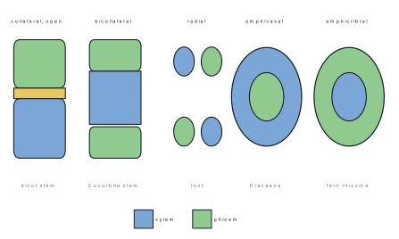
- Bundle words — radial (roots); conjoint collateral; bicollateral (Cucurbitaceae); amphivasal = xylem outside (Dracaena); amphicribral = phloem outside (ferns); open (cambium present) vs closed.
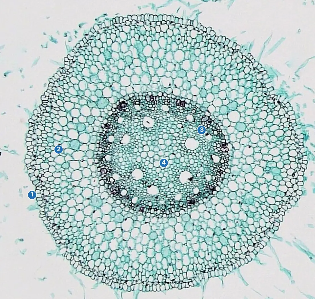
- Root sections — epiblema with root hairs, endodermis with Casparian strips and passage cells, radial exarch bundles; dicot diarch–hexarch, pith absent; monocot polyarch, big pith.
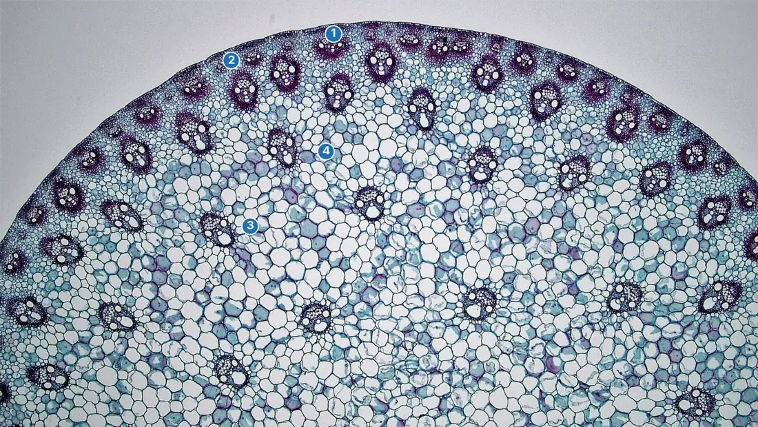
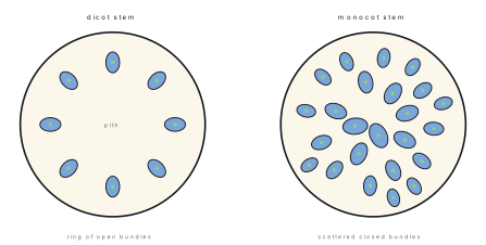
- Stem sections — dicot: ring of open bundles, collenchyma hypodermis, medullary rays; monocot: scattered closed bundles with sclerenchyma sheath, lysigenous cavity, undifferentiated ground tissue.
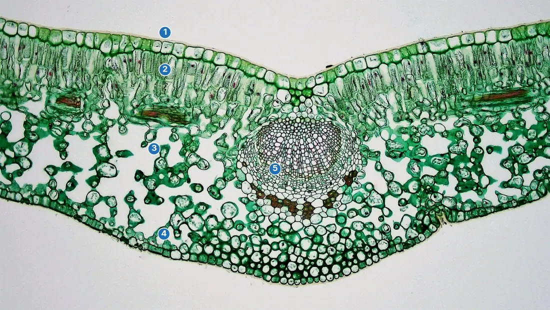
- Leaf sections — dorsiventral dicot (palisade above, spongy below, hypostomatic) vs isobilateral monocot (uniform mesophyll, bulliform/motor cells, dumb-bell guard cells).
10.1 Plant tissues: characters, classification, structure and functions
A tissue is a group of cells of common origin working together. Plant tissues divide first by one question — can the cells still divide? If yes, they are meristematic; if they have matured and stopped, they are permanent.
Meristematic tissues
Meristem cells are small and isodiametric, with thin cellulose walls, dense cytoplasm, a large central nucleus and no large vacuole, and they are packed together without intercellular spaces. They divide repeatedly by mitosis. Three classifications are asked:
By position. - Apical meristem — at root and shoot tips; causes primary growth in length. - Intercalary meristem — pieces of apical meristem left at the bases of internodes and leaves, famous in grasses (it is why a lawn regrows after mowing and grazing). - Lateral meristem — cylinders along the axis, the vascular cambium and cork cambium (phellogen); causes growth in thickness (secondary growth) in dicots and gymnosperms. Monocots generally lack lateral meristems.
By origin. The promeristem is the few initials at the very tip; primary meristems descend from them directly (apical, intercalary and the intrafascicular cambium); secondary meristems arise later when permanent cells regain the power to divide — dedifferentiation — as the cork cambium and interfascicular cambium do.
By function (Haberlandt). At an apex, protoderm makes the epidermis, procambium makes primary xylem, phloem and vascular cambium, and ground meristem makes cortex, endodermis, pericycle, pith and medullary rays.
Simple permanent tissues
Permanent cells arise from meristems by differentiation. Simple tissues are built of one cell type.
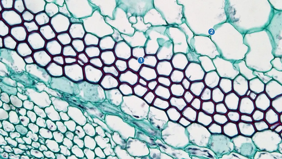
| Tissue | Cells | Walls | Function and notes |
|---|---|---|---|
| Parenchyma | living, isodiametric | thin, cellulose | the packing/storage tissue; with chloroplasts = chlorenchyma (photosynthesis); with big air spaces = aerenchyma (hydrophytes, buoyancy and aeration) |
| Collenchyma | living, elongated | unevenly thickened with pectin (angular, lamellar, lacunar types) | flexible mechanical support in still-growing organs — petioles, young stems, leaf midribs |
| Sclerenchyma | dead at maturity | thick, uniformly lignified, simple pits, narrow lumen | rigid mechanical support; long fibres (bundles) and short sclereids (stone cells) — grit in pear, hard seed coats and nutshells |
The collenchyma–sclerenchyma contrast is a perennial MCQ: collenchyma is living and plastic (grows with the organ), sclerenchyma is dead and rigid (only in organs that have finished elongating).
Complex permanent tissues
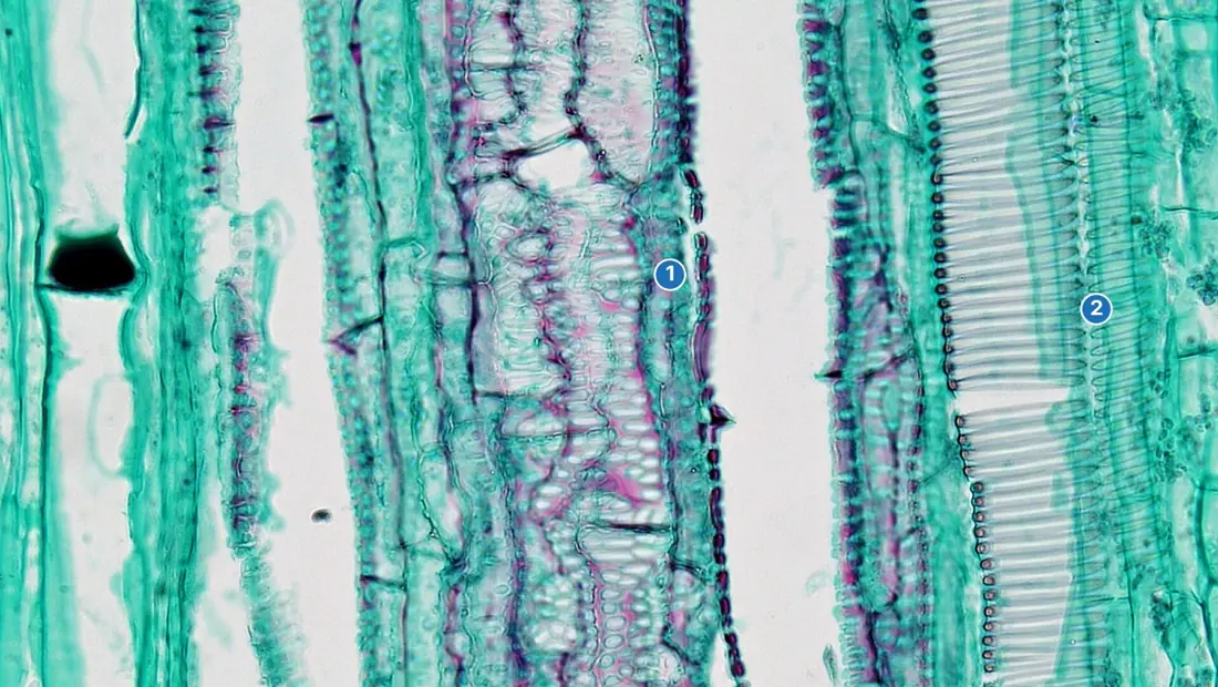
Xylem carries water and minerals upwards from root to leaf.
| Element | Living? | Structure and function |
|---|---|---|
| Tracheids | dead | elongated and tapering, lignified, water passes through bordered pits; the only conducting element of pteridophytes and gymnosperms; conduction + support |
| Vessels | dead | rows of cells with perforated end walls forming continuous wide pipes; the efficient conductors characteristic of angiosperms |
| Xylem fibres | dead | sclerenchymatous; support |
| Xylem parenchyma | living | storage and sideways conduction — the only living xylem element |
Phloem translocates prepared food (mainly sucrose) from source to sink, in either direction as needed.
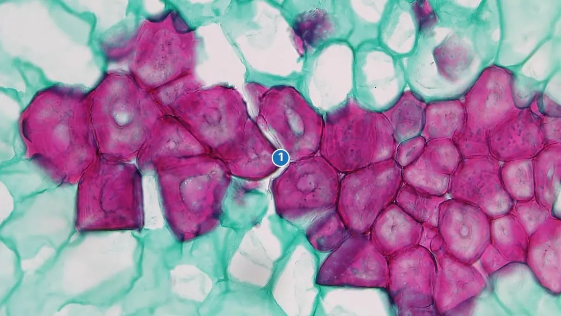
| Element | Living? | Structure and function |
|---|---|---|
| Sieve tube elements | living but enucleate | end-to-end tubes joined by perforated sieve plates; the conducting channel |
| Companion cells | living | narrow, dense cytoplasm, large nucleus; keep the sieve tube alive and load its sugar; confined to angiosperms |
| Phloem fibres (bast fibres) | dead | sclerenchymatous; support — jute, flax and hemp of commerce |
| Phloem parenchyma | living | storage; absent in most monocots — a standard named exception |
Xylem formed first (protoxylem, narrow annular/spiral elements that can stretch) differs from later metaxylem (wide, pitted). Their arrangement names the organ: exarch — protoxylem towards the periphery — in roots; endarch — protoxylem towards the centre — in stems. ("Roots are EXtroverts, stems are ENDoverts.")
Special secretory tissues — laticifers carrying latex (Euphorbia, Ficus, rubber), resin ducts (pines), oil glands and nectaries — complete the list.
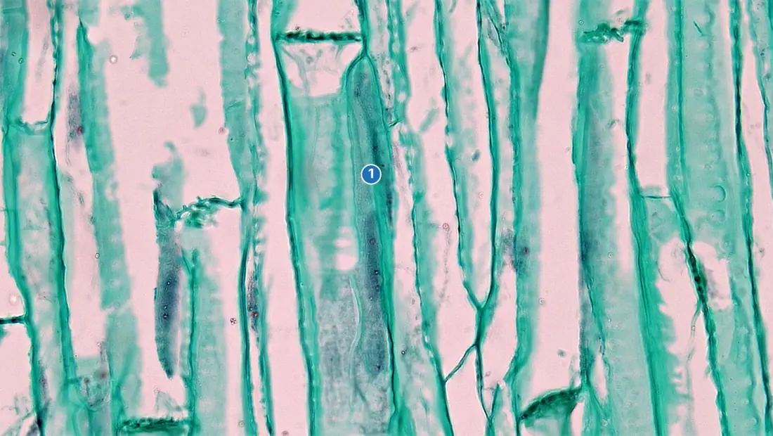
Problem. Four unlabeled transverse sections show: (i) bundles in a ring, each with cambium, xylem endarch, a starch sheath outside the bundles; (ii) bundles scattered in ground tissue, each closed with a sclerenchyma sheath; (iii) radial bundles with four xylem arms, a small pith, pericycle present; (iv) polyarch radial bundles around a large pith. Identify each. Solution. (i) Dicot stem — ring, open bundles, endarch xylem and the starch sheath are its signature. (ii) Monocot stem — scattered, closed, sheathed bundles in undifferentiated ground tissue. (iii) Dicot root — radial bundles, tetrarch (four arms), small pith; the cambium for its secondary growth arrives later. (iv) Monocot root — polyarch xylem and a large pith, no cambium ever. Four slides, four answers, each from two or three diagnostic features — exactly how the paper frames it.
10.2 Types of vascular bundles
A vascular bundle is a strand of xylem and phloem. The paper asks you to name bundles, so learn the vocabulary exactly:
- Radial — xylem and phloem in separate groups on different radii, alternating. The root arrangement.
- Conjoint — xylem and phloem together in one bundle (stems and leaves):
- Collateral — phloem on the outer side only, xylem inside (the usual stem bundle).
- Bicollateral — phloem on both sides of the xylem (Cucurbitaceae — Cucurbita, Luffa; also Solanum).
- Concentric — one tissue completely surrounds the other:
- Amphivasal (leptocentric) — xylem surrounds phloem (Dracaena, Yucca).
- Amphicribral (hadrocentric) — phloem surrounds xylem (ferns).
- Open — cambium present between xylem and phloem: secondary growth possible (dicot stems).
- Closed — no cambium: no secondary growth (monocot stems, all leaves).
Amphi-VAS-al: vascular tissue — wood, xylem — outside. Amphi-CRIB-ral: cribrum = a sieve — sieve tubes, phloem — outside.
10.3 T.S. of monocot and dicot root, stem and leaf
Roots — the shared ground plan
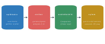
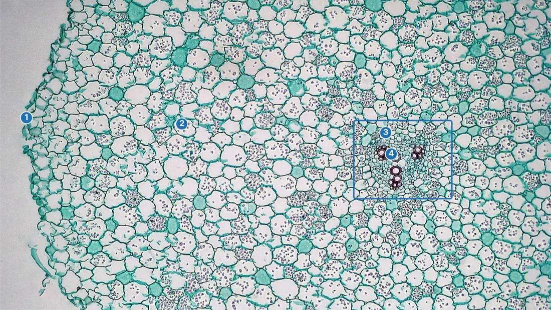
Work from outside in. Epiblema (rhizodermis): one layer of thin-walled cells, many extended into root hairs; no cuticle, no stomata — it absorbs. Cortex: broad parenchyma with air spaces. Endodermis: one ring of barrel-shaped cells whose radial and transverse walls carry a waterproof band of suberin and lignin, the Casparian strip, forcing water and minerals to cross a living membrane (the cell checks what enters the stele). A few endodermal cells opposite the protoxylem stay thin-walled — passage cells, letting water through quickly. Pericycle: one layer inside the endodermis; in roots it is the layer that gives rise to lateral roots (endogenous origin). Stele: xylem and phloem in separate alternating radial groups, xylem exarch, with conjunctive parenchyma between them.
| Feature | Dicot root (e.g. Ranunculus, gram) | Monocot root (e.g. maize) |
|---|---|---|
| Xylem groups | 2–6 — diarch, triarch, tetrarch … hexarch | many, more than 6 — polyarch |
| Pith | absent (xylem meets at the centre) | large, well-developed parenchyma |
| Secondary growth | occurs (cambium forms) | does not occur |
| Shared signs | both: root hairs, Casparian strips and passage cells, radial and exarch bundles |
Stems
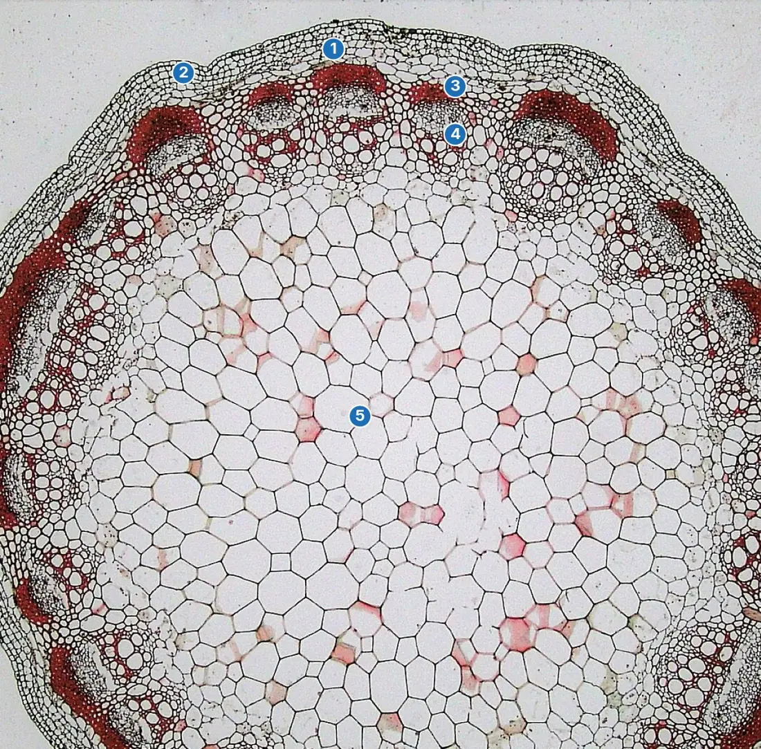
Dicot stem (Helianthus). Epidermis with cuticle, stomata and multicellular hairs; hypodermis of collenchyma; general cortex; endodermis full of starch grains (the starch sheath); pericycle with semilunar sclerenchyma caps over the bundles; then a single ring of vascular bundles (a eustele), uniform in size, each conjoint, collateral, open and endarch, separated by medullary rays; large central pith. The open bundles and ring layout are exactly what secondary growth needs: the cambium completes itself into a ring.
Monocot stem (Zea mays). Epidermis with thick cuticle; hypodermis of sclerenchyma; inside it, no cortex/endodermis/pith at all — one continuous parenchymatous ground tissue; vascular bundles scattered through it (an atactostele), numerous, smaller and denser towards the periphery, each conjoint, collateral and closed, wrapped in a sclerenchymatous bundle sheath, and carrying a water-filled lysigenous cavity (protoxylem lacuna) where the earliest protoxylem vessel broke down. Scattered closed bundles are why a maize stem can never thicken into a trunk.
Leaves
A horizontal dicot leaf is two-faced — dorsiventral. The upper epidermis has a thick cuticle and few or no stomata; the lower has many stomata (hypostomatic). The mesophyll is differentiated: palisade parenchyma above — elongated cells crammed with chloroplasts in the best light, the main photosynthetic layer — and spongy parenchyma below, loose cells and air spaces leading to the stomata. Vein bundles are conjoint, collateral and closed, with xylem towards the upper side and phloem towards the lower.
A vertical monocot (grass/maize) leaf is alike on both sides — isobilateral: stomata on both epidermes with dumb-bell-shaped guard cells (dicot guard cells are kidney-shaped); bulliform (motor) cells — big, thin-walled, watery cells in the upper epidermis that lose turgor in dry weather and roll the leaf inwards to cut water loss; mesophyll undifferentiated (no palisade/spongy split); bundles in a parallel row between the veins.
Problem. A stained transverse section shows: a cuticularised epidermis with stomata and hairs; a collenchymatous hypodermis; conjoint, collateral, endarch vascular bundles of nearly equal size arranged in one ring, each with cambium; medullary rays between the bundles; and a large central pith. Identify the section.
Solution. Eliminate in order. Root hairs and radial, exarch bundles are absent, and a cuticle with stomata is present — so it is not a root. The bundles are in a ring, not scattered — so it is not a monocot stem. Open bundles (cambium present), collenchyma hypodermis, medullary rays and a large pith complete the picture: a young dicot stem. Each decision used one feature the alternatives cannot have — that is how these marks are earned quickly.
Root or stem? Radial bundles + exarch xylem + root hairs = root; conjoint bundles = stem. Dicot or monocot root? Count the xylem groups and look for a pith. Dicot or monocot stem? Ring = dicot, scattered = monocot — then confirm with open vs closed. Leaf? Palisade present = dicot; bulliform cells and dumb-bell guard cells = grass.
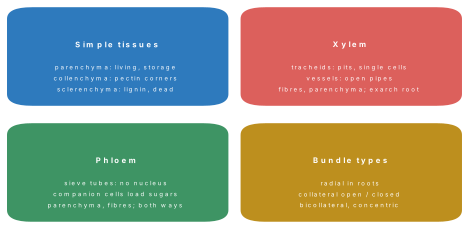
Four pairs the paper swaps: collenchyma is living with pectin corners, sclerenchyma dead with lignin — support that flexes vs support that stiffens; xylem is exarch in the root, endarch in the stem; the endodermis of a root carries Casparian strips, the endodermis of a dicot stem is a starch sheath without them; and companion cells belong to phloem only — xylem's dead pipes need no attendants.
Problem. A transverse section shows radial vascular bundles with exarch protoxylem and more than six xylem patches (polyarch) surrounding a large parenchymatous pith. Identify the organ and group.
Solution. Radial bundles + exarch protoxylem = root; polyarch condition (>6 xylem arches) + large persistent pith = monocot root (e.g., maize).
Formula and fact sheet
- Meristem characters: thin walls, dense cytoplasm, big nucleus, tiny vacuoles, no spaces. Apical = length; intercalary = grasses; lateral = thickness (vascular + cork cambium).
- Origins: protoderm → epidermis; procambium → vascular tissues; ground meristem → cortex, pith etc. Cork cambium = secondary meristem by dedifferentiation.
- Parenchyma: living, thin; chlorenchyma photosynthesises; aerenchyma aerates hydrophytes.
- Collenchyma: living, pectin-thickened corners, flexible. Sclerenchyma: dead, lignified — fibres + sclereids (pear grit, seed coats).
- Xylem: tracheids, vessels, fibres dead; parenchyma the only living element. Vessels = angiosperms; gymnosperms conduct by tracheids alone.
- Phloem: sieve tubes enucleate + companion cells; bast fibres = jute/flax; phloem parenchyma absent in most monocots.
- Exarch = root, endarch = stem; protoxylem narrow and early, metaxylem wide and later.
- Bundles: radial (root); collateral; bicollateral (Cucurbitaceae); amphivasal — xylem outside (Dracaena); amphicribral — phloem outside (ferns); open = cambium, closed = none.
- Root: epiblema + root hairs; Casparian strip in endodermis; passage cells opposite protoxylem; lateral roots from pericycle.
- Dicot root: 2–6 xylem groups, pith absent. Monocot root: polyarch, big pith.
- Dicot stem: collenchyma hypodermis, starch-sheath endodermis, ring of open bundles, medullary rays, pith. Monocot stem: sclerenchyma hypodermis, ground tissue, scattered closed bundles + sheath + lysigenous cavity.
- Leaves: dorsiventral — palisade up, spongy down, hypostomatic, kidney guard cells; isobilateral — uniform mesophyll, bulliform cells, dumb-bell guard cells, stomata both sides.
Rapid-revision tables and high-yield facts
| Feature | Diagnostic |
|---|---|
| Meristems | apical (length), intercalary (grass regrowth), lateral (girth) |
| Collenchyma / sclerenchyma | pectin corners, living / lignin, dead |
| Xylem conductors | tracheids (pits), vessels (perforated tubes) |
| Phloem | sieve tubes + companion cells; bidirectional |
| Root bundle | radial, exarch; dicot 2–6 arms, monocot polyarch |
| Stem bundle | conjoint collateral; dicot open in ring, monocot closed scattered |
| Leaf | dicot dorsiventral, monocot isobilateral + bulliform cells |
| C₄ leaf | Kranz bundle sheath with chloroplasts |
| Casparian strip | endodermis of root (suberin) |
| Lateral roots | arise from pericycle |
Chapter summary
- Tissues are meristematic (still dividing: apical, intercalary and lateral; primary and, by dedifferentiation, secondary) or permanent (simple and complex).
- Parenchyma packs and stores (aerenchyma in hydrophytes), living pectin-rich collenchyma supports growing organs flexibly, and dead lignified sclerenchyma (fibres and stone cells) supports finished ones rigidly.
- Xylem moves water up through dead tracheids and vessels, its parenchyma alone alive; phloem moves food through enucleate sieve tubes serviced by companion cells, with bast fibres for strength.
- Bundle names encode geometry: radial in roots; collateral and bicollateral conjoint bundles in stems; concentric amphivasal (xylem out) and amphicribral (phloem out); open bundles carry cambium, closed do not.
- Roots show epiblema with hairs, a Casparian-striped endodermis with passage cells, pericycle that births lateral roots, and exarch radial groups — few and pith-less in dicots, polyarch with a big pith in monocots.
- Dicot stems ring their open bundles round a pith (ready for secondary growth); monocot stems scatter closed, sheathed bundles with a lysigenous cavity through undifferentiated ground tissue.
- Dorsiventral dicot leaves split mesophyll into palisade and spongy layers and hide stomata below; isobilateral grass leaves keep mesophyll uniform, bear stomata and dumb-bell guard cells on both faces, and roll up by their bulliform cells.
Practice questions
Set A — Recall (25 questions)
- Cells of a meristem are characterised byathin walls, dense cytoplasm and a large nucleusbthick lignified wallsca huge central vacuoledabsence of a nucleus
- Primary growth in the length of root and shoot is caused by theavascular cambiumbcork cambiumcintercalary meristem onlydapical meristem
- Intercalary meristems, found at the bases of internodes, are characteristic ofagrassesbmango treescfernsdmushrooms
- Which of the following is a secondary meristem?acork cambium (phellogen)broot apical meristemcshoot apical meristemdprotoderm
- The tissue that forms the basic packing and storage tissue of plants isasclerenchymabcollenchymacparenchymadxylem
- Aerenchyma, parenchyma with large air spaces, is typically found inaxerophytesbmesophytescparasitesdhydrophytes
- Collenchyma differs from sclerenchyma in beingadead and lignifiedbwithout wallsca conducting tissuedliving with pectin-thickened walls
- Sclerenchyma cells arealiving and thin-walledbdead with thick lignified wallscenucleate and livingdfull of chloroplasts
- Stone cells responsible for the gritty texture of pear fruit areafibresbtracheidsccompanion cellsdsclereids
- Wide vessels with perforated end walls are characteristic of the xylem ofapteridophytesbgymnospermscangiospermsdbryophytes
- The only living element of xylem is theavesselbxylem parenchymactracheiddxylem fibre
- A sieve tube element at maturity lacks aanucleusbcell wallccytoplasmdsieve plate
- The jute and flax fibres of commerce areaxylem fibresbbast (phloem) fibrescsurface fibresdsclereids
- Phloem parenchyma is absent in mostamonocotsbdicotscgymnospermsdferns
- Exarch xylem (protoxylem towards the periphery) is found in theastembleafcrootdflower
- Bicollateral vascular bundles are characteristic of the familyaPoaceaebCucurbitaceaecLiliaceaedBrassicaceae
- In an amphivasal (leptocentric) bundle, as in Dracaena,aphloem surrounds the xylembxylem and phloem alternateccambium surrounds bothdxylem surrounds the phloem
- Radial vascular bundles, with xylem and phloem on different radii, occur inarootsbdicot stemscmonocot stemsdleaves
- A vascular bundle is called open when it possessesacambium between xylem and phloemba bundle sheathca lysigenous cavitydphloem on both sides
- Casparian strips are found in the cells of theaepiblemabpericyclecendodermisdhypodermis
- A well-developed pith is absent, the xylem meeting at the centre, in theamonocot rootbdicot rootcdicot stemdmonocot stem
- In a monocot stem the vascular bundles areain a single ring and openbscattered and closedcradial and exarchdbicollateral
- A lysigenous (water-containing) cavity is a feature of the vascular bundles ofasunflower stembgram rootcmaize stemdmango leaf
- Bulliform (motor) cells that roll the leaf in dry weather occur in the leaves ofamangobpeacgrassesdCucurbita
- In a dorsiventral (dicot) leaf, the mesophyll is differentiated intoacortex and stelebxylem and phloemcpalisade and bulliform tissuedpalisade and spongy parenchyma
Answers and explanations — Set A
1. (a) — Meristem cells are small, thin-walled and crowded with cytoplasm and nucleus; big vacuoles appear only as cells mature.
2. (d) — Apical meristems at the tips lengthen the axis; lateral meristems thicken it.
3. (a) — Grass nodes keep intercalary meristems, so lawns regrow after cutting.
4. (a) — The phellogen arises from permanent cortical cells that dedifferentiate — the definition of a secondary meristem.
5. (c) — Parenchyma fills, stores and (as chlorenchyma) photosynthesises.
6. (d) — Air cavities give hydrophytes buoyancy and a path for oxygen.
7. (d) — Collenchyma stays alive with plastic pectin thickenings and can stretch; sclerenchyma is dead and lignified.
8. (b) — Maturity brings a thick lignified wall, simple pits and loss of the protoplast.
9. (d) — Short, heavily thickened sclereids make pear gritty and seed coats hard.
10. (c) — True vessels are the angiosperm speciality; gymnosperms and ferns manage with tracheids.
11. (b) — Vessels, tracheids and fibres are all dead pipes and props; only xylem parenchyma lives.
12. (a) — The nucleus disintegrates; the companion cell's nucleus runs the sieve tube instead.
13. (b) — Bast fibres are the sclerenchyma of the phloem — jute, flax, hemp.
14. (a) — A standard named absence: monocot phloem usually lacks parenchyma.
15. (c) — Roots are exarch; stems are endarch.
16. (b) — Cucurbita and its relatives carry phloem on both faces of the xylem.
17. (d) — Amphivasal: vascular (xylem) tissue outside, phloem in the middle.
18. (a) — Alternate xylem and phloem poles on different radii diagnose a root.
19. (a) — "Open for (secondary) growth": the fascicular cambium is present.
20. (c) — Suberin–lignin bands in endodermal walls seal the stele against uncontrolled entry.
21. (b) — In dicot roots the few xylem arms meet in the middle; monocot roots keep a large pith.
22. (b) — Atactostele: bundles scattered in ground tissue, all closed.
23. (c) — The earliest protoxylem of the maize bundle breaks down into a water-filled lacuna.
24. (c) — Bulliform cells in the upper epidermis of grass leaves roll them against drought.
25. (d) — Palisade above for light capture, spongy below for gas exchange.
Set B — Understanding (15 questions)
- Meristematic cells lack a large central vacuole mainly because they areadead cellsbstoring foodcdividing repeatedly and packed with cytoplasmdlignified
- Collenchyma, rather than sclerenchyma, supports growing petioles because it isafull of air spacesbdead and rigidcliving and able to stretch as the organ growsdmade only of pectin-free walls
- Vessels conduct water more efficiently than tracheids because theiraend walls are perforated, forming continuous wide pipesbwalls are unlignifiedccells retain nucleidpits are bordered
- A sieve tube depends on its companion cell because the sieve tubeahas no wallbis deadchas lost its nucleus and most organellesdcannot make sugar flow downwards
- A closed vascular bundle cannot produce secondary tissues because it lacksaxylem vesselsbcambiumcphloem fibresda bundle sheath
- A section with radial bundles and exarch xylem must be a root, because in stems the bundles areaconjoint and the xylem endarchbabsentcalways bicollateraldwithout phloem
- Passage cells in the endodermis are important because they allowaeasy entry of water into the xylem opposite the protoxylembphotosynthesis in the rootcformation of root hairsdstorage of starch
- The ring arrangement of bundles in a dicot stem makes secondary growth possible because the bundle cambiaaare deadblie scatteredccan be joined by interfascicular cambium into a complete ringdproduce only phloem
- In a maize stem the vascular bundles are smaller and more crowded near the periphery, which is whereamechanical stress in bending is greatestbphotosynthesis is fastestcthe pith is largestdroot hairs form
- When bulliform cells lose water and become flaccid, the grass leafaopens flatbrolls inwards, reducing the exposed surface and transpirationcsheds its mesophylldcloses its stomata directly
- A hypostomatic leaf (stomata mostly on the lower surface) is an adaptation toaincrease photosynthesisbabsorb more CO₂cattract pollinatorsdreduce water loss
- Sclerenchyma is absent from the growing tip region because its dead, lignified cellsaconduct waterbdivide too fastcphotosynthesisedcannot stretch as the organ elongates
- Gymnosperms conduct water less efficiently than angiosperms because their xylem containsaonly vesselsbonly tracheids, water crossing end walls through pitsccompanion cellsdno lignin
- Palisade tissue lies just below the upper epidermis of a dorsiventral leaf becauseathe lower side has more lightbstomata are absent abovecit stores waterdthe upper side receives most light for photosynthesis
- The innermost cortical layer of a dicot stem is called the starch sheath because it is homologous to the endodermis and its cells are loaded withaoil dropsblatexccrystalsdstarch grains
Answers and explanations — Set B
1. (c) — Continual division keeps meristem cells small, dense and vacuole-poor; vacuolation comes with maturation.
2. (c) — Living pectin walls are plastic: support without imprisoning growth.
3. (a) — Perforations remove end-wall resistance; tracheid water must seep pit to pit.
4. (c) — Enucleate sieve elements are kept alive, and loaded, by their nucleate companions.
5. (b) — No fascicular cambium, no secondary xylem or phloem.
6. (a) — Stem bundles are conjoint with protoxylem towards the centre (endarch); the root pattern is the reverse.
7. (a) — Thin-walled passage cells short-circuit the Casparian seal right where the xylem poles lie.
8. (c) — Medullary-ray cells between bundles dedifferentiate into interfascicular cambium, closing the ring.
9. (a) — Sheathed peripheral bundles act like the steel near the surface of a beam.
10. (b) — The motor cells hinge the blade shut around its stomata in drought.
11. (d) — Shaded lower stomata lose less water than sun-struck upper ones would.
12. (d) — Rigid dead tissue in an elongating zone would tear or block extension.
13. (b) — Without vessels every water molecule must cross pit membranes between tracheids.
14. (d) — Chloroplast-packed columns are placed where light actually arrives.
15. (d) — The stem "endodermis" is recognised by its abundant starch rather than Casparian strips.
Set C — Application (10 questions)
- A stem section shows numerous closed bundles, each with a sclerenchymatous sheath and a water cavity, scattered in ground tissue. It is aadicot stembdicot rootcmonocot rootdmonocot stem
- A root section shows radial bundles with four xylem groups meeting at the centre and no pith. It is aamonocot rootbdicot rootcmonocot stemddicot stem
- A root section shows many (more than six) alternating xylem and phloem groups around a large central pith. It is aadicot rootbdicot stemcleafdmonocot root
- A stem section shows bicollateral bundles in two rings. The plant most likely belongs toaPoaceaebCucurbitaceaecLiliaceaedPinaceae
- A leaf section shows undifferentiated mesophyll, stomata on both surfaces and dumb-bell-shaped guard cells. It is aadicot leafbpine needlecfern fronddmonocot (grass) leaf
- The gritty particles felt while eating a pear are clusters ofaparenchymabcollenchymacsclereidsdvessels
- The commercial jute fibre is extracted from which tissue of the stem?abast (secondary phloem) fibresbxylem fibrescepidermal hairsdcortical parenchyma
- On a hot dry afternoon the leaves of grass roll into tubes. The cells mainly responsible are theapalisade cellsbguard cellscbulliform cellsdcompanion cells
- Root hairs, which absorb water and minerals, develop as outgrowths of theacortexbepiblemacendodermisdpericycle
- A bundle in which phloem occurs on both the outer and the inner side of the xylem is namedabicollateralbamphivasalcradialdamphicribral
Answers and explanations — Set C
1. (d) — Scattered closed bundles with sheath and lysigenous cavity: the monocot (maize) stem signature.
2. (b) — Tetrarch, pith-less: a dicot root.
3. (d) — Polyarch arrangement around a big pith: a monocot root.
4. (b) — Bicollateral bundles are the hallmark of Cucurbitaceae (Cucurbita, Luffa).
5. (d) — Isobilateral anatomy with dumb-bell guard cells belongs to grass-type monocot leaves.
6. (c) — Stone cells (sclereids) scattered in the soft parenchyma give the grit.
7. (a) — Jute is retted from the bast — the phloem fibres of Corchorus stems.
8. (c) — Flaccid bulliform (motor) cells fold the blade inwards against water loss.
9. (b) — Each root hair is a tubular extension of one epiblema (rhizodermis) cell.
10. (a) — Phloem flanking xylem on both sides defines the bicollateral conjoint bundle.