Chapter 8 · Genetics · 2 questions in the paper · 39 min read
Molecular Genetics: DNA, RNA and the Genetic Code
What the syllabus asks for 2
- Composition, structure and functions of DNA and RNA
- DNA replication; the central dogma; the genetic code
Molecular Genetics carries 2 of the 40 Botany questions, and they are the most number-dense two marks in the whole paper. One question is almost always a structural fact — the width of the helix, the base pairs per turn, which bases are purines, how many hydrogen bonds hold A to T — and the other is a piece of code arithmetic or a one-line statement about replication, the enzymes or the properties of the genetic code. No calculation here is longer than a division. Learn the numbers in the tables and the three classic experiments, and this unit pays two marks for very little work.
- The three experiments — Griffith (transformation, 1928), Avery–MacLeod–McCarty (DNase destroys the principle, 1944), Hershey–Chase (³²P enters the cell, ³⁵S does not, 1952).
- Nucleotide anatomy — phosphate + pentose + base; purines A and G (two rings), pyrimidines C, T, U (one ring); phosphodiester bond; 5′→3′ direction.
- The helix numbers — 2 nm wide, 3.4 nm per turn, 10 bp per turn, 0.34 nm rise, antiparallel, A=T with 2 hydrogen bonds, G≡C with 3.
- Chargaff's rule — A = T, G = C, purines = pyrimidines; arithmetic in one line.
- The three RNAs — mRNA (codons), tRNA (cloverleaf, anticodon, CCA end), rRNA (most abundant, catalytic).
- Semi-conservative replication — Meselson–Stahl and the ¹⁵N bands; helicase, primase, DNA polymerase III and I, ligase; leading versus lagging and Okazaki fragments.
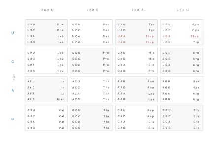
- The code — triplet, 64 codons, 61 sense, AUG start, UAA/UAG/UGA stop, degenerate but not ambiguous, non-overlapping, comma-less, universal.
- Translation — ribosome A, P and E sites; mRNA read 5′→3′; peptide bond made by rRNA.
8.1 Composition, structure and functions of DNA and RNA
The evidence that DNA is the genetic material
By 1940 most biologists assumed the genetic material was protein, because proteins are made of twenty different building blocks and DNA of only four. Three experiments destroyed that assumption. Each is a favourite of the paper, and each is worth one sentence in your memory.
Griffith, 1928 — transformation. Frederick Griffith worked with Streptococcus pneumoniae, the pneumococcus. The S strain has a polysaccharide capsule, forms smooth colonies and kills a mouse; the R strain has no capsule, forms rough colonies and is harmless. Griffith found that mice injected with heat-killed S bacteria plus live R bacteria died, and live S bacteria could be recovered from their blood. Something released by the dead S cells had permanently changed the harmless R cells into virulent S cells. He called it the transforming principle — but he did not know what it was. Note the logic carefully: neither component alone kills; the mixture does. That is the evidence.
Avery, MacLeod and McCarty, 1944 — the principle is DNA. Oswald Avery's group purified the transforming principle from heat-killed S cells and then destroyed each class of molecule in turn. Digested with protease (protein destroyed) the extract still transformed. Digested with ribonuclease (RNA destroyed) it still transformed. Digested with deoxyribonuclease (DNA destroyed) transformation stopped completely. The conclusion was that the transforming principle is DNA.
Hershey and Chase, 1952 — the label goes with the DNA. Alfred Hershey and Martha Chase used the T2 bacteriophage, which is nothing but a DNA molecule inside a protein coat. They grew one batch of phage in medium containing ³⁵S, which is built into the sulphur-containing amino acids of protein but not into DNA, and another in ³²P, which is built into the phosphate backbone of DNA but not into protein. Each batch was allowed to infect E. coli. A kitchen blender then sheared the empty coats off the bacterial surface, and centrifugation packed the bacteria into a pellet. Almost all the ³²P was inside the pellet with the bacteria; almost all the ³⁵S stayed in the supernatant with the coats. Since the injected material directs the making of new phage, the genetic material is DNA.
| Experiment | Organism | The decisive observation | Conclusion |
|---|---|---|---|
| Griffith (1928) | Streptococcus pneumoniae | heat-killed S + live R kills the mouse; live S recovered | a "transforming principle" exists |
| Avery, MacLeod, McCarty (1944) | S. pneumoniae extract | DNase stops transformation; protease and RNase do not | the principle is DNA |
| Hershey–Chase (1952) | T2 phage and E. coli | ³²P enters the bacterium, ³⁵S stays outside | DNA carries the instructions |
| Indirect evidence | all cells | DNA amount is constant for a species, halves in gametes, doubles in S phase; DNA absorbs ultraviolet at 260 nm, the most mutagenic wavelength | DNA is the hereditary material |
- Griffith did not identify DNA. He showed only that a principle existed; Avery's group named it.
- Hershey and Chase labelled protein with ³⁵S and DNA with ³²P — not the other way round. Sulphur is in cysteine and methionine; phosphorus is in the backbone.
- The blender does not break the bacteria open. It only removes the empty coats from the outside of intact cells; if the cells were broken, the experiment would prove nothing.
The nucleotide: sugar, phosphate and base
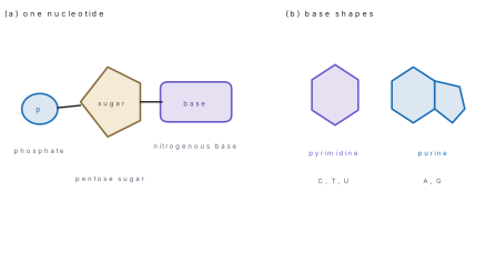
DNA and RNA are polynucleotides — chains of nucleotides. One nucleotide has three parts joined by two bonds:
- a pentose sugar — 2-deoxy-D-ribose in DNA (C₅H₁₀O₄), D-ribose in RNA (C₅H₁₀O₅);
- a phosphate group attached to carbon 5′ of the sugar;
- a nitrogenous base attached to carbon 1′ of the sugar.
A nucleoside is the sugar plus the base, without the phosphate. Adding the phosphate to a nucleoside gives the nucleotide: adenosine → adenosine monophosphate (AMP). Successive nucleotides are joined when the 3′-OH of one sugar makes a phosphodiester bond with the 5′-phosphate of the next. The result is a sugar–phosphate backbone with the bases projecting from it, and the chain has a chemical direction: a free phosphate at the 5′ end and a free hydroxyl at the 3′ end. Sequences are always written 5′ → 3′.
The two families of bases are told apart by their rings:
| Family | Ring structure | Members | Found in |
|---|---|---|---|
| Purines | two fused rings (a six-membered pyrimidine ring fused to a five-membered imidazole ring); 9 atoms | adenine (A), guanine (G) | DNA and RNA |
| Pyrimidines | one six-membered ring; 6 atoms | cytosine (C) | DNA and RNA |
| thymine (T) | DNA only | ||
| uracil (U) | RNA only |
Thymine is 5-methyluracil; RNA uses uracil in its place, and uracil lacks the methyl group. In double-stranded DNA the number of purines always equals the number of pyrimidines, because every purine is paired with a pyrimidine.
PURe As Gold — Adenine and Guanine are the purines (two rings, like a big word). CUT the PY — Cytosine, Uracil and Thymine are the pyrimidines (one ring). Purine pairs with pyrimidine so that every rung of the ladder is the same width.
The Watson–Crick double helix
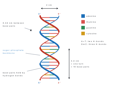
James Watson and Francis Crick published the model in Nature in 1953. They used three things: the X-ray diffraction photographs of Rosalind Franklin and Maurice Wilkins (Franklin's "Photograph 51" showed a helix), the Chargaff ratios, and the chemistry of the bases. The model has these features, and the paper asks for them one by one:
- Two polynucleotide chains wind round a common axis in a right-handed helix.
- The chains are antiparallel — one runs 5′ → 3′ upwards, the other 5′ → 3′ downwards.
- The sugar–phosphate backbones lie outside, and the bases point inwards, paired in the middle.
- Each base pair is one purine with one pyrimidine, held by hydrogen bonds: A pairs with T by two hydrogen bonds, G pairs with C by three.
- The two strands are complementary: the sequence of one fixes the sequence of the other. This is what makes faithful copying possible.
- The helix has a major groove and a minor groove, where proteins bind.
| Quantity | Value | Useful arithmetic |
|---|---|---|
| Diameter of the helix | 2 nm | — |
| Pitch (one complete turn) | 3.4 nm | turns = length ÷ 3.4 nm |
| Base pairs per turn | 10 | base pairs = turns × 10 |
| Rise per base pair | 0.34 nm | length = base pairs × 0.34 nm |
| Handedness | right-handed (B-DNA) | — |
| A=T hydrogen bonds | 2 | AT pairs × 2 |
| G≡C hydrogen bonds | 3 | GC pairs × 3 |
- A=T is two hydrogen bonds; G≡C is three. Students reverse them constantly. The consequence is the exam's favourite follow-up: a GC-rich DNA is more stable and melts at a higher temperature, because each GC pair has one more hydrogen bond than an AT pair. Heating DNA separates the strands — denaturation or melting — and the absorbance at 260 nm rises (the hyperchromic effect). Cooling lets the strands re-pair (annealing).
- 0.34 nm is the rise per base pair; 3.4 nm is the whole turn. A question that gives a length in millimetres and asks for the number of turns wants division by 3.4 nm, not by 0.34 nm.
Other forms of DNA. B-DNA is the Watson–Crick form and the one in living cells. A-DNA is also right-handed but shorter and fatter (11 bp per turn) and forms in dehydrated samples. Z-DNA is left-handed, with a zigzag backbone (12 bp per turn). Prokaryotes usually carry one circular DNA molecule; eukaryotes carry linear chromosomes, in which the DNA is wound round histone octamers into nucleosomes and then coiled into chromatin (Chapter 7).
Chargaff's rule and its arithmetic
Erwin Chargaff analysed DNA from many species in 1950 and found, without knowing the structure, that the bases are not present in equal amounts but are present in fixed pairs:
- %A = %T and %G = %C, so A + G = T + C — purines equal pyrimidines.
- The A + T to G + C ratio differs from species to species, but is constant within a species.
Every Chargaff question is solved in two lines. If one base is given, subtract its partner, halve the remainder, and remember that the four percentages add to 100.
Solution. A pairs with T, so T = 28 %. Then A + T = 56 %, leaving 44 % for G + C. Since G = C, each is 44 ÷ 2 = 22 %. In 200 bp there are 400 bases: A = T = 0.28 × 400 = 112, G = C = 88. Each AT pair has 2 hydrogen bonds and each GC pair has 3, and the pairs equal the counts of A and G: H = 112 × 2 + 88 × 3 = 224 + 264 = 488 hydrogen bonds.
Solution. A diploid cell has 2 × 3.2 × 10⁹ = 6.4 × 10⁹ bp. Length = 6.4 × 10⁹ × 0.34 nm = 2.18 × 10⁹ nm = 2.18 m (about 2 m — the figure to quote). Turns = base pairs ÷ 10 = 6.4 × 10⁹ ÷ 10 = 6.4 × 10⁸ turns. (Equivalently 2.18 m ÷ 3.4 nm gives the same number.)
Functions of DNA
- Storage of information. The base sequence is the information; a gene is a length of DNA that specifies one RNA or protein.
- Replication. Before a cell divides the DNA copies itself semi-conservatively, so both daughters receive a complete set.
- Control of the cell. Through transcription into RNA and then translation into protein, DNA decides the enzymes a cell makes and therefore its structure and behaviour.
- Mutation and variation. Rare copying errors change the sequence and provide the raw material of evolution.
- Inheritance. The gametes carry half the DNA of a body cell, so the information passes from parent to offspring.
RNA: the three types and their roles
RNA differs from DNA in four ways that the paper tests directly: it has ribose in place of deoxyribose, uracil in place of thymine, it is usually single-stranded (though it folds back on itself), and it is shorter and far less stable — the 2′-OH group of ribose makes RNA easy to hydrolyse, especially in alkali. Its single strandedness and its exposed bases let it fold into shapes and act as a catalyst.
| Feature | DNA | RNA |
|---|---|---|
| Sugar | 2-deoxy-D-ribose | D-ribose (2′-OH present) |
| Bases | A, G, C, T | A, G, C, U |
| Strands | two, antiparallel | usually one, folded |
| Size | very long (millions of bp) | short (70 – 10 000 nucleotides) |
| Location | mainly nucleus/chromosomes; also mitochondria and chloroplasts | nucleus and cytoplasm |
| Stability | stable | unstable, short-lived |
| Functions | store and transmit information | read the information and build protein; genetic material in some viruses |
| RNA | Share of cell RNA | Structure | Job |
|---|---|---|---|
| mRNA (messenger) | about 5 % | single strand, carries codons; in eukaryotes capped and poly-A tailed | carries the message from DNA to the ribosome — the only RNA that is translated |
| tRNA (transfer) | about 15 % | 75–90 nucleotides, folded into a cloverleaf; four arms — acceptor (with the CCA-3′ end that carries the amino acid), D arm, anticodon arm, T arm; L-shaped in three dimensions | the adaptor: reads the codon by its anticodon and delivers the matching amino acid |
| rRNA (ribosomal) | about 80 % — the most abundant | part of the ribosome; eukaryote 28S, 18S, 5.8S and 5S; prokaryote 23S, 16S and 5S | structural framework of the ribosome, and the peptidyl transferase that makes the peptide bond — a ribozyme |
Small nuclear RNA (snRNA) helps splice introns out of mRNA; micro-RNA regulates translation. In some viruses — tobacco mosaic virus, poliovirus, influenza, HIV — RNA is itself the genetic material, which is the exception to "DNA is the genetic material".
The anticodon of tRNA is complementary and antiparallel to the codon of mRNA, and the amino acid sits at the 3′-CCA end. One end reads the message, the other end carries the cargo. That is why tRNA is called an adaptor: it converts a three-letter nucleic-acid word into one amino acid.
8.2 DNA replication; the central dogma; the genetic code
Semi-conservative replication
Three models were possible. In conservative replication the parent double helix stays intact and an entirely new one is made. In semi-conservative replication the two strands separate and each acts as a template, so every daughter molecule has one old strand and one new one. In dispersive replication the old and new material is broken into pieces and mixed.
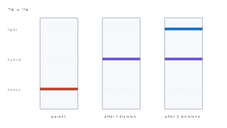
Meselson and Stahl, 1958 settled it with E. coli and a heavy isotope of nitrogen. Bacteria were grown for many generations in ¹⁵N (as ¹⁵NH₄Cl), so all their DNA was heavy. They were then transferred to ordinary ¹⁴N medium, and samples were taken after each division. DNA was extracted and spun in a caesium chloride density gradient, where DNA bands at the position matching its density.
- Before the transfer: one heavy band at the bottom.
- After one division: one band of intermediate (hybrid) density, in the middle. This rules out conservative replication, which would have given two bands, one heavy and one light.
- After two divisions: two bands in equal amounts, one hybrid and one light. This rules out dispersive replication, which would have given a single band drifting upwards.
Only semi-conservative replication predicts exactly this. The same result was shown in plant roots by Taylor, Woods and Hughes (1957) using tritiated thymidine in Vicia faba.
The machinery of replication
Replication starts at an origin (one in a bacterial chromosome, many in a eukaryotic chromosome), and proceeds bidirectionally, so a replication bubble opens with a fork at each end. In eukaryotes it happens in the S phase of the cell cycle. Every enzyme has one job, and the paper asks "which enzyme does this?":
| Protein | Job |
|---|---|
| DNA helicase | unwinds the double helix at the fork, breaking the hydrogen bonds, using ATP |
| Single-strand binding proteins | hold the separated strands apart so they cannot re-pair |
| Topoisomerase (DNA gyrase in bacteria) | relieves the supercoiling ahead of the fork |
| Primase | an RNA polymerase that lays down a short RNA primer |
| DNA polymerase III | the main enzyme: adds deoxyribonucleotides to the free 3′-OH, synthesising 5′ → 3′; proofreads with its 3′ → 5′ exonuclease activity |
| DNA polymerase I | removes the RNA primer (5′ → 3′ exonuclease) and fills the gap with DNA |
| DNA ligase | seals the nick between fragments, making the last phosphodiester bond; uses ATP |
DNA polymerase can only add a nucleotide to an existing free 3′-OH group; it cannot start a chain on a bare template. Primase therefore makes a short RNA primer first, which DNA polymerase I later removes and replaces with DNA. A question that says "DNA polymerase can synthesise DNA in the absence of a primer" is false.
Leading and lagging strands
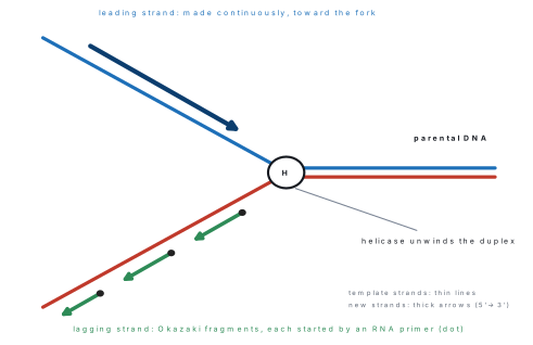
Because the two parental strands are antiparallel, and because DNA polymerase can only work 5′ → 3′, the two new strands cannot be made the same way.
- The leading strand is made continuously, in the same direction as the fork is moving, from a single primer.
- The lagging strand is made discontinuously, in short pieces that grow away from the fork, each started by its own primer. These pieces are the Okazaki fragments — about 100–200 nucleotides long in eukaryotes and 1000–2000 in bacteria. DNA polymerase I replaces their RNA primers and DNA ligase joins them into one strand.
So each fork produces one continuous and one fragmented strand. The lagging strand is the slower and more complicated of the two, and it is where the primers and the ligase are needed.
Transcription in outline
Transcription is the making of an RNA copy of one gene. RNA polymerase binds a promoter just upstream of the gene, unwinds a short stretch, and builds RNA 5′ → 3′ by reading the template strand in the 3′ → 5′ direction. The other strand, the coding (sense) strand, has the same sequence as the RNA except that T replaces U. No primer is needed, and only one strand of the gene is copied. The enzyme stops at a terminator.
In eukaryotes there are three polymerases: RNA polymerase I makes the large rRNA, RNA polymerase II makes mRNA, and RNA polymerase III makes tRNA and 5S rRNA. The mRNA is then processed: a 5′ cap of 7-methylguanosine, a 3′ poly-A tail, and splicing out of introns by the spliceosome.
| Point | Replication | Transcription |
|---|---|---|
| What is copied | the whole chromosome | one gene |
| Strands used as template | both | one |
| Product | DNA | RNA |
| Enzyme | DNA polymerase | RNA polymerase |
| Primer | needed | not needed |
| Base pairing with A | T | U |
| When | S phase | throughout the cell cycle, as the gene is needed |
The central dogma
Francis Crick stated the central dogma in 1958: information flows DNA → RNA → protein. Replication is DNA → DNA; transcription is DNA → RNA; translation is RNA → protein. The transfer is one-way in cells — protein sequence does not flow back into nucleic acid.
There are two real exceptions, and the paper likes them:
- Reverse transcription, RNA → DNA, catalysed by reverse transcriptase in retroviruses such as HIV. It is the basis of the enzyme used in the laboratory to make cDNA from mRNA.
- RNA replication, RNA → RNA, in some RNA viruses.
The genetic code
The code was broken between 1961 and 1966. Nirenberg and Matthaei made a synthetic mRNA of only uracil (poly-U) and got a polypeptide of only phenylalanine, so UUU codes for Phe. Khorana then made RNAs of known repeating sequence, and within five years all 64 codons had been assigned.
| Property | What it means | Consequence the paper tests |
|---|---|---|
| Triplet | three bases code for one amino acid | 4³ = 64 codons for 20 amino acids |
| 61 sense + 3 stop | UAA, UAG, UGA do not code for an amino acid | they end translation |
| Start codon AUG | also codes methionine | the first amino acid of nearly every protein |
| Degenerate (redundant) | most amino acids have more than one codon; Leu, Ser and Arg have six, Met and Trp have one | a base change may not change the protein |
| Unambiguous | one codon never specifies two amino acids | the message has only one reading |
| Non-overlapping | each base belongs to one codon only | a change affects one amino acid at most |
| Comma-less | no bases are wasted between codons | the reading frame runs continuously |
| Universal | the same in almost all organisms, from bacteria to humans | a human gene can be expressed in a bacterium |
These two words are the commonest confusion in the unit. Degenerate means many codons → one amino acid (Leu has six codons). Ambiguous would mean one codon → many amino acids, and that never happens. A question that says "the code is degenerate, so one codon can code for more than one amino acid" is false on its second half.
The wobble hypothesis (Crick, 1966) explains why a cell needs fewer than 61 tRNAs: the pairing between the third base of the codon and the first base of the anticodon is loose, so one tRNA can read several codons that differ only in that position. The first two bases pair strictly.
Translation
Translation turns the base sequence of mRNA into the amino acid sequence of a polypeptide. It happens on the ribosome — 70S (50S + 30S) in prokaryotes, 80S (60S + 40S) in eukaryotes — and it reads the mRNA 5′ → 3′.
- Activation. Each amino acid is joined to its tRNA by an aminoacyl-tRNA synthetase, using ATP. The amino acid is attached to the CCA-3′ end.
- Initiation. The small subunit binds the mRNA, the initiator tRNA carrying methionine pairs with the AUG start codon, and the large subunit joins. This tRNA sits in the P site.
- Elongation. The next codon lies in the A site; the matching aminoacyl-tRNA binds; peptidyl transferase — the rRNA of the large subunit — forms the peptide bond; then the ribosome translocates one codon along, so the tRNA that has given up its amino acid moves to the E (exit) site and leaves.
- Termination. A stop codon enters the A site, a release factor binds, and the polypeptide, mRNA and ribosomal subunits separate.
| Ribosomal site | What sits there |
|---|---|
| A (aminoacyl) | the incoming aminoacyl-tRNA, reading the new codon |
| P (peptidyl) | the tRNA holding the growing polypeptide chain |
| E (exit) | the tRNA that has lost its amino acid, about to leave |
Several ribosomes may travel along one mRNA at once, forming a polysome, so many copies of the protein are made from one message.
Solution. (i) The mRNA is complementary and antiparallel to the template, with U in place of T: 5′-AUG CCU AAG UGA-3′. Reading in triplets: AUG = Met, CCU = Pro, AAG = Lys, UGA = stop. The polypeptide is Met–Pro–Lys, three amino acids. (ii) Deleting the fifth base (the second G) gives the template 3′-TAC GAT TCA CT-5′ and the mRNA 5′-AUG CUA AGU GA…-3′. The codons are now AUG CUA AGU, that is Met–Leu–Ser, and there is no stop codon in the same place. This is a frameshift: one base lost shifts every later codon by one, so almost the whole protein changes. A substitution, by contrast, usually changes one codon only, and often not even that, because the code is degenerate.
Solution. Each amino acid needs one codon of three bases: 90 × 3 = 270. The stop codon adds three more: 270 + 3 = 273 nucleotides. (The start codon is already counted, because the first methionine is one of the 90.)
Solution. Two forks copy the chromosome at the same time, so the rate is 2 × 1000 = 2000 nucleotides per second. Time = 4.6 × 10⁶ ÷ 2000 = 2300 s = about 38 minutes.
Formula and fact sheet
| Item | Fact |
|---|---|
| Griffith | 1928, S. pneumoniae, heat-killed S + live R → live S; "transforming principle" |
| Avery, MacLeod, McCarty | 1944; protease and RNase no effect, DNase stops transformation → DNA |
| Hershey–Chase | 1952, T2 phage; ³²P → DNA, ³⁵S → protein; blender removes empty coats |
| Nucleotide | phosphate + pentose + base; nucleoside = sugar + base |
| Sugars | DNA 2-deoxyribose (C₅H₁₀O₄); RNA ribose (C₅H₁₀O₅) |
| Purines | A, G — two rings |
| Pyrimidines | C, T, U — one ring; T only in DNA, U only in RNA |
| Backbone bond | phosphodiester, 3′-OH to 5′-phosphate |
| Helix width | 2 nm |
| Pitch | 3.4 nm per turn |
| Base pairs per turn | 10 |
| Rise per base pair | 0.34 nm |
| Handedness | B-DNA right-handed; Z-DNA left-handed |
| Hydrogen bonds | A=T two, G≡C three; GC-rich DNA melts higher |
| Chargaff | A = T, G = C, A + G = T + C |
| Human genome | 3.2 × 10⁹ bp haploid; ≈ 1.09 m per haploid set, ≈ 2.18 m per diploid cell |
| RNA types | mRNA ≈ 5 % (codons); tRNA ≈ 15 % (cloverleaf, 75–90 nt, anticodon, CCA-3′); rRNA ≈ 80 % (peptidyl transferase) |
| Eukaryote rRNA | 28S, 18S, 5.8S, 5S; prokaryote 23S, 16S, 5S |
| Meselson–Stahl | 1958, E. coli, ¹⁵N, CsCl gradient; one hybrid band after one division |
| Replication enzymes | helicase unwinds; primase makes RNA primer; pol III extends 5′→3′; pol I replaces primer; ligase seals nicks |
| Okazaki fragments | 100–200 nt (eukaryote), 1000–2000 nt (bacteria) |
| Leading / lagging | continuous towards the fork / discontinuous away from it |
| Transcription | RNA polymerase, promoter, template strand read 3′→5′, RNA made 5′→3′, no primer |
| Eukaryote polymerases | I → rRNA; II → mRNA; III → tRNA and 5S rRNA |
| mRNA processing | 5′ cap (7-methylguanosine), 3′ poly-A tail, splicing of introns |
| Central dogma | DNA → RNA → protein; exceptions reverse transcription (HIV) and RNA → RNA |
| Codons | 64 total, 61 sense, 3 stop: UAA, UAG, UGA |
| Start codon | AUG, also methionine |
| Code properties | triplet, degenerate, unambiguous, non-overlapping, comma-less, universal |
| Wobble | loose pairing at the third base of the codon |
| Ribosome | 70S = 50S + 30S (prokaryote); 80S = 60S + 40S (eukaryote); sites A, P, E |
| Peptide bond made by | peptidyl transferase — the rRNA of the large subunit (a ribozyme) |
Chapter summary
- Griffith showed that a heat-killed virulent pneumococcus could transform a live avirulent one; Avery's group proved with DNase that the principle is DNA; Hershey and Chase proved with ³²P and ³⁵S that DNA, not protein, enters the bacterium.
- A nucleotide is phosphate + pentose + base; purines (A, G) have two rings and pyrimidines (C, T, U) one; nucleotides are joined by phosphodiester bonds into a 5′ → 3′ chain.
- The Watson–Crick helix is right-handed and antiparallel, 2 nm wide, 3.4 nm per turn, 10 bp per turn and 0.34 nm per base pair, with A=T (2 hydrogen bonds) and G≡C (3 hydrogen bonds) — so GC-rich DNA is more stable.
- Chargaff: A = T, G = C, so purines = pyrimidines; one given percentage fixes the other three.
- mRNA carries codons, tRNA is the cloverleaf adaptor with an anticodon and a CCA-3′ amino-acid end, and rRNA is the most abundant RNA and the catalyst that forms peptide bonds.
- Replication is semi-conservative: Meselson and Stahl's ¹⁵N experiment gave one hybrid band after one division and equal hybrid and light bands after two.
- Helicase unwinds, primase primes, DNA polymerase III extends 5′ → 3′, DNA polymerase I replaces the primer and ligase seals the nicks; the leading strand is continuous and the lagging strand is a series of Okazaki fragments.
- Transcription copies one gene with RNA polymerase and needs no primer; the central dogma is DNA → RNA → protein, with reverse transcription as the exception.
- The code is a triplet code of 64 codons — 61 sense, AUG start, UAA/UAG/UGA stop — degenerate, unambiguous, non-overlapping, comma-less and universal.
- Translation reads mRNA 5′ → 3′ on the ribosome through the A, P and E sites; a frameshift changes every codon after the mutation, a substitution usually changes one.
Practice questions
Set A — Recall (23 questions)
- Griffith's experiment with Streptococcus pneumoniae showed thataDNA is a double helixbheat-killed virulent bacteria can transform live avirulent onescprotein is the genetic materialdbacteria divide by binary fission
- In the Avery–MacLeod–McCarty experiment, transformation of R into S pneumococci stopped when the extract was treated withadeoxyribonucleasebproteasecribonucleasedamylase
- Hershey and Chase labelled the DNA of the T2 phage witha³⁵Sb¹⁵Nc³²Pd³H
- Which pair of bases are purines?aadenine and thyminebcytosine and guaninecthymine and uracildadenine and guanine
- A nucleoside consists ofaa sugar and a phosphateba sugar and a baseca base and a phosphatedtwo sugars and a base
- Two nucleotides in one polynucleotide strand are joined byaa hydrogen bondba glycosidic bondca phosphodiester bondda peptide bond
- The diameter of the DNA double helix isa2 nmb0.34 nmc3.4 nmd20 nm
- One complete turn of B-DNA containsa10 base pairs and is 0.34 nm longb20 base pairs and is 3.4 nm longc34 base pairs and is 2 nm longd10 base pairs and is 3.4 nm long
- In DNA, adenine is joined to thymine byaone hydrogen bondbthree hydrogen bondsctwo hydrogen bondsda covalent bond
- Chargaff's rule for double-stranded DNA states thataA + T = G + CbA = T and G = CcA = G and T = CdA + G = 2(T + C)
- The X-ray diffraction photographs used by Watson and Crick were made byaRosalind FranklinbErwin ChargaffcFrederick GriffithdOswald Avery
- The most abundant RNA in a cell isamRNAbtRNAcrRNAdsnRNA
- The three bases of tRNA that are complementary to a codon are calledacodonsbexonscintronsdanticodons
- Which of the following is a stop codon?aAUGbUGAcUGGdGUG
- The start codon AUG also codes foravalinebleucinecmethioninedtryptophan
- The total number of codons in the genetic code isa64b61c20d128
- Meselson and Stahl used which isotope to follow DNA replication?a³²Pb¹⁵Nc³⁵Sd¹⁴C
- Okazaki fragments are joined together byahelicasebprimasecDNA polymerase IIIdDNA ligase
- The enzyme that unwinds the double helix at the replication fork isaligasebprimasechelicasedtopoisomerase
- The central dogma of molecular biology isaDNA → RNA → proteinbprotein → RNA → DNAcRNA → DNA → proteindDNA → protein → RNA
- Reverse transcriptase synthesisesaRNA from a DNA templatebprotein from mRNAcDNA from a DNA templatedDNA from an RNA template
- In eukaryotes, mRNA is made byaRNA polymerase IbRNA polymerase IIcRNA polymerase IIIdDNA polymerase I
- The 5′ end of a polynucleotide chain carriesaa free hydroxyl groupban amino groupca free phosphate groupda nitrogenous base
Answers and explanations — Set A
1. (b) — Neither heat-killed S nor live R alone killed the mouse; the mixture did, and live S was recovered. Griffith did not identify the substance — that was Avery's group.
2. (a) — DNase destroys DNA, and transformation stopped; protease and RNase had no effect, so protein and RNA were excluded.
3. (c) — Phosphorus is in the DNA backbone, so ³²P labels DNA; ³⁵S labels the sulphur amino acids of the protein coat.
4. (d) — Adenine and guanine have the two-ring purine structure. Thymine, cytosine and uracil are single-ring pyrimidines.
5. (b) — A nucleoside is sugar + base. Adding a phosphate makes it a nucleotide.
6. (c) — The 3′-OH of one sugar bonds to the 5′-phosphate of the next through a phosphodiester bond; hydrogen bonds hold the two strands together, not the backbone.
7. (a) — B-DNA is 2 nm across. 0.34 nm is the rise per base pair and 3.4 nm the pitch of one turn.
8. (d) — Ten base pairs per turn, each rising 0.34 nm, gives 10 × 0.34 = 3.4 nm.
9. (c) — A=T has two hydrogen bonds; G≡C has three, which is why GC-rich DNA is harder to melt.
10. (b) — Every A is paired with a T and every G with a C, so the two are equal and purines equal pyrimidines.
11. (a) — Rosalind Franklin's photograph 51, with Wilkins's data, showed the helical pattern. Chargaff supplied the base ratios, not the diffraction data.
12. (c) — rRNA is about 80 % of the RNA of a cell because every ribosome is mostly rRNA. mRNA is only about 5 %.
13. (d) — The anticodon of tRNA is complementary and antiparallel to the mRNA codon; the codon itself is on mRNA.
14. (b) — UAA, UAG and UGA are the three stop codons. AUG is the start; UGG is tryptophan; GUG is valine.
15. (c) — AUG is both the start signal and the codon for methionine, so nearly every polypeptide begins with methionine.
16. (a) — Four bases in triplets give 4³ = 64 codons: 61 sense and 3 stop.
17. (b) — ¹⁵N makes DNA heavy, and the bands in the caesium chloride gradient show how the old and new strands are distributed.
18. (d) — DNA ligase makes the final phosphodiester bond between adjacent fragments. Primase only makes the RNA primer.
19. (c) — Helicase breaks the hydrogen bonds and separates the strands; topoisomerase only relieves the supercoiling ahead of it.
20. (a) — Information flows from DNA to RNA to protein: replication, transcription, translation.
21. (d) — Reverse transcriptase copies an RNA template into DNA; retroviruses such as HIV use it to make DNA from their RNA genome.
22. (b) — RNA polymerase II transcribes mRNA; I makes the large rRNA and III makes tRNA and 5S rRNA.
23. (c) — The 5′ end carries the phosphate and the 3′ end the free hydroxyl, which is why new nucleotides are always added at the 3′ end.
Set B — Understanding (14 questions)
- DNA is a better long-term store of information than RNA becauseaDNA contains uracil, which resists hydrolysisbDNA is single-stranded and therefore shortercDNA lacks the 2′-OH group, so it is chemically more stabledDNA is found only in the nucleus
- A DNA sample with a high GC content has a higher melting temperature becauseaeach GC pair is held by three hydrogen bonds instead of twobGC pairs are joined by covalent bondscguanine and cytosine are larger basesdGC pairs lie on the outside of the helix
- The Meselson–Stahl result after one generation in ¹⁴N medium ruled out conservative replication becauseatwo bands appeared, one heavy and one lightball the DNA remained heavycthe DNA was completely degradedda single band of intermediate density appeared
- The lagging strand is made in short fragments becauseaDNA polymerase can synthesise only in the 3′ → 5′ directionbDNA polymerase adds only to a 3′-OH end, opposite this templatecthe lagging strand has no primer at alldligase can join only short pieces
- DNA polymerase cannot start a new chain on its own becauseait needs a supply of ATPbit works only on single-stranded DNAcit can add nucleotides only to an existing free 3′-OH groupdit is inhibited by helicase
- "The genetic code is degenerate" means thatamore than one codon can specify the same amino acidbone codon can specify more than one amino acidcsome bases are never useddthe code differs from species to species
- A base substitution in the third position of a codon often does not change the protein becauseathe third base is not read by the ribosomebcode degeneracy lets the new codon specify the same amino acidcthe third base is removed during splicingdthe code is overlapping
- A single base deletion usually alters the whole protein, whereas a substitution alters at most one amino acid, becauseadeletions always remove more materialba deletion removes a whole codoncsubstitutions are repaired and deletions are notda deletion shifts the reading frame of every downstream codon
- tRNA is called an adaptor molecule becauseait carries the message out of the nucleusbit forms the structural core of the ribosomecone end binds an amino acid while its anticodon reads the codondit joins Okazaki fragments
- One tRNA can read more than one codon for the same amino acid becauseaits anticodon is four bases longbthe 3rd codon base wobbles with the 1st base of the anticodoncthe ribosome corrects the pairingdthe code is ambiguous
- RNA is more easily hydrolysed than DNA, especially in alkali, becauseaits ribose carries a 2′-OH groupbit contains uracilcit is single-strandeddit is much shorter
- In the Hershey–Chase experiment the blender was used toabreak open the bacteriabrelease the DNA from the phagecmix the two isotopes evenlydshear the empty phage coats off the bacterial surface
- Two cells of the same organism make different proteins mainly becauseathey contain different genesbdifferent genes are expressed in themctheir DNA contains different basesdtheir ribosomes are different
- The genetic code is described as non-overlapping becauseano base is used twice anywhere in the mRNAbthe codons are separated by punctuationceach base is read in one codon only, and the next codon follows at oncedno two codons share an amino acid
Answers and explanations — Set B
1. (c) — The 2′-OH of ribose makes RNA attack its own backbone, so RNA is short-lived. DNA has deoxyribose, no 2′-OH, and a second strand to check the sequence against.
2. (a) — Three hydrogen bonds per GC pair against two per AT pair means more energy is needed to separate the strands. Base size and position play no part.
3. (d) — Conservative replication would have kept the heavy parent intact and produced a separate light molecule, giving two bands. One hybrid band means each daughter molecule has one old and one new strand.
4. (b) — Polymerase works only 5′ → 3′, and on the lagging template that direction points away from the fork, so the new strand has to be restarted repeatedly.
5. (c) — It extends a chain; it cannot begin one. Primase therefore lays an RNA primer that supplies the first 3′-OH.
6. (a) — Degeneracy is several codons for one amino acid, as when four codons specify proline. One codon specifying several amino acids would be ambiguity, and the code is not ambiguous.
7. (b) — The third base is the wobble position and is often interchangeable, so UUU and UUC both give phenylalanine.
8. (d) — Codons are read continuously from the start, so losing one base moves every later triplet by one place and changes all the amino acids downstream.
9. (c) — Its anticodon reads a nucleic-acid codon and its CCA end carries the corresponding amino acid; it is the physical link between the two languages.
10. (b) — The wobble hypothesis: the third codon base pairs loosely with the first anticodon base, so one tRNA serves several codons.
11. (a) — The 2′-OH can attack the phosphodiester bond, so alkali breaks RNA quickly; DNA lacks this group and survives.
12. (d) — The bacteria were left intact; the blender only detached the empty coats so that they could be separated from the cells by centrifugation.
13. (b) — Every body cell has the same genes; what differs is which genes are switched on, so a leaf cell and a root cell make different proteins.
14. (c) — Each base is read once, in one triplet, and the next triplet follows immediately with no bases skipped — that is also why the code is comma-less.
Set C — Application (9 questions)
- A double-stranded DNA sample contains 20 % cytosine. The percentage of adenine isa20b40c60d30
- A DNA fragment of 100 base pairs contains 60 GC pairs. The total number of hydrogen bonds holding the two strands together isa200b260c300d240
- The haploid human genome contains 3.2 × 10⁹ base pairs. The total length of DNA in one diploid somatic cell is abouta2.2 mb1.1 mc22 md0.22 m
- A polypeptide contains 90 amino acids. The minimum number of nucleotides in the coding region of its mRNA, including the stop codon, isa90b180c273d270
- An mRNA reads 5′-AUG CCU AAG UGA-3′. The polypeptide formed from it containsafour amino acidsbtwo amino acidscone amino aciddthree amino acids
- The template strand of a gene reads 3′-TAC GGA TTC-5′. The mRNA transcribed from it isa5′-ATG CCT TTC-3′b5′-AUG CCU AAG-3′c5′-AUG GGA UUC-3′d5′-UAC GGA UUC-3′
- A DNA molecule is 1.36 mm long. The number of complete turns of the helix it contains isa4 × 10³b4 × 10⁶c4 × 10⁵d4 × 10⁴
- An E. coli chromosome has 4.6 × 10⁶ base pairs. Replication starts at one origin and each fork moves at 1000 nucleotides per second. The time taken is abouta38 minutesb77 minutesc19 minutesd230 minutes
- A DNA sample contains 30 % guanine. The percentage of thymine in it isa30b20c40d70
Answers and explanations — Set C
1. (d) — C = G = 20 %, so G + C = 40 % and A + T = 60 %; since A = T, A = 30 %.
2. (b) — 60 GC pairs × 3 = 180 and 40 AT pairs × 2 = 80; total 180 + 80 = 260 hydrogen bonds.
3. (a) — A diploid cell has 6.4 × 10⁹ bp, and 6.4 × 10⁹ × 0.34 nm = 2.18 × 10⁹ nm = 2.18 m. Option (b) is the haploid figure.
4. (c) — 90 codons × 3 = 270 nucleotides for the amino acids, plus 3 for the stop codon = 273.
5. (d) — AUG = Met, CCU = Pro, AAG = Lys, and UGA stops. Three amino acids are made; the stop codon adds none.
6. (b) — The mRNA is complementary and antiparallel to the template: T→A, A→U, C→G, G→C, giving AUG CCU AAG. Option (a) is a DNA copy, not RNA.
7. (c) — 1.36 mm = 1.36 × 10⁶ nm; dividing by the 3.4 nm pitch gives 4 × 10⁵ turns. Dividing by 0.34 nm would wrongly count base pairs.
8. (a) — Two forks copy at 2000 nucleotides per second in total, so 4.6 × 10⁶ ÷ 2000 = 2300 s ≈ 38 minutes. Option (b) forgets that there are two forks.
9. (b) — G = C = 30 %, so G + C = 60 % and A + T = 40 %; A = T = 20 %.