Chapter 7 · Cell Biology · 5 questions in the paper · 36 min read
Cell Biology and Cell Division
What the syllabus asks for 3
- Prokaryotic and eukaryotic cells; the cell theory
- Cell wall, cell membrane, mitochondria, chloroplasts, endoplasmic reticulum, Golgi body, lysosomes, ribosomes, nucleus, chromosomes, cilia, flagella and cell inclusions
- The cell cycle; amitosis, mitosis and meiosis and their significance
Cell Biology and Cell Division is the heaviest unit in the Botany paper — 5 of the 40 questions — and the most predictable. One question usually comes from the cell theory or the prokaryote/eukaryote comparison, two or three from organelle structure and function, and one or two from the cell cycle and the differences between mitosis and meiosis. The numbers are what examiners love: 70S and 80S, 9+2, 46 and 23, 2C and 4C. Learn the tables and count chromosomes carefully; this unit is where careless students lose marks they knew.
- Cell theory and its exceptions — Schleiden, Schwann, Virchow; viruses are acellular, coenocytic hyphae are multinucleate without cross walls.
- Prokaryote versus eukaryote — 70S versus 80S ribosomes, nucleoid versus nucleus, peptidoglycan versus cellulose, binary fission versus mitosis.
- Organelle structure and function — cristae, grana, cisternae, cis and trans faces, hydrolytic enzymes, the nucleolus, the nuclear pore.
- The wall in three layers — middle lamella (calcium pectate), primary wall, secondary wall (lignin).
- Chromosome vocabulary — chromatid, centromere, telomere, nucleosome (DNA + histone octamer).
- 9+2 — the microtubule arrangement of eukaryotic cilia and flagella; bacterial flagella are flagellin and have no 9+2.
- The cell cycle — G1, S, G2, M, G0; DNA replicates in S; checkpoints at G1/S and G2/M.
- Mitosis versus meiosis — one division versus two, 2n → 2n versus 2n → 4 × n, cell plate versus cleavage furrow, and the chromosome and DNA counts at each stage.
7.1 Prokaryotic and eukaryotic cells; the cell theory
How the cell was discovered, and the cell theory
| Year | Person | Contribution |
|---|---|---|
| 1665 | Robert Hooke | saw the empty "cells" (dead cork cell walls) and coined the word cell |
| 1674 | Antonie van Leeuwenhoek | first saw living cells — bacteria and protozoa in pond water |
| 1831 | Robert Brown | discovered the nucleus in orchid cells |
| 1838 | Matthias Schleiden | all plants are made of cells |
| 1839 | Theodor Schwann | all animals are made of cells; proposed the cell theory with Schleiden |
| 1855 | Rudolf Virchow | omnis cellula e cellula — every cell arises from a pre-existing cell |
| 1931 | Ernst Ruska | built the electron microscope, which made organelles visible |
The cell theory in its modern form says four things: the cell is the structural and functional unit of all living organisms; all organisms are made of one or more cells; new cells arise only by division of existing cells; and all cells are chemically similar, containing DNA, RNA, protein and a plasma membrane.
Exceptions. The theory has three standard exceptions:
- Viruses, viroids and prions are acellular — they have no cell, no cytoplasm and no ribosomes, and cannot reproduce outside a host.
- Coenocytic (aseptate) structures — the hyphae of Mucor and Rhizopus are a single continuous mass of cytoplasm with many nuclei and no cross walls; the latex vessels of plants and striated muscle fibres are also multinucleate. One "cell" holds many nuclei.
- Prokaryotes have no membrane-bound nucleus or organelles, so the classical statement "every cell has a nucleus" does not apply to them — though they are still cells.
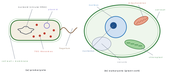
Prokaryotic and eukaryotic cells compared
| Feature | Prokaryotic cell | Eukaryotic cell |
|---|---|---|
| Typical size | 1–10 μm | 10–100 μm |
| Nucleus | absent; DNA lies in a nucleoid | present, with a double nuclear envelope and pores |
| DNA | circular, naked (no histone proteins in bacteria), one main chromosome | linear, bound to histones, many chromosomes |
| Extra DNA | plasmids | only inside mitochondria and chloroplasts |
| Ribosomes | 70S (50S + 30S) | 80S (60S + 40S) in the cytoplasm; 70S in mitochondria and chloroplasts |
| Membrane-bound organelles | none — no mitochondria, ER, Golgi, lysosome, chloroplast | present |
| Respiratory enzymes | on the plasma membrane and its infoldings (mesosomes) | on the inner mitochondrial membrane |
| Cell wall | peptidoglycan (murein); absent in Mycoplasma | cellulose in plants, chitin in fungi, absent in animals |
| Cell division | binary fission (amitosis), no spindle | mitosis and meiosis, with a spindle |
| Flagella | flagellin, single fibril, no 9+2, rotates | tubulin, 9+2 axoneme, bends |
| Other surface structures | pili, fimbriae, capsule | cilia, microvilli, glycocalyx |
| Cytoskeleton | poorly developed | microtubules, microfilaments, intermediate filaments |
| Examples | bacteria, cyanobacteria, mycoplasma, archaea | protists, fungi, plants, animals |
7.2 The cell wall, the membrane and the organelles
The cell wall
Only plants, fungi and bacteria have a wall; animal cells do not. A plant cell wall has three parts, laid down in this order:
| Layer | Position | Composition | Property |
|---|---|---|---|
| Middle lamella | outermost boundary between two neighbouring cells, formed first at cytokinesis | calcium pectate (and magnesium pectate) | cements the cells together; digested by pectinase when fruit softens |
| Primary wall | between the middle lamella and the plasma membrane | cellulose microfibrils in a matrix of hemicellulose, pectin and glycoprotein | thin, elastic, allows the cell to grow |
| Secondary wall | deposited inside the primary wall after growth stops | cellulose plus lignin (in xylem and sclerenchyma) | thick, rigid, three sub-layers (S1, S2, S3); gives strength and waterproofing |
Plasmodesmata are fine cytoplasmic channels that pass through the wall and connect the cytoplasm of adjacent cells. The wall's functions are support, protection, a limit to water uptake (which is why a plant cell does not burst in pure water), and transport through the plasmodesmata. Bacterial walls are peptidoglycan, fungal walls chitin — a fact the paper tests every year.
The plasma membrane
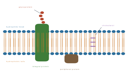
The membrane is a phospholipid bilayer about 7.5 nm thick. The phosphate heads face outwards into water; the fatty-acid tails face inwards, away from it. Proteins float in or through this bilayer, and cholesterol sits between the phospholipid tails. Because the proteins move about in a fluid lipid sea, Singer and Nicolson (1972) called it the fluid-mosaic model.
| Component | Role |
|---|---|
| Phospholipid | forms the bilayer; the barrier to water-soluble substances |
| Integral (intrinsic) protein | spans the bilayer; forms channels, pumps and carriers |
| Peripheral (extrinsic) protein | sits on one surface; enzymes, receptors, anchors |
| Glycoprotein and glycolipid | carbohydrate chains on the outer surface — cell recognition, ABO antigens, receptors |
| Cholesterol | keeps the membrane fluid at low temperature and stable at high temperature |
The membrane is selectively permeable: small non-polar molecules (O₂, CO₂) cross by simple diffusion, water by osmosis through channels called aquaporins, and ions and sugars through pumps and carriers, often against the gradient using ATP.
| Transport | Direction | Energy needed | Example |
|---|---|---|---|
| Simple diffusion | down the concentration gradient | none | O₂ and CO₂ across the bilayer |
| Osmosis | water moves down its water potential | none | water through aquaporins |
| Facilitated diffusion | down the gradient, through a carrier or channel | none | glucose into a red blood cell |
| Active transport | against the gradient, through a pump | ATP | Na⁺/K⁺ pump; mineral uptake by root hairs |
| Endocytosis / exocytosis | bulk movement in vesicles | ATP | phagocytosis by white cells; secretion of enzymes |
Mitochondria
A mitochondrion is 0.5–1 μm wide and 2–8 μm long, bounded by two membranes. The outer membrane is smooth; the inner one is folded into finger-like cristae that project into the jelly-like matrix. On the inner membrane sit the F₁ particles (oxysomes), which carry the enzymes of the electron transport chain and ATP synthase. The matrix holds the enzymes of the Krebs cycle, circular DNA, 70S ribosomes and phosphate granules.
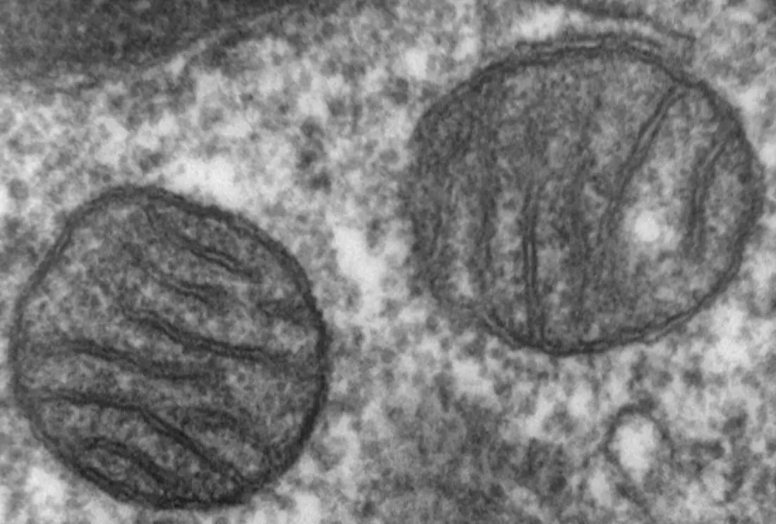
Because a mitochondrion has its own DNA and ribosomes it is called semi-autonomous: it makes some of its own proteins but depends on nuclear genes for the rest, and it divides on its own. Mitochondrial DNA is inherited from the mother in humans. The mitochondrion is the powerhouse — the site of aerobic respiration and oxidative phosphorylation. Cells that do heavy work (heart muscle, flight muscle, the tapetum of an anther) are packed with them.
Chloroplasts
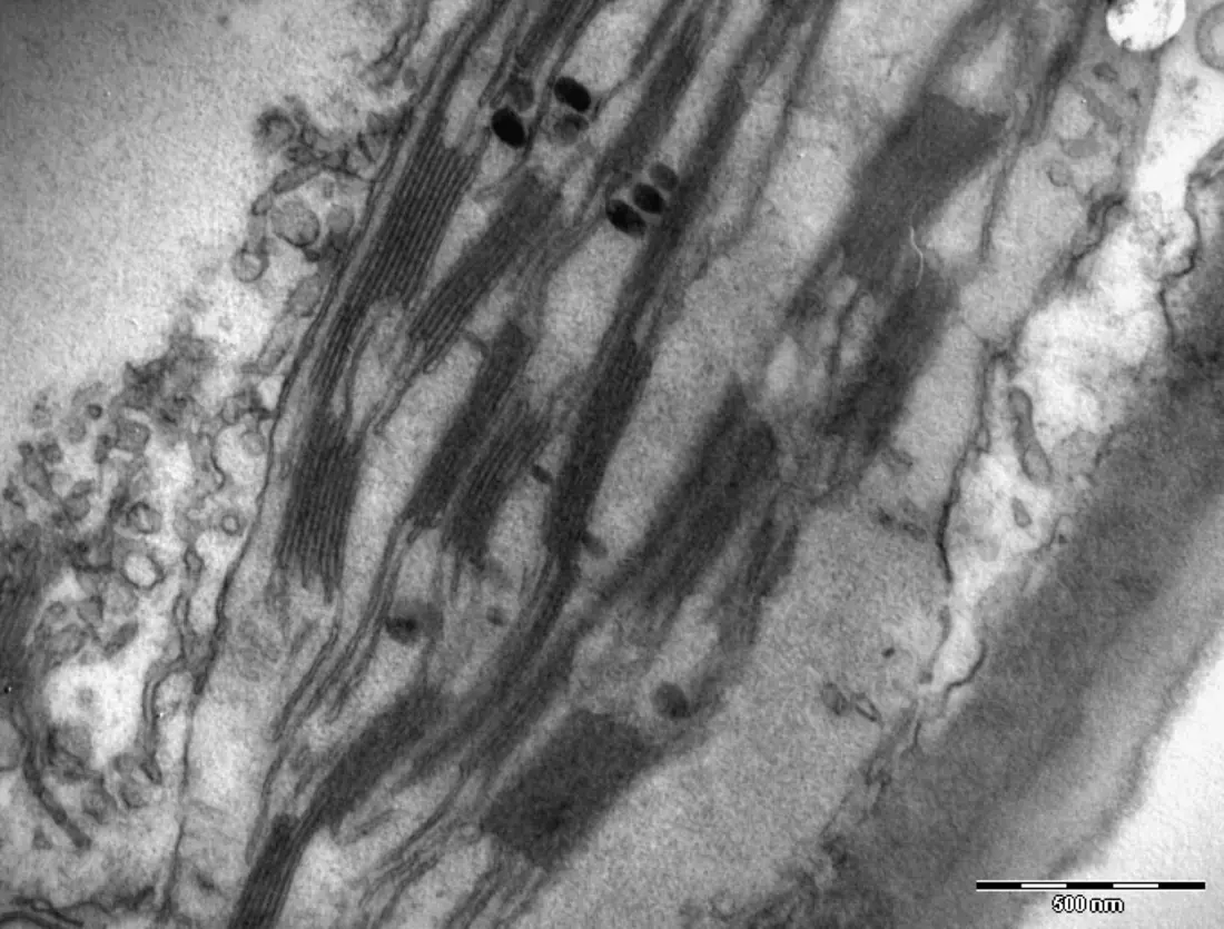
A chloroplast is a lens-shaped organelle 5–10 μm long, also bounded by two membranes. Inside is the stroma, a protein-rich fluid holding the enzymes of the Calvin cycle, starch grains, circular DNA and 70S ribosomes. From the stroma, flat membranous sacs called thylakoids arise; where they are stacked like coins they form a granum, and the unstacked thylakoids that join one granum to the next are the intergranal (stroma) lamellae. The chlorophyll and the photosystems sit in the thylakoid membrane, not in the stroma — a favourite trap. Algal chloroplasts carry a protein body, the pyrenoid, around which starch is laid down.
| Part | Function |
|---|---|
| Thylakoid membrane | light reactions — photosystems I and II, chlorophyll, electron transport |
| Grana | stacks of thylakoids; increase the surface area for light capture |
| Stroma | Calvin cycle (dark reaction); DNA and 70S ribosomes |
| Starch grain | the storage product of photosynthesis |
Endoplasmic reticulum
The ER is a network of membrane-bound tubules and flattened sacs (cisternae) continuous with the outer nuclear membrane.
- Rough ER carries ribosomes on its surface and is the site of protein synthesis and transport — abundant in cells that secrete enzymes (pancreas, salivary gland).
- Smooth ER has no ribosomes. It makes lipids and steroids, detoxifies drugs and poisons (liver), stores Ca²⁺ (sarcoplasmic reticulum of muscle) and carries out glycogen breakdown.
Golgi body
The Golgi body (Golgi apparatus, dictyosome in plants) is a stack of flattened, curved cisternae with swollen margins. It has two faces: the cis (forming) face receives vesicles from the ER, and the trans (maturing) face releases vesicles towards the membrane. Its jobs are to modify, sort, package and secrete — adding sugar chains to proteins (glycosylation), forming lysosomes, producing cell-wall material, and in plant cells laying down the cell plate at cytokinesis.
Lysosomes
A lysosome is a single-membrane vesicle, formed from the Golgi, filled with about 40 hydrolytic (acid hydrolase) enzymes — protease, lipase, nuclease, phosphatase — that work best at pH ≈ 5. They digest material taken in by the cell (heterophagy) and worn-out organelles of the cell itself (autophagy). If the lysosome membrane ruptures, its enzymes digest the cell's own contents; Christian de Duve, who discovered them, therefore called lysosomes the "suicidal bags" of the cell. They are common in animal cells and in fungi; in mature plant cells the large central vacuole does much of the same work.
Ribosomes
A ribosome is a tiny granule of rRNA and protein, with no membrane, first seen by George Palade. It is the site of protein synthesis. The prokaryotic ribosome is 70S, made of a 50S and a 30S subunit; the eukaryotic cytoplasmic ribosome is 80S, made of 60S and 40S subunits. Mitochondria and chloroplasts contain 70S ribosomes — evidence for their bacterial origin. Several ribosomes translating one mRNA form a polysome.
Nucleus
The nucleus is bounded by a double membrane, the nuclear envelope, perforated by nuclear pores that let RNA and proteins pass between nucleus and cytoplasm. Inside are the nucleoplasm, the chromatin (DNA with histones) and one or more nucleoli. The nucleolus is not an organelle with a membrane; it is the site where rRNA is made and ribosomal subunits are assembled. The nucleus holds the genetic material and controls the cell's activities.
Chromosomes
During division the chromatin condenses into chromosomes. A chromosome that has replicated has two identical chromatids joined at the centromere, a narrow region (the primary constriction) that carries the kinetochore to which spindle fibres attach. The ends of each chromatid are telomeres, which protect the DNA from being degraded or fused. Some chromosomes have a satellite beyond a secondary constriction.
The DNA itself is wrapped twice round an octamer of histones — two molecules each of H2A, H2B, H3 and H4 — with about 147 base pairs per turn, forming a nucleosome; the string of nucleosomes is coiled further to make the visible chromosome.
| By centromere position | Shape |
|---|---|
| Metacentric | centromere in the middle; two equal arms |
| Submetacentric | centromere slightly off-centre |
| Acrocentric | centromere near one end |
| Telocentric | centromere at the very end |
Cilia and flagella
Eukaryotic cilia and flagella have the same internal structure: a core, the axoneme, of nine pairs of microtubules arranged in a ring around a central pair — the 9+2 arrangement. The pairs are linked by dynein arms, which slide the microtubules past one another using ATP, so the structure bends. At the base is a basal body with a 9+0 arrangement, identical to a centriole. Cilia are short and numerous; flagella are long and few (the sperm tail, Chlamydomonas).
A bacterial flagellum is completely different: a single fibril of the protein flagellin, with no 9+2 and no membrane, that rotates like a propeller driven by a proton gradient. Do not mix the two.
Cell inclusions
Cell inclusions are non-living materials in the cytoplasm — they are not organelles and have no membrane of their own:
| Inclusion | Where |
|---|---|
| Starch grains | plant cells (storage) |
| Oil droplets and fat globules | seeds, adipose cells |
| Glycogen granules | liver and muscle |
| Calcium oxalate crystals (raphides) | Colocasia, Opuntia, many plant cells |
| Pigment granules | melanin in skin, anthocyanin in vacuoles |
| Vacuoles with cell sap, resins, gums, latex | plant cells |
| Secretory granules | gland cells |
Plant cells and animal cells compared
| Feature | Plant cell | Animal cell |
|---|---|---|
| Cell wall | present, of cellulose | absent |
| Shape | fixed, often angular | variable, rounded |
| Chloroplast | present in green parts | absent |
| Vacuole | one large central vacuole with sap | small and many, or none |
| Centriole | absent in higher plants | present |
| Cytokinesis | cell plate | cleavage furrow |
| Stored food | starch | glycogen |
| Lysosomes | few; the vacuole does much of the work | many |
| Cell junctions | plasmodesmata through the wall | gap junctions, desmosomes |
- Chlorophyll is in the thylakoid membrane, not in the stroma; the stroma holds the Calvin-cycle enzymes.
- The nucleolus makes rRNA, not DNA — DNA is made in the nucleoplasm during S phase.
- Lysosomes are single-membraned; mitochondria and chloroplasts are double-membraned.
- 70S ribosomes occur in prokaryotes, mitochondria and chloroplasts; 80S only in the eukaryotic cytoplasm.
- A plant cell has no centriole in higher plants, yet still forms a spindle — spindle fibres in plants are made without centrioles.
7.3 The cell cycle; amitosis, mitosis and meiosis
The cell cycle
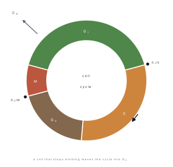
The cell cycle is the sequence of events by which a cell duplicates its contents and divides into two. It has two main phases:
| Phase | What happens | Share of a typical 24 h animal cell |
|---|---|---|
| Interphase | the cell grows and copies its DNA; no visible chromosomes | about 23 h |
| — G₁ | cell grows, makes proteins and organelles; the G₁/S checkpoint decides whether to divide | about 11 h |
| — S | DNA replication; each chromosome becomes two chromatids; histones are made | about 8 h |
| — G₂ | further growth, spindle proteins made; the G₂/M checkpoint checks the copied DNA | about 4 h |
| M phase | mitosis (karyokinesis) followed by cytokinesis | about 1 h |
A cell that leaves the cycle and stops dividing — a mature neuron, a muscle fibre — enters G₀, a resting state from which it may never return. The checkpoints are controlled by cyclins and cyclin-dependent kinases (CDKs); if DNA is damaged at the G₁/S checkpoint the cell arrests and repairs it, or kills itself (apoptosis). Loss of checkpoint control is a step towards cancer.
Amitosis
Amitosis (direct division) is division of the nucleus without spindle formation and without condensation of chromosomes: the nucleus simply constricts and divides into two. It occurs in bacteria (binary fission), in some yeasts and protozoa, and in damaged or ageing cells. It produces no variation and is not a substitute for mitosis in the cells of higher organisms.
Mitosis
Mitosis is the division of a nucleus into two genetically identical daughter nuclei with the same chromosome number. It is one continuous process, described in four stages:
| Stage | What happens |
|---|---|
| Prophase | chromatin condenses into visible chromosomes (each with two chromatids); the nucleolus disappears; spindle fibres begin to form from the poles; the nuclear envelope breaks down at the end of prophase |
| Metaphase | chromosomes line up at the equator (metaphase plate); spindle fibres from opposite poles attach to the kinetochore of each centromere |
| Anaphase | the centromeres split; the two chromatids of each chromosome are pulled to opposite poles, centromere first — so each pole receives one complete set |
| Telophase | chromosomes reach the poles and decondense; the nuclear envelope re-forms; the nucleolus reappears; the spindle disappears |
Cytokinesis follows. In an animal cell the membrane invaginates as a cleavage furrow (a contractile ring of actin and myosin) and pinches the cell in two. In a plant cell the rigid wall prevents pinching, so vesicles from the Golgi line up at the equator and fuse to form a cell plate, which becomes the middle lamella and then the new walls.

Significance of mitosis: it keeps the chromosome number constant (2n → 2n); it produces genetically identical cells, so it is the basis of growth, repair and replacement of tissues (skin, gut lining, blood cells), asexual reproduction (binary fission, budding, cuttings) and the vegetative propagation of plants.
Meiosis
Meiosis is a reduction division: one diploid cell gives four haploid cells after two divisions with only one round of DNA replication.
| Stage | What happens |
|---|---|
| Prophase I (longest) | Leptotene — chromosomes become visible as threads. Zygotene — homologous chromosomes pair up (synapsis) and form a bivalent held by the synaptonemal complex. Pachytene — crossing over between non-sister chromatids; segments are exchanged. Diplotene — the homologues begin to separate and the points of exchange are seen as chiasmata. Diakinesis — chromosomes condense fully, the envelope breaks down, the spindle forms. |
| Metaphase I | bivalents line up at the equator; the two homologues of each pair face opposite poles |
| Anaphase I | the homologues separate and move to opposite poles; centromeres do not divide |
| Telophase I | two haploid cells form (each chromosome still has two chromatids) |
| Meiosis II | like mitosis: sister chromatids separate; centromeres divide; four haploid cells result |
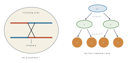
Significance of meiosis: it halves the chromosome number, so that fertilisation restores the diploid number and the species number stays constant across generations; it produces variation through crossing over and independent assortment, which is the raw material of evolution; and it shuffles alleles so that no two gametes are alike.
The two sources of variation. Crossing over (pachytene of prophase I) exchanges segments between non-sister chromatids of homologous chromosomes, producing recombinant chromatids. Independent assortment (metaphase I) is the random orientation of each bivalent: with n pairs, a gamete can receive 2^n different combinations of maternal and paternal chromosomes — 2^{23}, over eight million, in a human.
Solution. After S phase the DNA is doubled but the chromosome number is not. Mitosis metaphase: 8 chromosomes, 16 chromatids, 16 DNA molecules. Metaphase I: 8 chromosomes (4 bivalents), 16 chromatids, 16 DNA molecules. Metaphase II: 4 chromosomes, 8 chromatids, 8 DNA molecules — meiosis I has already halved the chromosome number.
Solution. G₁: 46 chromosomes, 2C. After S: 46 chromosomes, 4C — the DNA has doubled but the centromeres have not. Anaphase of mitosis: 92 chromosomes, 4C, because every centromere has split. After meiosis I: 23 chromosomes, 2C — the number is halved but each chromosome still has two chromatids. Sperm: 23 chromosomes, 1C. The rule is: count centromeres for chromosomes, count DNA molecules for C.
Mitosis and meiosis compared
| Feature | Mitosis | Meiosis |
|---|---|---|
| Divisions | one | two, one after the other |
| DNA replication | once | once |
| Products | 2 cells | 4 cells |
| Chromosome number of products | same as parent (2n → 2n) | half (2n → n) |
| Pairing of homologues (synapsis) | does not occur | occurs in prophase I |
| Crossing over | absent | present (pachytene) |
| Chiasmata | absent | present |
| Metaphase | single chromosomes at the equator | bivalents at the equator in meiosis I |
| Anaphase | centromeres divide, chromatids separate | meiosis I: homologues separate, centromeres do not divide; meiosis II: chromatids separate |
| Genetic result | daughter cells identical to parent | daughter cells genetically different |
| Where | all somatic cells, meristems | reproductive organs: anther, ovule, testis, ovary |
| Role | growth, repair, asexual reproduction | gamete formation, variation, evolution |
- Meiosis II is equational, not reductional. It separates sister chromatids exactly as mitosis does; the reduction happens in meiosis I.
- DNA content and chromosome number are not the same thing. After S phase a human cell still has 46 chromosomes but 92 DNA molecules. At anaphase of mitosis the centromeres split, so the cell briefly has 92 chromosomes.
- Plant cells make a cell plate, animal cells a cleavage furrow. A question about "cytokinesis by a furrow" is about an animal cell.
- In anaphase I the centromeres do not divide — the whole chromosome, with both chromatids, travels to the pole.
- Crossing over happens in pachytene, but the chiasmata that show where it happened are seen in diplotene.
Problem. How many mitotic divisions are required to produce 64 cells from a single meristematic cell, and how many total meiotic divisions are required to form 100 wheat grains (2n = 42)?
Solution. Each mitotic division adds one cell to the count (N - 1 total mitoses, in \log_2 64 = 6 generations): 63 mitotic divisions. For n seeds in an angiosperm, 1 microspore mother cell makes 4 pollen grains (n/4 meioses) and 1 megaspore mother cell makes 1 functional egg (n meioses), so n + n/4 = \frac{5}{4}n = \frac{5}{4}(100) = \mathbf{125\text{ meiotic divisions}}.
Formula and fact sheet
| Item | Fact |
|---|---|
| Cell discovered | Robert Hooke, 1665, in cork |
| First living cells | Leeuwenhoek, 1674 |
| Nucleus | Robert Brown, 1831 |
| Cell theory | Schleiden (1838, plants) + Schwann (1839, animals) |
| Omnis cellula e cellula | Virchow, 1855 |
| Exceptions | viruses, viroids, prions (acellular); coenocytic hyphae (Mucor); multinucleate muscle fibres |
| Prokaryote ribosome | 70S = 50S + 30S |
| Eukaryote ribosome | 80S = 60S + 40S; 70S in mitochondria and chloroplasts |
| Prokaryote wall / plant wall / fungal wall | peptidoglycan / cellulose / chitin |
| Cell wall layers | middle lamella (calcium pectate) → primary (cellulose) → secondary (cellulose + lignin) |
| Membrane model | fluid mosaic, Singer and Nicolson, 1972; bilayer about 7.5 nm |
| Mitochondrion | double membrane, cristae, matrix, own circular DNA, 70S ribosomes, F₁ particles; maternal inheritance |
| Chloroplast | double membrane, stroma, thylakoids, grana, intergranal lamellae, own DNA, 70S ribosomes; chlorophyll in the thylakoid membrane |
| Rough ER | ribosomes, protein synthesis and transport |
| Smooth ER | lipid and steroid synthesis, detoxification, Ca²⁺ store |
| Golgi | cis (forming) and trans (maturing) faces; secretion, glycosylation, lysosome and cell-plate formation |
| Lysosome | single membrane, acid hydrolases, pH ≈ 5; "suicidal bag" (de Duve) |
| Nucleolus | rRNA synthesis and ribosome assembly |
| Nucleosome | 147 bp DNA round a histone octamer (2 each of H2A, H2B, H3, H4) |
| Chromosome parts | chromatid, centromere (kinetochore), telomere, satellite |
| Chromosome types | metacentric, submetacentric, acrocentric, telocentric |
| 9+2 | eukaryotic cilia and flagella; basal body 9+0; dynein + ATP |
| Bacterial flagellum | flagellin, no 9+2, rotates |
| Bacterial surface | capsule, peptidoglycan wall, plasma membrane, mesosome, pili, flagellum |
| Organelle membranes | double: nucleus, mitochondrion, chloroplast; single: ER, Golgi, lysosome, vacuole, peroxisome; none: ribosome, centriole |
| Cell inclusions | starch, oil, glycogen, calcium oxalate crystals, pigments, vacuolar sap |
| Cell cycle | G₁ → S → G₂ → M; DNA replicates in S; G₀ = resting |
| Cell-cycle duration | human cell about 24 h: G₁ 11, S 8, G₂ 4, M 1 |
| Checkpoints | G₁/S (restriction point), G₂/M, spindle assembly; cyclins and CDKs |
| Mitosis stages | prophase, metaphase, anaphase, telophase |
| Cytokinesis | animal: cleavage furrow; plant: cell plate from Golgi vesicles |
| Meiosis prophase I | leptotene, zygotene (synapsis), pachytene (crossing over), diplotene (chiasmata), diakinesis |
| Meiosis products | 4 haploid cells from one diploid cell |
| Variation | crossing over (prophase I) + independent assortment (metaphase I), 2^n combinations |
| Amitosis | direct division, no spindle, no chromosome condensation; bacteria |
| Human numbers | somatic 46 (2n), gamete 23 (n) |
Chapter summary
- The cell theory (Schleiden 1838, Schwann 1839, Virchow 1855) has exceptions: viruses are acellular and coenocytic hyphae are multinucleate without cross walls.
- Prokaryotes have no nucleus or membrane-bound organelles, 70S ribosomes, a peptidoglycan wall and binary fission; eukaryotes have a nucleus, 80S cytoplasmic ribosomes and mitosis.
- The plant wall is middle lamella (calcium pectate), primary wall (cellulose) and secondary wall (cellulose plus lignin); plasmodesmata connect neighbouring cells.
- The plasma membrane is a fluid mosaic of phospholipid bilayer and proteins (Singer and Nicolson, 1972), selectively permeable.
- Mitochondria (cristae) and chloroplasts (grana, thylakoids, stroma) are double-membraned and semi-autonomous, with their own DNA and 70S ribosomes; chlorophyll sits in the thylakoid membrane.
- Lysosomes are single-membraned bags of acid hydrolases — the "suicidal bags"; the Golgi packages and secretes and forms the cell plate.
- Chromatin is DNA wound round histone octamers into nucleosomes; a replicated chromosome has two chromatids joined at the centromere, with telomeres at the ends.
- Eukaryotic cilia and flagella are 9+2 microtubules moved by dynein and ATP; bacterial flagella are flagellin and rotate.
- The cell cycle is G₁, S, G₂, M with checkpoints; DNA replicates only in S, so chromosome number and DNA content must be counted separately.
- Mitosis gives two identical diploid cells (growth, repair); meiosis gives four haploid cells in two divisions, with crossing over in pachytene and independent assortment at metaphase I providing the variation.
Practice questions
Set A — Recall (30 questions)
- The cell theory was proposed byaHooke and LeeuwenhoekbSchleiden and SchwanncVirchow and PasteurdBrown and Purkinje
- "Omnis cellula e cellula" — every cell arises from a pre-existing cell — was stated byaSchwannbSchleidencHookedVirchow
- The cell was first discovered byaRobert Hooke in corkbLeeuwenhoek in pond watercRobert Brown in orchiddSchwann in cartilage
- Which of the following is absent from a prokaryotic cell?aribosomesbplasma membranecmembrane-bound nucleusdcell wall
- The ribosomes of a prokaryotic cell are of the typea80Sb60Sc40Sd70S
- The genetic material of a bacterium lies in theanucleoidbnucleuscmitochondriondchloroplast
- The middle lamella of a plant cell wall is made mainly ofacellulosebligninccalcium pectatedchitin
- The fluid-mosaic model of the plasma membrane was proposed byaDanielli and DavsonbSinger and NicolsoncRobertsondPalade
- The inner membrane of a mitochondrion is folded intoacristaebgranacthylakoidsdcisternae
- A stack of thylakoids in a chloroplast is called aacristabstromaccisternadgranum
- Rough endoplasmic reticulum differs from smooth ER in bearingaDNAbribosomescchlorophylldhydrolytic enzymes
- The organelle that modifies, packages and secretes cell products is thealysosomebribosomecGolgi bodydperoxisome
- Lysosomes are called "suicidal bags" because theyacontain DNAbmake ATPcdigest fooddhold enzymes that can digest the cell's own contents
- The 80S ribosome of a eukaryote is made of the subunitsa60S and 40Sb50S and 30Sc40S and 30Sd60S and 50S
- The nucleolus is the site ofaDNA replicationbrRNA synthesis and ribosome assemblycATP synthesisdprotein digestion
- In a nucleosome, DNA is wound aroundaa single histonebnon-histone protein onlycan octamer of histonesdRNA
- The region at which the two chromatids of a chromosome are joined is theacentromerebtelomerecchiasmadsatellite
- The 9+2 arrangement of microtubules is found inabacterial flagellabprokaryotic ciliaccentriolesdeukaryotic cilia and flagella
- Which of these is a non-living cell inclusion?amitochondrionbribosomeccalcium oxalate crystaldGolgi body
- The longest phase of the cell cycle isaM phasebinterphasecanaphasedtelophase
- DNA replicates duringaG₁ phasebG₂ phasecM phasedS phase
- The stage of mitosis in which chromosomes line up at the equator isametaphasebanaphasecprophasedtelophase
- In plant cells, cytokinesis takes place by formation of aacleavage furrowbcontractile ringccell platedchitin septum
- Synapsis and crossing over occur inamitosisbprophase I of meiosiscprophase II of meiosisdanaphase I
- The chromosome number is halved duringameiosis Ibmeiosis IIcmitosisdS phase
- Meiosis II is called equational because itahalves the chromosome numberbdoubles the DNAcpairs the homologuesdseparates sister chromatids without changing the number
- Direct division of the nucleus without spindle formation isamitosisbamitosiscmeiosisdendomitosis
- Chiasmata are first clearly visible inaleptotenebzygotenecdiploteneddiakinesis
- Colchicine arrests a dividing cell ataprophasebanaphasectelophasedmetaphase
- A human somatic cell has 46 chromosomes. The number in a gamete isa23b46c92d12
Answers and explanations — Set A
1. (b) — Schleiden (plants, 1838) and Schwann (animals, 1839) gave the cell theory; Virchow added the third tenet later.
2. (d) — Virchow (1855) stated that new cells come only from existing cells. Schwann and Schleiden believed cells formed from a shapeless matrix.
3. (a) — Hooke saw the empty cell walls of cork in 1665 and named them cells; Leeuwenhoek first saw living cells nine years later.
4. (c) — Prokaryotes have no nuclear envelope; the DNA lies free in the nucleoid. They do have ribosomes, a membrane and usually a wall.
5. (d) — 70S (50S + 30S). 80S is the eukaryotic cytoplasmic ribosome.
6. (a) — The single circular chromosome lies in the nucleoid region, not in a nucleus. Plasmids are separate small circles.
7. (c) — Calcium pectate cements adjacent cells; pectinase digests it when fruit ripens and softens.
8. (b) — Singer and Nicolson (1972). Danielli and Davson had proposed the earlier sandwich model.
9. (a) — Cristae increase the area of the inner membrane, where the electron transport chain and ATP synthase sit.
10. (d) — A granum is a stack of thylakoids; the stroma is the fluid around them.
11. (b) — Rough ER carries ribosomes and makes protein; smooth ER makes lipid and steroid.
12. (c) — The Golgi modifies, sorts and packages; the lysosome only digests.
13. (d) — The hydrolytic enzymes, if released, digest the cell itself. The membrane normally keeps them safely inside.
14. (a) — 80S = 60S + 40S. The 50S + 30S pair makes the 70S ribosome of prokaryotes.
15. (b) — The nucleolus makes rRNA and assembles ribosomal subunits; DNA replication happens in the nucleoplasm.
16. (c) — About 147 bp of DNA wind round two each of H2A, H2B, H3 and H4.
17. (a) — The centromere carries the kinetochore for the spindle; the telomeres are the ends.
18. (d) — Nine doublets around a central pair, with dynein arms. Bacterial flagella are flagellin and have no 9+2.
19. (c) — Crystals are ergastic, non-living inclusions. Mitochondria, ribosomes and the Golgi are organelles.
20. (b) — Interphase takes about 23 h of a 24 h cycle; M phase about 1 h.
21. (d) — S phase is the synthesis phase; each chromosome becomes two chromatids.
22. (a) — At metaphase the chromosomes lie on the metaphase plate with spindle fibres attached to their kinetochores.
23. (c) — Golgi vesicles fuse at the equator to make a cell plate, which becomes the middle lamella.
24. (b) — Synapsis and crossing over are confined to prophase I; meiosis II has no pairing.
25. (a) — Homologues separate in anaphase I, so each daughter nucleus gets n chromosomes.
26. (d) — Meiosis II separates chromatids, like mitosis, and leaves the chromosome number unchanged.
27. (b) — Amitosis is direct division, seen in bacteria and some protozoa; no spindle or condensed chromosomes are involved.
28. (c) — Crossing over happens in pachytene but the chiasmata become visible as the homologues pull apart in diplotene.
29. (d) — Colchicine blocks spindle formation, so chromosomes cannot move to the poles and the cell arrests in metaphase — the basis of karyotyping.
30. (a) — Meiosis halves 46 to 23; fertilisation restores 46.
Set B — Understanding (18 questions)
- Viruses are an exception to the cell theory because theyahave a nucleusbare prokaryotescare acellular and have no celldlack nucleic acid
- A coenocytic hypha of Mucor is an exception because itais one mass of cytoplasm with many nuclei and no cross wallsbhas no nucleuscis a virusdhas a cellulose wall
- A plant cell placed in pure water does not burst becauseait has no membranebit has a contractile vacuolecit lacks a cell walldthe rigid wall resists the turgor pressure
- Mitochondria and chloroplasts are called semi-autonomous because theyalack DNAbhave their own DNA and 70S ribosomes but depend on nuclear genescdivide only with the nucleusdhave 80S ribosomes
- The lysosome membrane is important because itakeeps the hydrolytic enzymes away from the cytoplasmbmakes ATPcsynthesises proteindcontains DNA
- A cell that secretes large amounts of digestive enzyme would have manyachloroplastsblysosomescrough ER and Golgi bodiesdcilia
- The nuclear pore allowsafree passage of ribosomesbexchange of RNA and proteins between nucleus and cytoplasmcATP synthesisdDNA replication
- During anaphase of mitosis, a human cell containsa23 chromosomesb46 chromosomesc46 pairs of chromosomesd92 chromosomes
- Meiosis is important for evolution mainly because itaproduces new gene combinations by crossing over and independent assortmentbdoubles the chromosome numbercmakes identical cellsdrepairs tissues
- Mitosis in a haploid cell producesahaploid gametes onlybdiploid cellsctwo haploid cells identical to the parentdfour haploid cells
- Spindle fibres attach to a chromosome at theatelomerebsatellitecchiasmadkinetochore of the centromere
- In a cell with 2n = 8, the number of bivalents at metaphase I isa8b4c16d2
- A cell in G₀ahas left the cycle and is not dividingbis in S phasechas no DNAdis undergoing meiosis
- Colchicine-treated cells show a doubled chromosome number because colchicineabreaks the DNAbprevents DNA replicationcprevents spindle formation, so the nucleus re-forms undividedddissolves the cell wall
- The significance of crossing over is that itahalves the chromosome numberbexchanges segments between non-sister chromatidscseparates sister chromatidsddoubles the DNA
- A plant cell differs from an animal cell at cytokinesis because the plant cellahas no spindlebdivides by binary fissioncforms a cleavage furrowdforms a cell plate from Golgi vesicles
- The enzyme used to separate cells by digesting the middle lamella isapectinasebcellulasecligasedamylase
- If the DNA content of a cell in G₁ is 2C, the content at metaphase of mitosis isa2CbCc4Cd8C
Answers and explanations — Set B
1. (c) — Viruses have no cell, cytoplasm or ribosomes; they are obligate parasites and cannot reproduce alone.
2. (a) — The aseptate hypha is a continuous cytoplasm with many nuclei, so "one cell, one nucleus" fails.
3. (d) — The wall resists the pressure; an animal cell without a wall would swell and lyse.
4. (b) — They carry their own circular DNA and 70S ribosomes, yet most of their proteins are coded by nuclear genes.
5. (a) — The membrane isolates the acid hydrolases; if it leaks, the cell digests itself.
6. (c) — Secretory proteins are made on rough ER and packed by the Golgi. Chloroplasts and cilia have nothing to do with secretion.
7. (b) — mRNA leaves and ribosomal proteins enter through the pore; ribosomes are assembled in the cytoplasm, not exported whole.
8. (d) — The centromeres split, so 46 chromosomes become 92 daughter chromosomes before cytokinesis.
9. (a) — Crossing over and independent assortment generate new combinations; mitosis makes identical cells.
10. (c) — Mitosis conserves the number, so a haploid parent gives two haploid daughters.
11. (d) — The kinetochore, a protein plate on the centromere, is the attachment point.
12. (b) — Eight chromosomes make four homologous pairs, hence four bivalents.
13. (a) — G₀ is the resting state outside the cycle; the DNA is intact but not being replicated.
14. (c) — Without a spindle the chromosomes cannot separate, so the nuclear envelope re-forms around a doubled set.
15. (b) — Non-sister chromatids of homologous chromosomes exchange segments, producing recombinant chromatids.
16. (d) — Golgi vesicles line up at the equator and fuse into a cell plate; the animal cell pinches in.
17. (a) — Pectinase digests the calcium pectate of the middle lamella, releasing single cells — the basis of protoplast and tissue-culture work.
18. (c) — DNA doubles in S phase, so a 2C cell reaches 4C by metaphase, though the chromosome number is unchanged.
Set C — Application (12 questions)
- A human cell with 46 chromosomes completes meiosis. The number of cells and the chromosome number in each area2 and 46b4 and 23c4 and 46d2 and 23
- A cell has 2n = 24. At anaphase of mitosis, the number of chromosomes in the cell isa12b24c36d48
- An organism has 2n = 6. Ignoring crossing over, how many different combinations of maternal and paternal chromosomes can a gamete receive?a3b6c8d64
- A cell in G₁ has 10 chromosomes and 10 DNA molecules. At metaphase I of meiosis it hasa10 chromosomes and 20 DNA moleculesb20 chromosomes and 20 DNA moleculesc10 chromosomes and 10 DNA moleculesd5 chromosomes and 10 DNA molecules
- After meiosis I, each daughter of a 2n = 10 cell hasa10 chromosomes and 10 DNA moleculesb5 chromosomes and 5 DNA moleculesc10 chromosomes and 20 DNA moleculesd5 chromosomes and 10 DNA molecules
- A drug blocks formation of the cell plate. Which cells are affected?aanimal cells onlybplant cells onlycbacterial cells onlydboth plant and animal cells
- An onion root-tip cell has 2n = 16. At anaphase of mitosis it containsa8 chromosomesb16 chromosomesc32 chromosomesd64 chromosomes
- Two genes lie on different chromosomes. The gametes of a heterozygote AaBb are produced in the ratioa1 : 1 : 1 : 1b3 : 1c9 : 3 : 3 : 1d1 : 2 : 1
- A cell has 2n = 46. After meiosis II, the DNA content of each cell compared with G₁ isathe samebhalfcdoubledfour times
- A moss gametophyte has 12 chromosomes in each cell. The sporophyte cells havea6b12c48d24
- Crossing over occurs between two linked genes in 20 % of meioses. The recombination frequency isa10 %b20 %c40 %d5 %
- A cell with 2n = 8 undergoes meiosis. The number of chromatids per cell at metaphase II isa4b16c8d2
Answers and explanations — Set C
1. (b) — Two divisions give four cells, and the chromosome number is halved to 23.
2. (d) — At anaphase the 24 centromeres split, giving 48 daughter chromosomes in the cell before cytokinesis.
3. (c) — 2^n with n = 3 gives 8 combinations.
4. (a) — After S phase the DNA is doubled (20 molecules) but the chromosome number is still 10; the 10 chromosomes form 5 bivalents.
5. (d) — Meiosis I halves the chromosome number to 5, but each chromosome still has two chromatids, so 10 DNA molecules.
6. (b) — Only walled cells build a cell plate; animal cells use a cleavage furrow.
7. (c) — The 16 centromeres split, so 32 daughter chromosomes are present at anaphase.
8. (a) — Independent assortment of two unlinked pairs gives four gamete types in equal numbers.
9. (b) — Meiosis halves the DNA: a 2C cell yields 1C gametes.
10. (d) — The sporophyte is diploid, so 2 × 12 = 24.
11. (a) — Each meiosis with a crossover yields 2 recombinant chromatids out of 4, so 20 % × ½ = 10 %.
12. (c) — After meiosis I each cell has 4 chromosomes, each with 2 chromatids: 8 chromatids at metaphase II.